![]()
![]()
![]()
Use LEFT and RIGHT arrow keys to navigate between flashcards;
Use UP and DOWN arrow keys to flip the card;
H to show hint;
A reads text to speech;
33 Cards in this Set
- Front
- Back
|
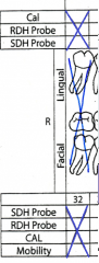
What method of charting do we use? |
Anatomical Charting, same form as Periodontal Assessment |
|
|
What color are existing conditions charted in? What is charted? |
|
|
|
What Color is pathology diagnosed by the dentist charted in? What pathology is charted? |
|
|
|
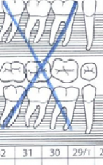
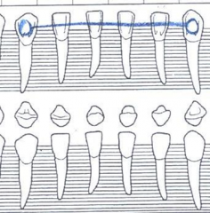
How are missing teeth charted? |
|
|
|
How are multiple missing teeth charted? |
|
|
|
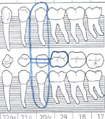
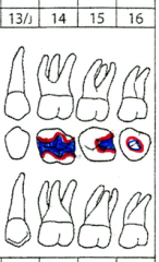
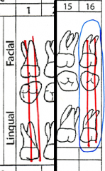
How are impacted teeth charted?
|
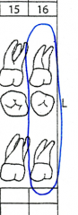
Impacted teeth are charged by drawing a blue circle around all aspects of the tooth
Fully imparted teeth have not erupted through the bone or tissue- only detectable in radiographs |
|
|
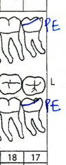
How are partially erupted teeth charted? |
|
|
|
How big are restorations? what are they made of? How are restorations found? |
|
|
|
Blacks classification of Caries: I |
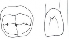
Pits and Fissures |
|
|
Blacks classification of Caries: II |
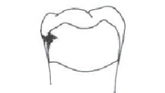
Interproximal (posterior) |
|
|
Blacks classification of Caries: III |
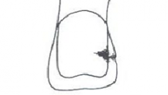
Interproximal (anterior)
Does Not Involve Incisal Edge |
|
|
Blacks classification of Caries: IV |
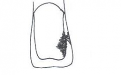
Interproximal (anterior)
Involves Incisal Edge |
|
|
Blacks classification of Caries: V |
Cirvical Third of Crown |
|
|
Blacks classification of Caries: VI |

Cusp Tips |
|
|
How do you chart Amalgam fillings? Composite? Gold onlays or inlays? |
|
|
|
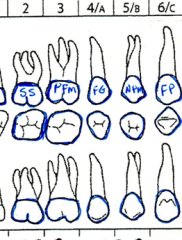
How are sealants charted? |
|
|
|
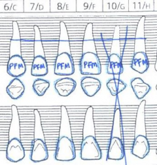
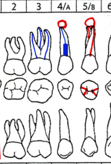
How are crowns charted? |
|
|
|
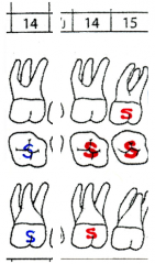
How are bridges charted? |
|
|
|
How are Maryland Bridges charted? |
|
|
|
How are Dentures Charted? |
|
|
|
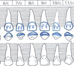
How are Porcelain Veneers charted? |
|
|
|
How are implants charted? |
|
|
|
How are space maintainers charted? |
|
|
|
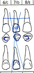
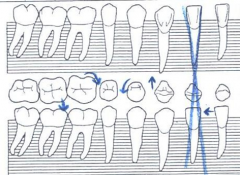
How are malposed teeth charted? |
|
|
|
How is a lingual bar charted? |
|
|
|
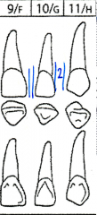
How are Open contacts and Diastemas charted? |
|
|
|
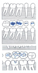
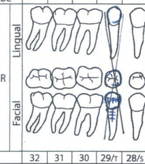
How are Carious Lesions charted on Molars? |
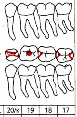
Dot on occlusal for occlusal only
|
|
|
How are recurrent caries charted? |
|
|
|
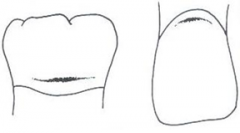
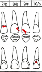
How are anterior caries or fracture Charted? |
|
|
|
How are endodontic treatments, Post and core, and lesions charted? |
|
|
|
How are extractions charted? |
|
|
|
How is attrition charted? |
|
|
|
Who's job is it to make sure charting is done accurately? |
|