Reading...
![]()
Play button
![]()
Play button
![]()
Use LEFT and RIGHT arrow keys to navigate between flashcards;
Use UP and DOWN arrow keys to flip the card;
H to show hint;
A reads text to speech;
78 Cards in this Set
- Front
- Back

Identify the structure labeled "A"
|
supraglenoid tubercle (tuberosity) of the scapula
|
|
|
What muscle originates from the supraglenoid tuberosity?
|
biceps brachii
|
|
|
Name one clinically relevant reason for knowing the supraglenoid tuberosity.
|
biceps tendinopathy in large breed dogs
|
|
|
What muscle lies parallel and immediately adjacent and cranial to the scapular spine?
|
infraspinatus m.
|
|
|
What is the innervation of the infraspinatus muscle?
|
supraspinous nerve
|
|
|
Where does the infraspinatus muscle insert?
|
lateral aspect of humeral head
|
|
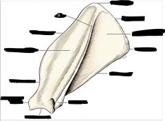
Identify the prominence labeled "B"
|
scapular spine
|
|
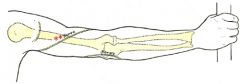
What nerve is labeled with two red asterisks?
|
radial nerve
|
|
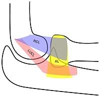
The structures in blue and pink make up what structure?
|
the lateral collateral ligament of the elbow joint
|
|
|
What group of muscles has been reflected to reveal the lateral collateral ligament of the elbow?
|
forearm extensors
|
|
|
What is the insertion point(s) for the lateral collateral ligament of the elbow?
|
radial head [cranial crura] and adjacent ulna [caudal crura]
|
|
|
Name the specific muscles in the forearm extensors that are reflected to reveal the lateral collateral ligament of the elbow.
|
extensor carpi radialis, common digital extensor, lateral digital extensor, ulnaris lateralis [aka – extensor carpi ulnaris]
|
|
|
Name a surgical procedure where you would incise and reflect the anconeus muscle
|
removal of ununited anconeal process
|
|
|
What is the function of the extensor carpi radialis?
|
extend the carpus
|
|
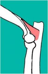
What is the name of this muscle?
|
anconeus muscle
|
|
|
What tendon is obliquely crossing the extensor carpi radialis?
|
abductor pollicis longus
|
|
|
What is the insertion of the superficial gluteal muscle?
|
third trochanter
|
|
|
What is the function of the superficial gluteal muscle?
|
abduction of hip
|
|
|
What is the function of the semitendinosus muscle?
|
extension of hip, flexion of stifle
|
|
|
What is the innervation of the semitendinosus muscle?
|
sciatic nerve
|
|
|
What course does the sciatic nerve take from pelvis to stifle?
|
It crosses the pelvis at the lesser ischiatic notch caudal to acetabulum and courses caudally down the thigh until the tibial and peroneal branches split at the level of the stifle. The tibial branch continues caudolateral and the peroneal branch continues craniolateral.
|
|
|
Where is the origin of the pectineus muscle?
|
prepubic tendon and pubic bone
|
|
|
What vascular structure lies adjacent to the pectineus muscle?
|
femoral artery
|
|
|
What is the function of the long digital extensor tendon?
|
extension of the digits
|
|
|
What muscle does the long digital extensor tendon run beneath?
|
cranial tibial muscle
|
|
|
What is the innervation of the cranial tibial muscle?
|
peroneal nerve
|
|
|
What muscle group is associated with the patellar tendon?
|
quadriceps muscle group
|
|
|
What is the innervation of the quadriceps muscle group?
|
femoral nerve
|
|
|
Where do each of the muscles in the quadriceps group originate?
|
rectus femoris – rectus tuberosity cranial to acetabulum, v. lateralis – base of greater trochanter, v. medialis – base of femoral neck and intertrochanteric ridge, v. intermedius – lateral part of proximal femure adjacent to v. lateralis.
|
|
|
What nervous structure runs in close proximity to the gastrocnemius fabellae?
|
peroneal nerve
|
|
|
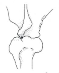
What is the function of the cranial cruciate ligament?
|
prevents excessive internal rotation, hyperextension, and cranial translocation of the tibia
|
|
|
What is the clinical significance of the medial saphenous vein, the cranial saphenous artery, and the cutaneous branch of saphenous nerve?
|
encountered in medial approach to tibia
|
|
|
What inserts on the greater tubercle of the humerus?
|
supraspinatus muscle
|
|
|
What is the medial portion of the humeral condyle called?
|
the trochlea
|
|
|
What role in elbow stability does the anconeal process play?
|
stabilizes elbow in extension
|
|
|
What muscle inserts on the olecranon process?
|
triceps brachii muscle
|
|
|
Is the medial coronoid process larger or smaller than the lateral coronoid process?
|
larger
|
|
|
What maintains the radial head in proper alignment to the ulna?
|
the annular ligament
|
|
|
What ligamentous structure attaches to the ulnar styloid process?
|
ulnar collateral ligament
|
|
|
What is the largest carpal bone?
|
radial carpal bone
|
|
|
What is the 2nd largest carpal bone?
|
ulnar carpal bone
|
|
|
Why is the ungual crest of clinical significance?
|
growth of claw occurs from germinal cells in crest
|
|
|
What common clinical procedure in cats occurs at the distal interphalangeal joint?
|
disarticulation of the joint [declawing]
|
|
|
What is the wing of the ilium a good source of?
|
cancellous bone for grafting
|
|
|
What inserts on the pubis?
|
the prepubic tendon
|
|
|
What muscle originates from the ischiatic tuberosity?
|
semitendinosus muscle
|
|
|
What type of tissue lines the lunate surface (concave surface) of the acetabulum?
|
articular cartilage
|
|
|
What inserts on the lesser trochanter?
|
iliopsoas muscle
|
|
|
What originates at the base of the greater trochanter?
|
the vastus lateralis
|
|
|
What ligament attaches the femur to the acetabulum?
|
ligament of the head of the femur
|
|
|
What muscles insert at the linea aspera?
|
adductor magnus and brevis
|
|
|
What is the purpose of the trochlear ridges in regards to the patella?
|
maintain alignment of the patella
|
|
|
What inserts on the fibular head?
|
lateral collateral ligament of the stifle
|
|
|
What originates from the tibial malleolus?
|
medial collateral ligament of the hock
|
|
|
What developmental orthopedic disease occurs at the trochlea of the femur?
|
OCD (osteochondrosis dessicans)
|
|
|
What muscles insert on the calcaneus?
|
gracilis, semitendinosus, biceps femoris, and gastrocnemius
|
|
|
What is the proper term for the joint between 4th tarsal bone and the talus?
|
taloquartal joint
|
|
|
Where is the insertion of the superficial digital flexor?
|
proximal interphalangeal [PIP] joint
|
|
|
Determine the spatial relationship of the greater tubercle to the acromion.
|
greater tubercle craniodistal to acromion
|
|
|
Determine the spatial relationship of the radial head to the lateral humeral epicondyle.
|
distal and in same parasagittal plane
|
|
|
Determine the spatial relationship of the gastrocnemius fabellae to the patella to the tibial crest.
|
fabellae caudal to patella and proximocaudal to tibial crest
|
|
|
Determine the spatial relationship of the iliac crest to the ischiatic tuberosity to the greater trochanter.
|
shallow “V” with line from iliac crest to trochanter longer than line from trochanter to ischiatic tuberosity
|
|
|
Determine the normal range of motion and start and stop points for each of the following joints: (Flexion: Extension)
SHOULDER |
Shoulder (60-70/65-75)
|
|
|
Determine the normal range of motion and start and stop points for each of the following joints: (Flexion: Extension)
ELBOW |
Elbow (70-75/70-75)
|
|
|
Determine the normal range of motion and start and stop points for each of the following joints: (Flexion: Extension)
CARPUS |
Carpus (155-160/20-30)
|
|
|
Determine the normal range of motion and start and stop points for each of the following joints: (Flexion: Extension)
Metacarpophalangeal |
Metacarpophalangeal (35/85)
|
|
|
Determine the normal range of motion and start and stop points for each of the following joints: (Flexion: Extension)
PIP/DIP |
PIP/DIP (45/45)
|
|
|
Determine the normal range of motion and start and stop points for each of the following joints: (Flexion: Extension)
HIP |
Hip (70-80/80-90)
|
|
|
Determine the normal range of motion and start and stop points for each of the following joints: (Flexion: Extension)
STIFLE |
Stifle (65-75/65-75)
|
|
|
Determine the normal range of motion and start and stop points for each of the following joints: (Flexion: Extension)
HOCK |
Hock (90-110/65-75)
|
|
|
Determine the normal range of motion and start and stop points for each of the following joints: (Flexion: Extension)
Metatarsophalangeal |
Metatarsophalangeal (35/85)
|
|
|
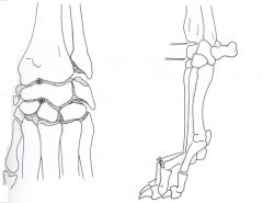
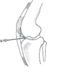
What landmarks would you use for proper needle placement for arthrocentesis of the carpus?
|
|
|
|
What landmarks would you use for proper needle placement for arthrocentesis of the elbow?
|
|
|
|
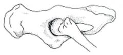
What landmarks would you use for proper needle placement for arthrocentesis of the hip?
|
|
|
|
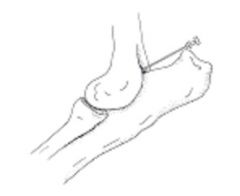
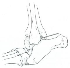
What landmarks would you use for proper needle placement for arthrocentesis of the hock?
|
|
|
|
What landmarks would you use for proper needle placement for arthrocentesis of the hock?
|
|
|
|
What landmarks would you use for proper needle placement for arthrocentesis of the stifle?
|
|
|
|
What landmarks would you use for proper needle placement for arthrocentesis of the shoulder?
|

|