![]()
![]()
![]()
Use LEFT and RIGHT arrow keys to navigate between flashcards;
Use UP and DOWN arrow keys to flip the card;
H to show hint;
A reads text to speech;
22 Cards in this Set
- Front
- Back
|
3 fluid compartments |
interstitial (between cells)
intravascular
intracellular (inside cells) |
|
|
Edema definition |
an accumulation of excess fluid in either the interstitial space or the body cavities |
|
|
2 sites of fluid imbalance
Which one manifests as edema? |
1. Imbalance between the intracellular and interstitial compartments (leads to cell shrinking or swelling)
2. Imbalance between the interstitial and intravascular compartments. Usually manifests as edema. |
|
|
Forces that move fluids between blood vessels and extravascular tissue |
hydrostatic pressure - fluid is forced out of blood and into tissues
intravascular osmotic pressure - plasma proteins pull fluid back into bloodstream
lymphatic absorption - excess interstitial fluid is absorbed and returned to systemic circulation via a large vein |
|
|
Causes of Edema (4) |
1. Increased Vascular Permeability 2. Increased Intravascular Hydrostatic Pressure 3. Decreased Plasma Osmotic Pressure 4. Decreased Lymphatic Drainage |
|
|
mechanism and edema location of Increased Vascular Permeability |
- general mechanism is endothelial reaction to inflammation. - many inflammatory mediators are vasoactive. This causes endothelial cell contraction, creating wider inter-endothelial gaps. This increases vessel permeability and allows fluid leakage - can be localized or generalized |
|
|
mechanism and edema location of increased intravascular hydrostatic pressure edema |
- mechanism is increased blood in the microcirculation. Due to congestion of blood either by venous obstruction or heart failure - venous obstruction = localized edema - heart failure = generalized edema |
|
|
mechanism and edema location of decreased plasma osmotic pressure edema |
- mechanism is decreased albumin in the circulation. albumin is the most abundant plasma protein and when it's lacking there's not a lot of "pull" to bring fluid back into bloodstream - albumin shortage either due to reduced synthesis in the liver caused by malnutrition, liver disease, or intestinal malabsorption of protein; or because of increased albumin loss from the gut, kidney, or skin - generalized only, never localized |
|
|
mechanism and edema location of decreased lymphatic drainage causing edema |
- mechanism is blockage or compression of lymphatic vessels. - compression from tumors, nearby inflammatory swelling - blockage by lymphatic vessel inflammation and fibrosis, tumours, or clots - localized |
|
|
appearance of edema fluid |
clear or slightly yellow fluid with a small amount of protein (transudate) |
|
|
gross appearance of edema in the interstitium, lung, peritoneal cavity, and pleural cavity |
interstitium - gelatinous expansion of organ. If in subcutis there can be "pitting edema" lung - pulmonary edema. lungs are heavy, wet, ooze fluid when squeezed peritoneal cavity - ascites or hydroperitoneum pleural cavity - hydrothorax |
|
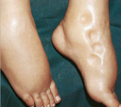
what kind of edema is this? |
pitting edema |
|
what kind of edema is this? how can you tell? |
subcutaneous edema.
has gelatinous expansion of the subcutaneous tissues/subcutis. |
|
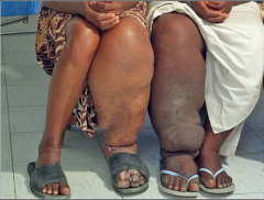
which one of the 4 causes of edema made this? |
decreased lymphatic drainage.
Filarial parasites from mosquitoes damage the lymphatic system so it can't drain properly. Causes what's called "lymphatic filariasis" aka elephantiasis. |
|
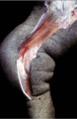
what kind of edema is this? |
pulmonary edema |
|
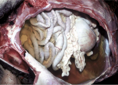
what's does this dog have? (had?) |
Ascites
- slightly yellow fluid (transudate) in the peritoneal cavity. |
|
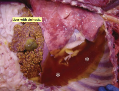
what does this dog have? |
hydrothorax.
yellow transudate in the pleural cavity. |
|
|
Hyperemia vs. Congestion |
both mean increased volume of blood at a site
hyperemia = active. more blood delivered because of arteriolar dilation due to exercise or inflammation congestion = passive. blood flow from a tissue is impaired due to venous obstruction or heart failure. Blood backs up |
|
|
which colour do tissues turn with hyperemia vs tissues with congestion? |
hyperemia = red
congestion = red-blue |
|
|
2 phases of congestion |
Acute - tissues are red and swollen. may be edematous
Chronic - more common. Capillaries may rupture, creating multifocal hemorrhage. Parenchymal cells undergo atrophy or death, creating contraction and scarring. Lungs and liver most commonly affected |
|
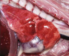
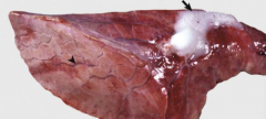
what type of congestion is this? |
acute congestion.
lung is red because of congestion of pulmonary vasculature |
|
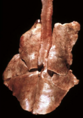
what kind of congestion is this one? |
chronic |