![]()
![]()
![]()
Use LEFT and RIGHT arrow keys to navigate between flashcards;
Use UP and DOWN arrow keys to flip the card;
H to show hint;
A reads text to speech;
45 Cards in this Set
- Front
- Back
|
What is Integumentary System? |
This is a body system composed of the skin, hair oil, and sweat glands, nails and sensory receptors that helps maintain body temperature, protects the body, and provides sensory information |
|
|
List and describe the functions of the integumentary system |
1. Regulates body temperature 2. Stores blood 3. Protects body from external environment 4. Detects cutaneous sensations 5. Excretes and absorbs substances 6. Synthesizes vitamin D |
|
|
Describe the two major components of the integument and the associated structures
|
Cutaneous membrane and accessory structures 1. Cutaneous Membrane (skin): involves the Epidermis (superficial epithelium) which has thinner layer. It's composed of epithelial tissue, and it's avascular b. Dermis (underlying area of connective tissue) It has a thicker layer, It's composed of connective tissue and vascular. 2. Accessory Structures: Involves hair, nails and exocrine glands (Multicellular). The sebaceous (oil) glands produces sebum (oil). Ceruminous glands produces cerumen (wax) and Sudoriferous (Sweat) glands produces perspiration (sweat) |
|
|
List and describe each of the layers of human skin
|
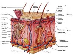
Sectional view of subcutaneous layer Skin has three layers: 1. Epidermis: Is the outer layer of skin, provides a waterproof barrier and creates our skin tone. Also the superficial, thinner layer of skin, composed of keratinized stratified squamous epithelium. It's avascular 2. Dermis: Is beneath the epidermis, contains tough connective tissue, hair follicles, and sweat glands. Also its a layer of dense irregular connective tissue lying deep to the epidermis. It's vascular 3. Hypodermis: Is made of fat and connective tissue. It's acontinous sheet of areolar connective tissue and adipose tissue between the dermis of the skin and the deep fascia of the muscles. |
|
|
List the characteristics of the epidermis and describe/ diagram the cell types, the layers of the epidermis and types of skin. |
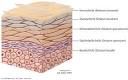
There are five layers of the epidermis 1. Stratum Corneum: It's few to fifty or more rows of dead, flat keratinocytes that contain mostly keratin. 2. Stratum Lucidum: Its present only in skin of fingertips, palms, and soles; it consist of four to six rows of clear, flat, dead keratinocytes with large amounts of keratin. 3. Stratum Granulosum: There are three to five rows of flattened keratinocytes, in which organelles are beginning to degenerate; cells contain the protein keratohyalin (converts keratin intermediate filaments into keratin) and lamellar granules (release lipid-rich, water-repellent secretion). 4. Stratum Spinosum: They are eight to ten rows of many-sided keratinocytes with bundles of keratin intermediate filaments; contains projections of melanocytes and intraepidermal macrophages. 5. Stratum Basale: This is the deepest layer, composed of single row or columnar karatinocytes that contain scattered keratin intermediate filaments (tonofilaments); stem cells undergo cell division to produce new keratinocytes; melanocytes and tactile epithelial cells associated with tactile discs are scattered among keratinocytes. |
|
|
Images of Epidermis layers |
|
|
|
Describe the function of the different epidermal cell types.
|
1. Keratinocytes: This is the most numerous of the epidermal cells; that produces keratin. They're arranged in four or five layers. Keratin is a tough, fibrous protein that helps the skin and underlying tissues from abrasions, heat, microbes and chemicals. 2. Melanocytes: This is a pigmented cell, located between or beneath cells of the deepest layer of the epidermis, that synthesizes melanin. Melanin is a yellow-red or brown-black pigment that contribute to skin color and absorbs damaging ultraviolet light. 3. Intraepidermal Macrophages or Langerhans cells: this is an epidermal dendritic cell that arise from red bone marrow and migrate to the epidermis, where they constitute a small fraction of the epidermal cells. They participate in immune responses mounted against microbe that invade the skin, and are easily damaged by ultraviolet light. 4. Tactile Epithelial Cells or Merkel cells: This is a type of cell that are the least numerous of the epidermal cells. They are located in the deepest layer of the epidermis, where they contact the flattened process of a sensory neuron (nerve cell), a structure called a tactile disc or merkel disc. Tactile epithelial cells and their associated tactile discs detect touch sensation. |
|
|
Explain the formation of the pigments carotene and melanin, give the function for each and explain how each can determine skin color.
|
Carotene: This is an orange-yellow pigment that normally accumulates in epidermal cells, and fatty tissues (adipose tissues) in the deep dermis and subcutaneous layer. b. It can be converted to vitamin A (required for maintenance of epithelia and synthesis of photoreceptor pigments in the eye) Melanin: This a brown, or black pigment produced by melanocytes. b. It's a transfer of melanin containing intracellular vesicles called melanosomes from melanocyte to keratinocyte c. The number of melanocytes distributed among the keratinocytes vary depending on region of the body. E.g: Cheeks, forehead, nipples, scrotum of males and labia majora of females have a higher concentration of melanocytes versus most other areas of the body. c. Melanin protects the cutaneous membrane form harmful effects of ultraviolet radiation, but melanin synthesis accelerates slowly. d. the skin pigmentation depends on melanin production, not number of melanocytes. |
|
|
Describe how the epidermis grow, including the role of EGF and the process of keratinization.
|
Epidermal grow factor: It's a powerful peptide growth factor b. It's produced by glands (salivary and duodenum) c. It's used in laboratories to grow skin grafts. Functions of EGF a. It promotes division of germinative (basal) cells b. It accelerates keratin production c. It stimulates epidermal repair d. It stimulates glandular secretion. |
|
|
Describe the structure and function of the two layers of the dermis and its accessory structures, and explain how the dermis and epidermis are held together.
|
Two layers of the skin are Papillary layer and Reticular layer Papillary Layer (superficial layer): It consist of areolar tissue. It contain smaller capillaries, lymphatics, and sensory neurons. It also has dermal papillae projecting between epidermal ridges. Reticular Layer (deeper layer): It consists of irregular connective tissue (containing both collagen and elastic fibers). It also contain larger blood vessels, lymphatic vessels, nerve fibers, hair follicles, sebaceous glands The Epidermal ridges create a strong bond between the epidermis and dermis in a region of high mechanical stress. It also increase the surface area of the epidermis and thus increase the grip of the hand or foot by increasing friction. Finally, it greatly increase surface area, which increases the number of corpuscles of touch and thus increase tactile sensitivity. |
|
|
Describe the difference between thick and thin skin |
Thin skin: a. It has hairy skin (except for lips and external genitalia) b. It makes up the majority of superficial epithelium c. It's composed of 4 layers (strata pl): (stratum, sing) d. The dermis is thicker e. It does contain hair, sebaceous gland, some areas have apocrine sweat glands, but fewer eccrine sweat glands than thick skin. f. It lack epidermal ridges due to poorly developed, fewer and less well organized dermal papillae g. Hair follicles and arrector pili muscles are present h. Sudoriferous glands are fewer, and sensory receptors are sparser. Thick Skin: a. They're are found in the fingertips, toes, soles of the feet & palms of hands. b. It's composed of 5 strata c. The dermis is thinner d. It does not contain hair, sebaceous glands, or apocrine sweat e. Epidermal ridges are present due towel developed and more numerous dermal papillae organized in parallel rows f. Sudoriferous glands are more numerous, and sensory receptors are denser. |
|
|
Describe the blood supply to the integument
|
There are two Plexus that supply blood to the Dermal: a. Cutaneous plexus: has blood vessels in the subcutaneous that branch into the reticular layer b. It supply both the adipose tissue of the subcutaneous and tissues of the integument. Papillary Plexus: has capillary network from small arteries in papillary layer. b. It follows the contours of the epidermis-dermis boundary. Contusion: It damage to blood vessels resulting in "black and blue bruising" |
|
|
Describe the hypodermis
|
The hypodermis is known as subcutaneous layer or superficial fascia: It lie below the integument (not part of the integument) b. It stabilizes the skin and allows separate movement. c. Its consist of elastic areolar and adipose tissues d. Its connected to the reticular layer of integument by interwoven connective tissue fibers e. Few capillaries and no vital organs ( only the superficial region contains large arteries and veins) f. Subcutaneous fat provides extra insulation, reduces heat loss, and an energy depot g. the site of subcutaneous injections using hypodermic needles. |
|
|
Define: Cleavage lines/tension line, and describe their importance in human surgery.
|
Cleavage lines (tension line) is a collagen and elastic fibers in the dermis. It's arranged in parallel bundles and it's resist force in a specific direction. Importance in human surgery: a. A parallel cut remains shut, heals well b. a cut across (right angle) pulls open and scars |
|
|
List the various types of skin cancer and their describe their relative severity, and the ABCDs.
|
There are 3 types of skin cancer: a. Basal cell carcinomas: This account for about 78% of skin cancers. The tumors arise from cells in the stratum basale of the epidermis and rarely metastasize. b. Squamous cell carcinomas: This account for about 20%of all skin cancers, arise from the stratum spinosum of the epidermis, and they have a variable tendency to metastasize. Basal and squamous cell carcinomas are together known as nonmelanoma skin cancer. c. Malignant melanomas: This arise from melanocytes and account for about 2% of all skin cancers. The estimated lifetime risk of developing melanoma is now 1 in 75, double the risk only 20 years ago. ABCDE: A-Asymmetry; malignant melanomas tend to lack symmetry. This means that they have irregular shapes, such as two very different looking halves. B-Border; malignant melanomas have irregular-notched, indented, scalloped, or indistinct-borders. C-Color; malignant melanomas have uneven coloration and may contain several colors. D-Diameter; ordinary moles typically are smaller than 6mm (0.25 in), about the size of a pencil eraser. E-Evolving; malignant melanomas change in size , shape, and color. once a malignant melanoma has the characteristics of A,B, and C it is usually larger than 6mm. |
|
|
Diagram and describe the structures which make up hair follicles and nails, and explain how these structures grow and how hair gets its color.
|
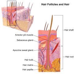
Hair follicle is located deep in dermis. It produces nonliving hairs. Its wrapped in a dense connective tissue sheath and the base is surrounded by sensory nerves (root hair plexus) The arrector pili involuntary smooth muscle, it causes hairs to stand up and produces goose bumps. Sebaceous glands lubricate the hair and control bacteria |
|
|
Hair Production
|
Hair begins at the base of a hair follicle, deep in the dermis. The hair papilla contains capillaries and nerves. The hair bulb produces hair matrix, a layer of dividing basal cells. It produces hair structure and it pushes hair up and out of skin HOW HAIR GET ITS COLOR Hair is produced by melanocytes at the hair papilla and it's determined by genes |
|
|
Explain the hair growth cycle
|
Growing hair is firmly attached to matrix. The club hair is not growing and it's attached to an inactive follicle. The new hair growth cycle: Follicle becomes active. It produces new hair and club hair is shed.
|
|
|
Describe the types of hairs found on a human.
|
We have the Vellus hairs and Terminal hairs. Vellus hairs is soft, and fine. It cover body surface Terminal hairs is heavy and pigmented. The head, eyebrows and eyelashes. And other parts of the body after puberty. |
|
|
Describe in detail the glands of the integumentary system, their function and their products
|
Sebaceous glands (oil glands): Largely in lips, glans penis, labia minora, and tarsal glands; small in trunk and limbs; absent in palms and soles. Location: The location of the secretory portion is the dermis The termination of the excretory duct is mostly connected to hair follicle. Secretion: It secretes sebum(mixture of triglycerides, cholesterol, proteins and inorganic salts.) Function: It prevents hairs from drying out, prevent water loss from skin, keep skin soft, inhibit growth of some bacteria. Onset of Function: Relatively inactive during childhood; activated during puberty |
|
|
Eccrine sweat glands
|
It's throughout skin of most regions of body, especially skin of forehead, palms and soles. Location: Location of the secretory portion is mostly in deep dermis (sometimes in upper subcutaneous layer) The termination of excretory duct is surface of epidermis Secretion: The perspiration which consists of water, ions, urea, uric acid, ammonia, amino acids, glucose, and lactic acid. Function: The regulation of body temperature, waste removal, stimulated during emotional stress Onset of function: Soon after birth. |
|
|
Apocrine Sweat glands (Sudoriferous glands)
|
Skin of axillae, groin, areolae, bearded regions of face, clitoris and labia minora. Location: Location of secretory portionis mostly in deep dermis and upper subcutaneous layer Termination of excretory duct is hair follicles Secretion: Perspiration, which consists of same components as eccrine sweat glands plus lipids and proteins. Function: Stimulated during emotional stress and sexual excitement. Onset of Function: puberty |
|
|
Ceruminous Glands
|
It's the external auditory canal. Location: Location of secretory portion is subcutaneous layer Termination of excretory duct is surface of external auditory canal or into ducts of sebaceous glands. Secretion: Cerumen, a waxy material Function: Impede entrance of foreign bodies and insects into external ear canal, prevents microbes from entering cells Onset of function: Soon after birth. |
|
|
compare and contrast sensible and insensible perspiration and list the ways that the human integumentary system functions to regulate body temperature.
|
Sensible Perspiration: This is when the sweat that is excreted in larger amounts and is seen as moisture on the skin. Water is excreted by sweat glands
Insensible Perspiration: This is the sweat that evaporates from the skin before it is perceive as moisture. There is interstitial fluid lost by evaporation through the stratum corneum. Dehydration results from damage to stratum corneum (e.g burns & blisters), and from immersion in hypertonic solution (e.g sea water). Thermoregulation is the homeostatic regulation of body temperature through sweating and adjustment of blood flow in the dermis. The role of eccrine sweat glands in helping the body to achieve thermoregulation is know as thermoregulatory sweating. |
|
|
Describe the repair process of the Integument |
In a deep wound of an integument: Bleeding occurs Mast cells trigger inflammatory response A scab stabilizes and protects the area Germinative cells migrate around the wound Macrophages clean the area Fibroblasts and endothelial cells move in, producing granulation tissue Fibroblasts produce scar tissue Inflammation decreases, clot disintegrates Fibroblasts strengthen scar tissue A raised keloid may form. 1. Bleeding occurs at the site of injury immediately after the injury, and mast cells in the region trigger an inflammatory response. 2. After several hours, a scab has formed and cells of the stratum basale are migrating along the edges of the wound. Phagocytic cells are removing debris, and more of these cells are arriving via the enhanced circulation in the area.Clotting around the edges of the affected area partially isolates the region. 3. One week after the injury, the scab has been undermined by epidermal cells migrating over the meshwork produced by fibroblast activity. Phagocytic activity around the site has almost ended, and the fibrin clot is breaking up 4. After several weeks, the scab has been shed, and the epidermis is complete. A shallow depression marks the injury site, but fibroblasts in the dermis continue to create scar tissue that will gradually elevate the overlying epidermis. |
|
|
Know the effects of aging |
a. Epidermal thinning b. Decrease numbers of dendritic (Langerhans) cells c. Decreased vitamin D3 production d. Decreased melanocyte activity e. Decreased glandular activity (sweat and oil glands) |
|
|
Explain how burns are classified, and describe the major complications of serious burns. (also define the rule of nines) |
Burns are classified into first, second and third degree burn 1. First degree burn: This involves only the epidermis. It is know by mild pain and erythema but no blisters. Skin function remain intact. Healing may occur 3 to 6 days. 2. Second-degree burn: This destroys the epidermis and part of the dermis. some skin functions are lost. There's redness, blister formation, edema and pain. In a blister the epidermis seperates from the dermis due to the accumulaton of tissue fluid between them. Hair follicles, sebaceous glands, and sweat glands, are not injured. Healing occurs without skin grafting in about 3 to 4 weeks, but scar may result 3. Third-degree burn or full thickness burn: This destroy the epidermis, dermis, and subcutaneous layer. Most skin functions are lost. Such burns vary in appearance from marble-white to mahogany colored to charred, dry wounds. Edema is marked, the burn region is numb because sensory nerve have been destroyed. Regeneration occurs slowly, and much granulation tissue forms before been covered by epithelium. |
|
|
Complications of serious burns (Also define the rules of nines) |
1. There is a large loss of water, plasma, and plasma proteins, which causes shock; 2. Bacteria infection 3. Reduced circulation of blood; 4. Decreased production of urine; 5. Diminished immune responses. Rules of Nines 1. count 9% if both the anterior(Anterior 41/2%) and posterior(Posterior 41/2%) surfaces of the head and are affected. 2. Count 9% for both the anterior(anterior 9%) and posterior(posterior 9%) surfaces of each upper limb (total of 18% for both upper limbs) 3. Count 4*9, or 36%, for both the anterior and posterior surfaces of the trunk, including the buttocks. 4. Count 9% of the anterior and 9% of the posterior surfaces of each lower limb as far up as the buttocks (total of 36% for both lower limbs) 5. Count 1% of the perineum. Total of 100% |
|
|
Define Tinea Corposis (Ringworm) |
This is a fungal infection characterized by scaling, itching, and sometimes painful lesions that may appear on any part of the body; also known as ringworm. Fungi thrive in warm, moist places such as skin folds of the groin, where it is known as tinea cruris |
|
|
Psoriasis |
This a common and chronic skin disorder in which keratinocytes divide and move more quickly than normal from the stratum basale to the stratum corneum. As a result, the surface cells never get a chance to cycle into the later keratinizing stages. The surface cells are shed immaturely and on the scalp are called dandruff. |
|
|
Nevus or Mole |
This is a round, flat or raised area that represents a benign localized overgrowth of melanocytes and usually develops in childhood or adolescence. |
|
|
Furuncle |
It's a boil. A boil is a bacteria or fungal infection of hair follicle. the infected hair can be on any part of your body it is not limited to your scalp |
|
|
Eczema (to boil over) |
This is an inflammation of the skin characterized by patches of red, blistering, dry, extremely itchy skin. It occurs mostly in skin creases in the wrist, backs of the knees, and fronts of the elbows. It typically begins in infancy and many children outgrow the condition. The cause is unknown but is linked to genetics and allergies. |
|
|
Vitilgo |
This is a condition in which the pigment is lost from areas of the skin, causing whitish patches, often with no clear cause. white patches develop on the skin and location on the body can be affected. |
|
|
Carbuncle |
This is a severe abscess or multiple boil in the skin, typically infected with staphylococcus bacteria. It's a red, swollen and painful cluster of boils that are connected to each other under the skin. |
|
|
Decubitus ulcers |
It's also know as pressure ulcers, or bed sores, it's caused by a constant deficiency of blood flow to tissue. Typically the affected tissue overlies a bony projection that has been subjected to prolonged pressure against an object such as a bed, cast or splint. If the pressure is relieved in a few hours, redness occurs but no lasting tissue damaged results. |
|
|
Impetigo |
It's a contagious bacterial skin infection forming pustules and yellow, crusty sores. It usually occurs on the face, neck, and hands of young children and infants. Children who wear diapers tent to get it around the diaper area. |
|
|
Onycholysis |
This is a common nail disorder. It is the separation of a fingernail from its nail bed. It usually starts at the tip of the nail and progresses back. it can be a sign of skin disease, or the result of injury, which occurs mostly with women with long fingernails. |
|
|
Jaundice |
This is a condition characterized by yellowness of the skin, the white of the eyes, mucous membranes, and body fluids because of a buildup of bilirubin. |
|
|
Erthyema Stretch Marks/ Striae
|
Stretch marks (Striae gravidarum)is a form of internal scarring, can result from the internal damage to this layer that occurs when the skin is stretched too much. When the skin is overstretched, the lateral bonding between adjacent collagen fibers is disrupted and small dermal blood vessels rupture. This is why it appear as reddish streaks at these sites. Erythema is redness of the skin, is caused by engorgement of capillaries in the dermis with blood due to skin injury, exposure to heat, infection, inflammation, or allergic reactions. |
|
|
Lamellar
|
This release water repellent sealant that decreases water entry and loss and inhibits the entry of foreign materials.
|
|
|
Ichthyosis
|
This is a congenital skin condition that causes the epidermis to become dry and rough like fish scales.
|
|
|
Wart
|
It's produced by uncontrolled growth of epithelial skin cells; caused by a papillomavirus. Most warts are noncancerous.
|
|
|
Pallor
|
It's paleness of the skin, may occur in conditions such as shock and anemia. All skin color changes are observed most readily in people with light-colored skin and may be more difficult to discern in people with darker skin
|
|
|
Lamellar Ichthyosis
|
this is an autosomal recessive disorder that is apparent at birth and is present throughout life. The new born is born encased in a collodion membrane that sheds within 10-14 days. The shedding of the membrane reveals generalized scaling with variable redness of the skin
|