Reading...
![]()
Play button
![]()
Play button
![]()
Use LEFT and RIGHT arrow keys to navigate between flashcards;
Use UP and DOWN arrow keys to flip the card;
H to show hint;
A reads text to speech;
53 Cards in this Set
- Front
- Back
|
What is the most common cause of dwarfism?
|
Achondroplasia
|
|
|
What causes achondroplasia?
|
It’s a hereditary failure of endochondral bone formation
|
|
|
Long bone findings in achondroplasia on imaging?
|
Femurs and humeri are more profoundly affected than the other long bones, although the entire skeleton is abnormal
The long bones are short but have normal width, giving them a thick appearance |
|
|
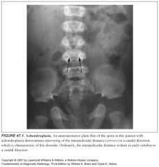
Spine findings in achondroplasia on imaging?
|
The spine typically has narrowing of the interpedicular distances in a caudal direction, the opposite of normal, in which the interpedicular distances get progressively wider as one proceeds down the spine.
|
|
|
Define avascular necrosis:
|
Lack of blood supply with subsequent bone death and ensuing bony collapse in an articular surface
|
|
|
Name some causes of AVN:
|
-Trauma
-Steroids -Collagen vascular diseases -Alcoholism -Idiopathic -Infection |
|
|
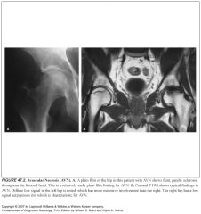
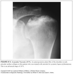
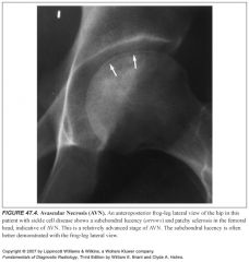
What does AVN look like on imaging?
|
It ranges from patchy sclerosis to articular surface collapse and fragmentation
Just before collapse, a subchondral lucency is occassionally seen (this is inconsistent) |
|
|
What is the best modality to evaluate AVN?
|
MR
|
|
|
What is the long name for clubbing of the fingers?
|
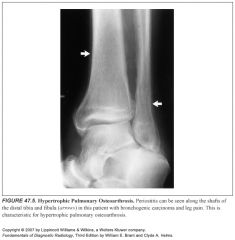
Hypertrophic pulmonary osteoarthorpathy
|
|
|
Who gets clubbing?
|
Most commonly, lung cancer, but also seen in bronchiectasis, GI disorders, and liver disease.
|
|
|
What is the mechanism of clubbing?
|
Unknown
|
|
|
Differential for periostitis in a long bone without underlying bony abnormality:
|
-Hypertrophic pulmonary osteoarthropathy
-Venous stasis -Thyroid acropachy -Pachydermoperiostosis -Trauma |
|
|
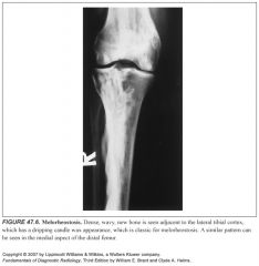
What is melorheostosis?
|
Rare, idiopathic disorder characterized by thickened cortical new bone that accumulates near the ends of long bones, usually only on one side of the bone
Appearance has been term “dripping candle wax” |
|
|
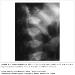
What is Morquio syndrome?
|
Abnormal storage and excretion of keratan sulfate
|
|
|
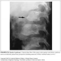
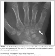
What is Hurler syndrome?
|
Abnormal storage and excretion of heparan sulfate
|
|
|
What does Morquio syndrome look like on imaging?
|
-Shortened spines
-Platyspondyly (generalized flattening of the vertebral bodies) -Central anterior projection or “beak” off the vertebral body on lateral films -Wide, flared iliac wing and broad femoral necks |
|
|
What do Hunter and Hurler syndrome look like on imaging?
|
-Shortened spines
-Platyspondyly (generalized flattening o the vertebral bodies) -Anteroinferiorly positioned beak off the vertebral body on lateral films -Wide, flared iliac wing and broad femoral necks -The hands have a pointed proximal fifth metacarpal base that has a notched appearance to the ulnar aspect. |
|
|
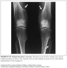
What is multiple hereditary exostosis also known as?
|
Diaphyseal aclasia
|
|
|
What are the findings in multiple hereditary exostosis?
|
Multiple osteochondromas or exostoses
|
|
|
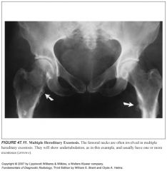
What is an osteochondroma?
|
Cartilage-capped bone outgrowth that may be pedunculated or sessile
Undertubulation (a widened diameter of the bone) is invariably present at the site of the exostosis |
|
|
Which areas are typically involved in multiple hereditary exostosis?
|
The knees are virtually always involved
The proximal femurs are frequently involved |
|
|
What is the rate of malignant degeneration in multiple hereditary exostosis?
|
It’s extremely rare
|
|
|
Which lesions in multiple hereditary exostosis undergo malignant degeneration?
|
The more axially situated the lesions, the more they are prone to undergo malignant degeneration
|
|
|
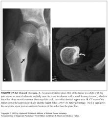
How does osteoid osteoma present?
|
Painful bone lesion in patients younger than 30.
|
|
|
What do osteoid osteomas look like?
|
-Cortically based sclerotic lesion in a long bone that has a small lucency within it called the nidus
-If the nidus is located in the medullary rather than the cortical portion of a bone, or if it is located in a joint, there is much less reactive sclerosis present -Rarely, it will be in the periosteum, causing exuberant periostitis. -the nidus is often lucent, but can develop calcification within it. If it calcifies completely, it blends into the surrounding sclerosis. -Therefore, diagnosis should not depend on visualization of a nidus. |
|
|
How is osteoid osteoma treated?
|
-Surgical excision or thermal ablation
-It is the nidus that cuases the pain and surrounding reactive sclerosis -If the nidus is removed or ablated, the pain stops. -The natural history of this disease is eventual spontaneous regression |
|
|
What is the imaging of choice for osteoid osteoma?
|
CT is often helpful in demonstrating the exact location of the nidus
|
|
|
What is osteoid osteoma often confused with? Why?
|
Osteomyelitis
The nidus is usually lucent, but often develops some calcification within it. IT then has the appearance of a sequestrum |
|
|
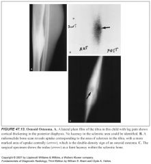
How can you tell the difference between an osteoid osteoma and osteomyelitis?
|
Nuclear bone scans—the nidus avidly accumulates radiotracer relative to the uptake of the surrounding reactive sclerosis. This is called the “double-density sign”
In contrast, osteomyelitis has a photopenic area corresponding to the avascular focus of purulent material |
|
|
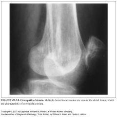
Define osteopathia striata:
|
-Also known as Voorhoeve disease
-Multiple 2-3 mm thick linear bands of sclerotic bone aligned parallel to the long axis of a bone -It usually affects multiple long bones and is asymptomatic, so it’s usually just an incidental finding |
|
|
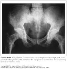
Define osteopoikilosis:
|
Hereditary asymptomatic multiple small (3-10mm) sclerotic bony densities affecting primarily the ends of long bones and the pelvis
|
|
|
What is the clinical significance of osteopoikilosis?
|
There is none, except that it can be confused for diffuse osteoblastic metastases.
|
|
|
Define pachydermoperiostosis
|
-Rare familial disease
-Thickening of the skin of the extremities and face, clubbing of the fingers, and widespread periostitis. -More common in black patients. -The periosteal reaction looks like hypertrophic pulmonary osteoarthropathy, but it is only occassionally painful |
|
|
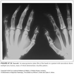
What does sarcoid look like in the bones?
|
-The hands are the most often affected
-It causes a characteristic lacelike pattern of body destruction in the hands -Multiple phalanges are typically affected in either one or both hands |
|
|
What is the differential diagnosis for sarcoid-like changes of the hands?
|
There is none; it is very characteristic
|
|
|
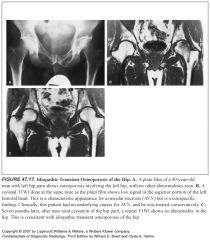
Describe transient osteoporosis of the hip:
|
-Findings of osteoporosis limited to a single, painful, hip
-Some think that this is early AVN, but this has not been proven -Invariably self-limited with full resolution -Tends to occur more in men |
|
|
1
|
|
|
2
|
|
|
3
|
|
|
4
|
|
|
5
|
|
|
6
|
|
|
7
|
|
|
8
|
|
|
9
|
|
|
10
|
|
|
11
|
|
|
12
|
|
|
13
|
|
|
14
|
|
|
15
|
|
|
16
|
|
|
17
|