![]()
![]()
![]()
Use LEFT and RIGHT arrow keys to navigate between flashcards;
Use UP and DOWN arrow keys to flip the card;
H to show hint;
A reads text to speech;
52 Cards in this Set
- Front
- Back
|
What is the vascular response of inflammation? |
-Transudate produces localized edema -Blood vessels constrict to reduce blood loss -platelet aggregation -activated platelets release chemical mediators Vasodialation -exudate(mass of cells and fluid that leak out) formation -histamine release -prostaglandin release |
|
|
What are the cells involved in the inflammatory phase? |
-Platelets -PMNs -Fibroblasts -Macrophages -Mast cells |
|
|
What are the functions of the cells involved in the inflammatory phase? |
k |
|
|
What is the proliferative phase of wound healing? |
Angiogenesis Granulation tissue Formation Wound contraction Epithelialization |
|
|
What are the cells that are involved in the proliferative phase? |
-angioblasts -Fibroblast -myofibroblasts -keratinocyte |
|
|
What are the functions of the cells involved in the proliferative phase? |
-Forms new blood vessels (angioblasts) -builds granulation tissue (fibroblast) -causes wound contraction (myofibroblast) -reepithelializes wound surface (keratinocyte) |
|
|
What occurs in the maturation and remodeling phase of wound healing? |
New collagen synthesis Old collagen is broken down by collagenases Reorientation of collagen fibers May continue up to 2 years after wound closure |
|
|
Compare and contrast absence of inflammation and chronic inflammation |
k |
|
|
Why does absence of inflammation occur? |
k |
|
|
Why does chronic inflammation occur? |
-presence of foreign body in wound bed -repetitive mechanical trauma -cytotoxic agents -senescent cells -higher levels of MMPs -lower levels of TIMPs -greater numbers of inflammatory cytokines and chronic wound cells -arrested current of injury |
|
|
What are the interventions that may improve wound healing for absence of inflammation? |
k |
|
|
What are the interventions that may improve wound healing for chronic inflammation ? |
k |
|
|
What is hypogranulation? |
Nonadvancing wound edge -hypogranular wound -epibole (rolled edge of wound before it is closed) -do not granulate may be a vascular issue |
|
|
What is hypergranulation? |
-hypergranular wound |
|
|
Why does hypogranulation occur? |
-may have less granulated cells -may not have enough vascularization |
|
|
Why does hypergranulation occur? |
k |
|
|
What are the interventions for hypogranulation to improve wound healing? |
k |
|
|
What are the interventnions for hypergranulation to improve wound healing? |
k |
|
|
What are the differences between (hypertrophic scaarring, keloids, contractures, and wound dehiscence)? |
k |
|
|
Why would hypertrophic scarring occur? |
is a cutaneous condition characterized by deposits of excessive amounts of collagen which gives rise to a raised scar, but not to the degree observed with keloids. Like keloids, they form most often at the sites of pimples, body piercings, cuts and burns |
|
|
Why would keloids occur? |
an area of irregular fibrous tissue formed at the site of a scar or injury. |
|
|
why would contractures occur? |
a condition of shortening and hardening of tissue, often leading to deformity and rigidity of joints |
|
|
Why would wound dehiscence occur? |
not a doctors fault -failure of cells to catch one another? - release of material by splitting open of an organ or tissue; the natural bursting open at maturity of a fruit or other reproductive body to release seeds or spores or the bursting open of a surgically closed wound |
|
|
What are the interventions for hypertrophic scarring? |
k |
|
|
What are the interventions for keloids? |
k |
|
|
What are the interventions for contractures? |
|
|
|
What are the interventions for wound dehiscence? |
k |
|
|
starting slide 23 |
k |
|
|
What are the phases or wound healing? |
Inflammation Proliferation Maturation & Remodeling (overlapping phases) |
|
|
What are the cardinal signs of inflammation? |
-swelling -redness -warmth -pain -decreased function |
|
|
What is the function of platelets in inflammation phase? |
-form platelet plug -secrete growth factors and chemotactic agents |
|
|
What is the function of polymophonuclear leukocytes in inflammation phase? |
-margination( free-flowing leukocytes exit the central blood stream,) diapedesis(blood cells passing through walls), chemotaxis -first cells to site of injury -scavengers -kill bacteria -clean wound -secrete inflammatory mediators and MMPs |
|
|
What is the function of the macrophages in the inflammation phase? |
-directs repair -assists with killing bacteria and cleaning wound -secretes growth factors and MMPs |
|
|
What is the function of the mast cells in the inflammation phase? |
-secretes enzymes -secretes inflammatory mediators |
|
|
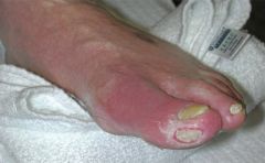
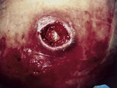
Picture of wound in inflammatory phase: (edema and erythema from wound and around, wound bed covered with yellow slough, slightly warm, toe is tender to touch) |
|
|
|
What is the function of the angioblast in proliferation phase? |
forms new blood vessels |
|
|
What is the function of the fibroblast in proliferation phase? |
builds granulation tissue |
|
|
What is the function of the myofibroblast in proliferation phase? |
causes wound contraction |
|
|
What is the function of the keratinocyte in proliferation phase? |
reepithelializes wound surface |
|
|
How does the granulation tissue formation occur? |
-Matrix metalloproteases (MMPs) -Granulation tissue -Integrins (protein) (fibroblasts) |
|
|
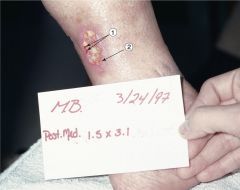
Picture of proliferative phase of wound healing: (1) small endothelial buds: indicating angiogenesis, (2) pale pink epithelial cells at wound edge:evidence of epithelialization |
|
|
|
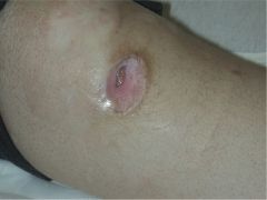
Picture of wound contraction in proliferative phase of wound healing: The perimeter is remodeling near the wound edge, less granulation tissue has been formed adn the scar is less mature and more pink in color, farther out scar is closer to pt. natural skin color. |
|
|
|
What are some key growth factors and cytokines? |
•EGF •KGF •FGF •PDGF •GM-CSF •TGF-β •TNF-α •IL-1 •VEGF •Insulin-likegrowth factor |
|
|
What is primary closure? |
-wound is cleaned of foreign material and edges are approximated -wound should be closed in 1-14 days -also known as healing by primary intention -closure of all tissue levels during the original surgery, regardless of the presence of wires, wicks, drains, or other devices or objects extruding through the incision |
|
|
What is secondary closure? |
-wound repair follows 3 phases of normal wound healing -signs of progression through phases acute wound: within 2 weeks chronic wound: within 30 days -also known as healing by secondary intention -closure by secondary intention,the skin edges of the wound are not sutured together; the wound is left “open.” Dressings are applied regularly to keep the wound clean, and the wound gradually closes and heals on its own |
|
|
What is a delayed primary closure? |
-wound is cleanded and observed for signs of infection -then closed with sutures -wound should be closed within 1-2 wks or suturing -also known as healing by tertiary intention |
|
|
What is an epibole? |
rolled edge of wound before it is closed) |
|
|
What may occur if the rate of synthesis remains greater than collagen lysis during the maturation and remodeling phase? |
pt. develops hypertrophic scarring or keloids |
|
|
Pic of tunneling wound: |
|
|
|
Pic of stage III pressure ulcer over greater trochanter: |
|
|
|
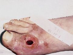
Pic of Type 1 diabetes sore: |
|
|
|
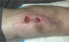
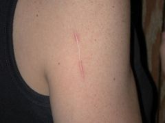
Pic irregular scar maturation and remodeling: |
|