Reading...
![]()
Play button
![]()
Play button
![]()
Use LEFT and RIGHT arrow keys to navigate between flashcards;
Use UP and DOWN arrow keys to flip the card;
H to show hint;
A reads text to speech;
263 Cards in this Set
- Front
- Back
|
Central Nervous System
|
Composed of the brain and spinal cord
|
|
|
Surface anatomy of the brain
|
-cerebral hemispheres, cerebellum, and brain stem
|
|
|
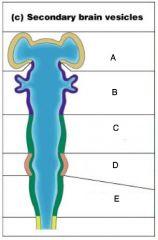
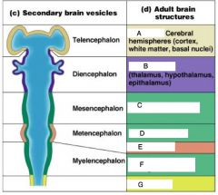
A) Telencephalon
B) Diencephalon C) Mesencephalon D) Metencephalon E) Myelencephalon |
|
|
A) Cerebrum
B) Diencephalon C) Brain Stem; midbrain D) Brain Stem; pons E) Cerebellum F) Brain Stem; Medulla onlongata H) Spinal Cord |
|
|
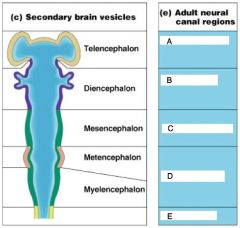
Telencephalon
|
Lateral Ventricles
|
|
|
Diencephalon
|
Third Ventricles
|
|
|
Mesenecephalon
|
Cerebral aqueduct
|
|
|
A) Lateral Ventricles
B) Third Ventricles C) Cerberal Ventricles D) Fourth Ventricles E) Central Canal |
|
|
Know
|
|
|
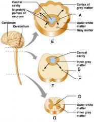
Spinal Cord
|
External to which is white matter composed of myelinated fiber tracts
|
|
|
Brain
|
-Similar to spinal cord but with more grey matter
-Cerebellum has gray matter in nuclei |
|
|
A) Inner gray matter
B) Outer white matter C) Gray Matter D) Central Cavity E) Region of cerebellum F) Brain Stem G) Spinal Cord |
|
|
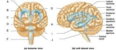
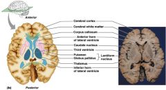
Ventricles of the Brain
|
Arise from the expansion of the lumen of the neural tube
|
|
|
The ventricles are (3)
|
-The paired C-shaped lateral ventricles
-The third ventricles found in the diencephalon -The fourth ventricle found in the hindbrain dorsal to the pons |
|
|
A) Lateral ventricle
B) Septum pellucidum C) Third ventricle D) Cerebral aqueduct E) Fouth Ventricle F) Central Canal G) Anterior Horn H) Interventricular foramen I) Inferior horn J) Lateral Seperture |
|
|
The cerebral hemipsheres contain ______ and shallow ______
|
Ridges (gyri)
Grooves (sulci) |
|
|
Fissures
|
Deep groves in the cerberal hemispheres
|
|
|
Cerebral hemipsheres are seperatted by the
|
longitudinal fissure
|
|
|
The Cerebral hemipsheres have three basic regions
|
Cortex, white matter and basal nuclei
|
|
|
Deep sulci divide the hemipsheres into five lobes
|
-Frontal
-Parietal -Temporal -Occipital -Insula |
|
|
Central Suculus
|
Seperates the frontal and parietal lobes
|
|
|
Parieto-occipital suculus
|
Seperates the parietal and occipital lobes
|
|
|
Lateral sulcus
|
seperates the parietal and temporal lobes
|
|
|
What two structures border the central sulcus
|
precentral and postcentral gyri
|
|
|
Cerebral cortex
|
superficial gray matter, that accounts for 40% of the mass of the brain. It enables sensation, communication, memory, understanding and voluntary movements
|
|
|
Each hemisphere in the cerebral cortex acts
|
contralaterally: controls opposite sides of the body
|
|
|
What is different about the hemispheres of the cerebral cortex
|
they are not equal in function
|
|
|
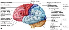
Motor Areas
|
control voluntary movement
|
|
|
Sensory areas
|
conscious awareness of sensation
|
|
|
Association areas
|
integrate diverse information
|
|

|
Know
|
|
|
Know
|
|
|
Primary motor cortex
|
loacted in the precentral gyrun
|
|
|
What is the primary motor cortex composed of
|
pyramidal cells whos axons make up the corticospinal tracts
|
|
|
What does the primary motor complex allow
|
conscious control of precise, skilles, voluntary movements
|
|
|
Motor homunclulus
|
Caricature of relative amounts of cortical tissue devoted to each motor function
|
|
|
Know
|
|
|
premotor cortex
|
located anterior to the precentral gyrus
|
|
|
What does premotor cortex control
|
learned, repititous, and patterned motor skills
|
|
|
Brocas Area
|
-Located anterior to the inferior region of the premotor area
-present in one hemisphere |
|
|
What role does brocas area play
|
-motor speech area that directs muscles of the tongue
-activates as one prepares to speak |
|
|
Frontal eye field
|
Located anterior to the premotor cortex and superior to brocas area
|
|
|
What does the frontal eye field control
|
voluntary eye movement
|
|
|
What are the sensory areas of the brain
|
-Primary somatosensory cortex
-Somatosensory association cortex -Visual and auditory areas -Olifactory, gustatory, and vestibular cortices |
|
|
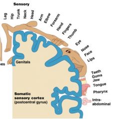
Primary somatosensory cortex
|
Located in the postcentral gyrus
|
|
|
What role does the primary somatosensory cortex play
|
-recieves information from the skin and skeletal muscles
-exhibits spatial discrimination |
|
|
Somatosensory homunculus
|
-caricature of relative amounts of cortical tissue devoted to each sensory function
|
|
|
Know
|
|
|
Know
|
|
|
Somatosensory association cortex
|
located posterior to the primary somatosensory cortex
|
|
|
What is the role of the somatosensory association cortex
|
Intergrates sensory information
|
|
|
Primary visual (striate) cortex
-Location -Function |
-seen on the extreme posterior tip of the occipital lobe
-Recieves visual information from the retinas |
|
|
Visual association area
-Location -Function |
-Surrounds the primary visual cortex
-Interprets visual stimuli (color, form, and movement) |
|
|
Primary Auditory cortex
-Location -Function |
-Located at the superior margin of the temporal lobe
-Revieves information related to pitch, rythm, and loudness |
|
|
Auditory association area
-Location -Function |
-Located posterior to the primary auditory cortex
-Stores memories of sounds and permits preception of sounds |
|
|
Where is wernickes area located
|
Auditory association area
|
|
|
What did carl wernicke describe about the brain
|
That people who suffer neurophysiological damage to this area are unable to understand the content word while listening, and unable to produce meaningful sentence; there speech has grammatical structures but no meaning
|
|
|
What are the association areas (4)
|
-prefrontal cortex
-Language areas -General interpretation area -Visceral association |
|

|
Know
|
|
|
Prefrontal cortex
-Location -Function |
-in the anterior portion of the frontal lobe
-Involved with intellect, cognition, recall, and personality |
|
|
The prefrontal cortex is necessary for
|
judgement, reasoning, persistance, and conscience
|
|
|
Which cortex is linked to the limbic system
|
Prefrontal cortex
|
|
|
Where are the language areas located
|
-large areas surrounding the left lateral sulcus
|
|
|
Wernickes area (language area)
|
involved in sounding out unfamilary words
|
|
|
Brocas area (language area)
|
Speech preperation and production
|
|
|
Lateral prefrontal cortex (language area)
|
Language comprehension and word analysis
|
|
|
Lateral and ventral temporal lobe (language area)
|
coordinate auditory and visual aspects of language
|
|
|
General Interpretation Area
-Location -Function |
-Found in one hemisphere, usually the left
-Integrates incoming signals into a single though -Involved in processing spatial relationships |
|
|
A) Motor Cortex
B) Wernickes area C) Angular gyrus D) Visual cortex E) Auditory Cortex F) Brocas Area |
|
|
Lateralization
|
each hemisphere has the abilities not shared with its partner
|
|
|
Cerebral dominance
|
-designates the hemisphere dominant for language
|
|
|
Left Hemisphere
|
-Controls language, math and logic
|
|
|
Right Hemisphere
|
-controls visual-spatial skills, emotion, and artistic skills
|
|
|
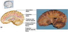
Cerebral White Matter
|
consists of deep myelinated fibers and their tracts
|
|
|
Cerebral white matter is responsible for the communication between
|
The cerebral cortex and lower CNS center, and areas of the cerebrum
|
|
|
Commissures
|
Connect corresponding gray areas of the two hemispheres
|
|
|
TEST QUESTION!!!!
|
|
|
TEST QUESTION!!!
|
|
|
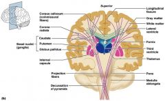
Basal Nuclei/ganglia
|
Masses of gray matter found deep within the cortical white matter
|
|
|
The corpus stratum is composed of three parts
|
-Caudate nucleus
-Lentiform nucleus -Fibers of internal capsule |
|
|
Lentiform Nucleus
|
Composed of the putamen and the globus pallidus
|
|
|
Know
|
|
|
What are the functions of Basal Nuclei (4)
|
-Influence muscualr activity
-Regulate attention and cognition -Regulare intensity of slow or sterotyped movements -Inhibit antagonistic and unnecessary movement |
|
|
Diencephalon
|
-Central core of the forebrain
|
|
|
What three paired structures does the diencephalon consists of
|
-Thalamus
-Hypothalamus -Epithalamus |
|
|
What structure enlcoses the third ventricle
|
-Dienchephalon
|
|
|
Know
|
|
|
Thalamus
|
Paired, egg-shaped masses that form the superolateral walls of the third ventricle
|
|
|
The thalamus is connected to the midline by the
|
intermediate mass
|
|
|
All inputs ascending to the cerebral cortex pass through the (TQ)
|
Thalamus
|
|
|
Thalamus plays a key role in (5)
|
mediating sensation, motor activities, cortical arousal, learning, and memory
|
|
|
Hypothalamus
|
Located below the thalamus, it caps the brainsetm and forms the inferolateral walls of the third ventricle
|
|
|
Mammillary Bodies
|
small, paired nuclei bulging anteriorly from the hypothalamus
|
|
|
The mammillary bodies are the relay station for (TQ)
|
Olfactory pathways
|
|
|
Infundibulum
|
Stalk of the hypothalamus; connects to the pituitary gland
|
|
|
What is the functions of the hypothalamic function
|
-blood pressue
-rate and force of heartbeat -digestive tract motility, -rate and depoth of breathing, and many other visceral activities |
|
|
Hypothalamic is involved with perception of (3)
|
-pleasure
-fear -rage |
|
|
The hypothalamic controls mechanisms needed to maintain
|
normal body temperature
|
|
|
The hypothalamic regulates feelings of
|
hunger and satiety
|
|
|
The hypothalamic regulates
|
sleep and the sleep cycle
|
|
|
Endocrine functions of the hypothalamus
|
Releasing hormones control secreation of hormones by the anterior pituitary
|
|
|
The supraoptic and paraventricular nuclei produce
|
ADH and oxytocin
|
|
|
Epithalamus
|
Most dorsal portion of the diencephalon; forms roof of the third ventricle
|
|
|
Pienal gland
|
extends from the posterior border and secreats melatonin
|
|
|
Melatonin
|
a hormone involved with sleep regulation, sleep-wake cycles, and mood
|
|
|
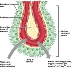
Choroid plexus
|
a structure that secreats cerebral spinal fluid
|
|

|
Know
|
|
|
The brain stem consists of three regions
|
-midbrain
-pons -medulla oblongata |
|
|
The brain stem is similar to the spinal cord by contains
|
embedded nuclei
|
|
|
The brain stem controls (TQ)
|
automatic behaviors necessary for survival
|
|
|
The brain stem is associated with (TQ)
|
10 of the 12 pairs of cranial nerves
|
|
|
Know
|
|
|
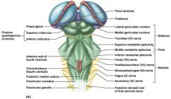
Midbrain
|
located between the diencephalon and the pons
|
|
|
Cerebral aqueduct
|
hollow tube that connects the third and fourth ventricles
|
|
|
Midbrain nuclei control cranial nerves _____ and _____
|
III(oculomotor) IV(trochlear)
|
|
|
Corpora quadrigemina
|
four domelike protrusions of the dorsal midbrain
|
|
|
Superior colliculi (TQ)
|
Visual reflex centers
|
|
|
Inferior colliculi (TQ)
|
Auditory replay centers
|
|
|
Substantia nigra
|
functionally linked to basal nuclei
|
|
|
Pons
|
Forms part of the anterior wall of the fourth ventricle
|
|
|
The fibers of the pons relay
|
impulses between the motor cortex and the cerebellum
|
|
|
pons are the origin of the cranial nerves ___, _____ and ____
|
V (trigeminal)
VI (abducens) VII (facial) |
|
|
pons contain nuclei of the
|
reticular formation
|
|
|
Cardivascular control center
|
adjust force and rate of heart contractions
|
|
|
Respiratory centers
|
control rate and depth of breathing
|
|
|
The cerebellum provides
|
precise timing and appropriate patterns of skeletal muscle contraction
|
|
|
The cerebellar activity occurs
|
subconciously
|
|
|
Folia
|
transversely oriented gyri
|
|
|
Arbor vitae
|
distinctive treelike pattern of the cerebellar white matter
|
|
|
Cerebellum Anatomy
|
two bilaterally symmetrical hemispheres connected medially by the vermis
|
|
|
cerebellum recieves impulses of the intent
|
to initiate voluntary muscle contraction
|
|
|
Proprioceptors and visual signals inform
|
the cerebellum of the bodys condition
|
|
|
the cerebellum cortex calculates
|
the best way to perfrom a movement
|
|
|
A "blueprint" of coordinated movement is sent
|
to the cerebral motor cortex
|
|
|
Two functional brain systems
|
-Limbic system
-Reticular formation |
|
|
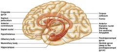
Limbic system
|
structures on the medial acpects of cerebral hemispheres and diencephalon
|
|
|
Limbic system includes (3)
|
-rhinencephalon
-amygadala -hypothalamus |
|
|
Amygadala
|
deals with anger, danger, and fear responses
|
|
|
Which system puts emotional responses to odors
|
Limbic System
|
|
|
Know
|
|
|
Reticular Activating System
|
sends impulses to the cerebral cortex to keep it conscious and alert
|
|
|
The Reticular Activating System filters out (TQ)
|
repetitive and weak stimuli
|
|
|
Electroencephalogram (EEG)
|
records brain wave activity
|
|
|
Alpha Waves
|
regular and rhythmic, low amplitude, slow, synchronous waves indicating an "idiling" brain
|
|
|
Beta Waves
|
rhythmic, more irregular waves occurring during the awake and mentall alert state
|
|
|
Theta Waves
|
more irregular than alpha waves; common in children but abnormal in adults
|
|
|
Delta Waves
|
high-amplitude waves seen in deep sleep and when reticular activating system is damped
|
|
|
A flat EEG is evidence of
|
Death
|
|
|
Epilepsy is not associated with, nor does it cause
|
intellectual impairments
|
|
|
Absence seizures, or petit mal
|
mild seizures seen in young children where the expresson goes blank.
|
|
|
grand mal seizures
|
victim loses consciousness, bones are often broken due to instesive convulsions, loss of bowel and bladder control, and severe biting of the tounge
|
|
|
What is used to control epilepsy
|
valproic acid, a nonsedating drug, enhances GABA and is a drug of choice
|
|
|
Clinical Consciousness
|
defines on a continuum that grades levels of behavior-alertness, drowsiness, stupor
|
|
|
How many states of NREM does one pass through during the first 30-24 min
|
four
|
|
|
When does REM occour
|
after this fourth NREM stage has bee achieved
|
|
|
Stage 1 NREM
|
eyes are closed and relaxiation begins; the EEG shows alpha waves; one can be easily aroused
|
|
|
Stage 2 NREM
|
EEG pattern is irregular with sleep spindles (high voltage wave bursts); arousal is more difficult
|
|
|
Stage 3 NREM
|
sleep deepnes; theta and delta waves appear; vital signs decline; dreaming is common
|
|
|
Stage 4 NREM
|
EEG pattern is dominated by delta waves; skeletal muscles are relaxed; arousal is difficult
|
|
|
What is a characterisics of skeletal muscles in REM sleep
|
are inhibited
|
|
|
When does most dreaming take place
|
in the REM stage
|
|
|
Know
|
|
|
what is presumed to be the restorative stage
|
Slow wave sleep
|
|
|
Narcolepsy
|
lapsing abruptly into sleep from the awake state
|
|
|
Insomnia
|
chronic inability to obtain the amount or quality of sleep needed
|
|
|
sleep apnea
|
temporary cessation of breathing during sleep
|
|
|
What are the three principles of memory
|
-storage, processing, memory traces
|
|
|
Storage
|
occurs in stages and its continually changing
|
|
|
Processing
|
accomplished by the hippocampus and surrounding structures
|
|
|
Memory traces
|
Chemical or structural changes that encode memory
|
|
|
How long does shorts term memory last and how much information is it limited to
|
-seconds to hours
-7 or 8 pieces of information |
|
|
Emotional State
|
we learn best when we are alert, motivated, and aroused
|
|
|
Rehersal
|
repeating or rehersing materal enchances memory
|
|
|
Association
|
associating new information with old memories in LTM enchances memory
|
|
|
What are the two categories of memory
|
fact memory and skill memory
|
|
|
Name the 3 traits of fact memory
|
-Entails learning explicit information
-Is related to our conscious thoughts nd out language ability -Is stored with the contex in which it was learned |
|
|
Skill memory
|
-Is aquried through practice
|
|
|
What is the difference from skill memory to fact memory
|
-Skill memories do not retain the contex in which they were learned
|
|
|
In memory neuronal ______ content is altered
|
RNA
|
|
|
In memory dendritic _______ change shapes
|
Spine
|
|
|
Extracellular protines are deposited at synapses involed in _____
|
Long Term Memory
|
|
|
More _______ is releases by presynaptic neurons
|
neurotransmitter
|
|
|
In memory new ______ neurons appear
|
hippocampal
|
|
|
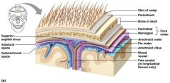
The brain is protected by
|
-Bone, meninges and cerebrospinal fluid
|
|
|
Harmful substances are shielded from the brain by the ____-______ _____
|
Blood-brain barrier
|
|
|
What are the three connective tissue membranes that lie external to the CNS
|
-Dura matter
-arachnoid mater -pia mater |
|
|
What are the four functions of the meninges
|
-Cover and protect the CNS
-Protect blood vessels and enclose venous sinuses -Contain cerebrospinal fluid -Form partitions within the skull |
|
|
Know
|
|
|
Dura mater
|
leathery, strong meninx composed of two fibrous connective tissue layers
|
|
|
The two layers of dura mater seperate
|
in certian areas and from dural sinuses
|
|
|
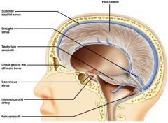
What are the three dural septa extend inward and limit excwessive moevement of the brain
|
-Falx cerebri
-Falk cerebelli -Tentorium cerebelli |
|
|
Falx cerebri
|
-fold that dips into the longitudinal fissure
|
|
|
Falx cerebelli
|
-runs along the vermis of the cerebellum
|
|
|
-Tentorium cerebelli (TQ)
|
-horizontal dural fold extends into the transverse fissure
|
|
|
Know
|
|
|
Arachnoid Mater
|
-middle menix, which forms a loose brain covering
|
|
|
________ ______ protude superiorly and permit CSF to be absorbed into venous blood
|
Arachnoid Villi
|
|

|
Know
|
|
|
Pia Mater
|
-Deep matrix composed of delicate connective tissue that clings tightly to the brain
|
|
|
Cerebrospinal Fluid
|
watery solution that prevents the brain from crushing under its own weight
|
|
|
What function foes cerebrospinal fluid play
|
nourishes the brain and carries chemical signals throughout it
|
|
|
Know
|
|
|
Blood-Brain Barrier
|
protective mechanism that helps maintain a stable enviroment for the brain
|
|
|
Bloodborn substances are seperated from neurons by (3)
|
-Continuous endothelium of capillary walls
-Relatively thick basal lamina -Bulbous feet of astrocytes |
|
|
What is the function of a blood-brain barrier
|
allows nutrients to pass freely
|
|
|
What is the blood-brain barrier innefective against
|
substances that can diffuse through plasma membranes
|
|
|
Transient ischemic attacks (TIAs)
|
temporary episodes of reversible cerebral ischemia
|
|
|
Tissue plasminogen activator (TPA)
|
is the only approved treatment for stroke
|
|
|
Alzheimers Disease
|
a progressive degenerative disease of the brain that results in dementia
|
|
|
Perkinson Disease
|
degeneration of the dopamine-releasing neurons of the substantia nigra
|
|
|
Huntingtons disease
|
a fatal hereditary disorder caused by accumulation of the protien huntingtin that leads to degeneration of the basal nuclei
|
|
|
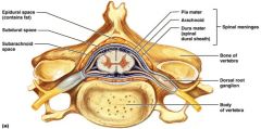
CNS tissue is enclosed within the vertebral column from the foramen magnum to
|
L1
|
|
|
Epidural Space
|
space between the vertebrae and the dural sheath (dura mater) filled with fat and a network of veins
|
|
|
Know
|
|
|
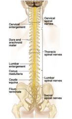
Conus medullaris
|
terminal portion of the spinal cord
|
|
|
Filum terminale
|
fibrous extension of the pia mater; anchors the spinal cord to the coccyx
|
|
|
Denticulate ligaments
|
delicate shelves of pia mater; attach the spinal cord to the vertebrae
|
|
|
Spinal nerves
|
31 pairs attach to the cord by paired roots
|
|
|
Cervical and lumbar enlargements
|
sites where nerves serving the upper and lower limbs emerge
|
|
|
Cauda equina
|
collection of nerve roots at the inferior end of the vertebral canal
|
|
|
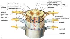
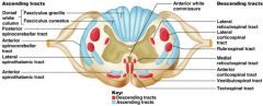
Anterior median fissure
|
separates anterior funiculi
|
|
|
Posterior median sulcus
|
divides posterior funiculi
|
|
|
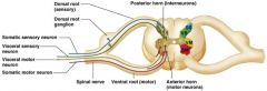
What does gray matter consists of
|
-soma
-unmyelinated -processes -neuroglia |
|
|
Grey commissure
|
connects masses of gray matter; encloses central canal
|
|
|
Posterior (dorsal) horns
|
interneurons
|
|
|
Anterior (ventral) horns
|
interneurons and somatic motor neurons
|
|
|
Lateral horns
|
contain sympathetic nerve fibers
|
|
|
Know
|
|
|
Know
|
|
|
TEST QUESTION!!
|
|
|
TEST QUESTION!!!
|
|
|
(Gray matter) Dorsal half
|
sensory roots and ganglia
|
|
|
(Gray matter) ventral half
|
Motor roots (anterior horn cells)
|
|
|
_______ and ______ roots fuse laterally to form spinal nerves
|
-dorsal
-ventral |
|
|
Fibers from touch and pressure receptors form collateral _______ with interneurons in the ______ horns
|
-Synapses, Dorsal
|
|
|
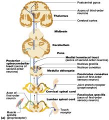
the _______ tracts send impulses to the cerebellum and do not contribute to sensory preception
|
spinocerebellar
|
|
|
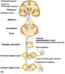
Lateral spinothalamic tract (TQ***)
|
nonspecific pathway for pain, temperature, and crude touch
|
|
|
TEST QUESTION
|
|
|
KNOW
|
|
|
Motor pathways
|
decending tracts delieve efferent impulses from the brain to the spinal cord, and are divided into two groups
|
|
|
Motor pathways involve
|
two neurons (upper and lower)
|
|
|
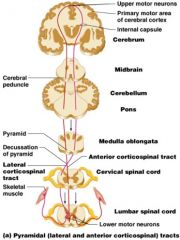
What do direct pathways originate with
|
the pyramidal neurons in the precentral gyri
|
|
|
Impulses from the direct pyramidal syatem are sent through the _________ tracts and ______ in the anterior horn
|
-corticospinal, synapse
|
|
|
What activates skeletal muscles
|
stimulation of anterior horn neurons
|
|
|
What are the two parts of the direct pathway called
|
-corticobullar tracts
-innervate cranial nerve nuclei |
|
|
Know
|
|
|
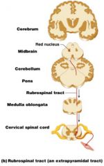
What parts are included in the indirect system
|
-rubrospinal, vestibulospinal, reticulospinal, and tectospinal tracts
|
|
|
Motor pathways are complex and multisynaptic, and regulate (3)
|
-axial muscles that maintain balance and posture
-Muscles controlling coarse movements of the proximal portions of limbs -Head, neck and eye movement |
|
|
Know
|
|
|
Recticulospinal tracts
|
maintain balance
|
|
|
Rubrospinal tracts
|
control flexor movements
|
|
|
Superior colliculi and tectospinal tracts
|
mediate head movements
|
|
|
Paralysis
|
loss of motor function
|
|
|
Flaccid paralysis
|
sever damage to the ventral foor or anterior horn
|
|
|
Spastic paralysis
|
only upper motor neurons of the primary motor cortex are damages
|
|
|
in spastic paralysis spinal neurons are intact and muscles are
|
stimulated irregulary
|
|
|
In spastic paralysis there is no
|
voluntary control of muscles
|
|
|
Paraplegia
|
transection between T1 and L1
|
|
|
Quadriplegia
|
Transection in the cervical region
|
|
|
Poliomeylitis
|
destruction of the anterior horn motor neurons by the poliovirus
|
|
|
Lou Gehrigs Disease
|
neuromuscualr condition involving destruction of anterior horn motor neurons and fibers of the pyramidal tract
|
|
|
When is CNS established (TQ)
|
during the first month of development
|
|
|
What can harm a fetus developing CNS
|
maternal exposure to radiation, drugs
|