![]()
![]()
![]()
Use LEFT and RIGHT arrow keys to navigate between flashcards;
Use UP and DOWN arrow keys to flip the card;
H to show hint;
A reads text to speech;
59 Cards in this Set
- Front
- Back
|
Site where 2 or more bones meet |
Articulations (joints) |
|
|
Functions of joints |
1. Give skeleton mobility 2. Hold skeleton together |
|
|
Classifications of joints |
1. Functional 2. Structural |
|
|
Functional classifications are based on___ |
Amount of movement joint allows |
|
|
Functional classifications |
1. Synarthroses (immovable joints) 2. Ampiharthroses (slightly movable) 3. Diarthroses (freely movable joints) |
|
|
Most common functional classification |
Diarthroses |
|
|
Structural classification of joints are based on |
- material binding bones together - presence/ absence of joint cavity |
|
|
Structural classification |
1. Fibrous joints 2. Cartilaginous joints 3. Synovial joints |
|
|
Fibrous joints |
- Joined by fibrous connective tissue - NO joint cavity - most synarthrotic ( NOT movable) |
|
|
Types of fibrous joints |
Sutures Syndesmoses Gomphoses |
|
|
Sutures |
Fibrous joints: Rigid, interlocking. Immovable joints for protection of brain Contain short connective fibers Allow for growth during youth In mid age, sutures ossify and fuse (synostoses) |
|
|
Syndesmoses |
FIBROUS JOINTS: - Bones connected by ligaments Fiber length and movement vary
|
|
|
When sutures ossify and fuse |
Synostoses |
|
|
If fibers of Syndesmoses are short. Is there much movement of joints? |
Little or NO movement (EX: tibula and fibula) |
|
|
If fibers of Syndesmoses are long. Is there any joint movement? |
Yes, large amt of movement is possible (EX: radius and ulna) |
|
|
Gomphoses |
FIBROUS JOINTS; - peg-in-socket joints of teeth in alveolar sockets -Fibrous connection is the periodontal ligament |
|
|
Cartilaginous joints |
- Bones United by cartilage - NO JOINT CAVITY - NOT highly movable |
|
|
Types of cartilagous joints |
1. Synchondroses 2. Symphyses |
|
|
Synchondroses. Examples |
CARTILAGOUS JOINTS; - bar/plate of hyaline cartilage unites bones - EXAMPLE: -temporary epiphyseal plate joints - cartilage of 1st rib and the manubrium ALL SYNARTHROTIC |
|
|
Symphyses. Examples |
CARTILAGOUS JOINTS; - fibrocartilage unites bone - hyaline cartilage present as articular cartilage - strong, flexible ampiarthroses Example: pubic symphysis |
|
|
Synovial joints |
- Bones separated by fluid-filled joint cavity -all are diarthrotic -ex: limb joints, most joints of body. |
|
|
Distinguishing features of synovial joints |
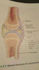
1. Articular cartilage: hyaline cartilage 2. Joint (synovial) cavity 3. Articular (joint) capsule 4. Synovial fluid 5. Reinforcing ligaments 6. Nerves and blood vessels |
|
|
Explain the articular cartilage and joint cavity feautures |
1.articular cartilage: Prevents crushing bone ends 2. Joint cavity: Small, fluid-filled potential space |
|
|
Layers of articular (joint) capsule |
1. External fibrous layer; Dense Irregular connective tissue 2. Inner synovial membrane; Loose connective tissue makes synovial fluid |
|
|
Synovial fluid features |
- viscous, slippery filtrate of plasma and hyaluronic acid - lubricates and nourishes articular cartilage - contains phagocytotic cells (Viscous =thick) |
|
|
Different types of reinforcing ligaments |
1. Capsular: thickened part of fibrous layer 2. Extracapsular: outside the capsule 3. Intracapsular: deep to capsule; covered by synovial membrane |
|
|
Nerves and blood vessels (synovial joints feautures) |
- nerve fibers - capillary beds |
|
|
Fatty pads |
Protects against trauma and injury. Found in some synovial joints like the hip and knee joint. |
|
|
Articular discs also called__. |
Menisci Separate articular cartilage - improve fit between articulating bone ends - stabilize joints -reduce wear and tear |
|
|
Sacs joined with synovial membrane. Function |
Bursae; reduce friction |
|
|
Tendon sheaths |
Elongated bursa wrapped completely around tendon subjected to friction |
|
|
Stability factors of synovial joints |
1. Shapes of articular surfaces (minor role) (Determine how joint can move) 2. Ligament number and location (limited role) ( the more ligaments the stronger) 3. Muscle tendons that cross joint (most important ) |
|
|
What keeps muscle taut? |
Muscle tone |
|
|
Types of synovial joints |
1. Planar joint 2. Hinge joint 3. Pivot joint 4. Condyloid joint 5. Saddle joint 6. Ball-and-socket joint |
|
|
Planar joint example |
Joint surface flat and slightly curved. Allows back n forth and side to side movement.
Ex: intercarpal joints |
|
|
Hinge joint |
Convex surface of a bone fits into concave surface of another. An open and closing action, like a hinge
Ex: Elbow joints |
|
|
Pivot joint |
Allows rotation around longitutional axis
Ex: atlantoaxial joint |
|
|
Condyloid joint |
Oval-shaped projections of one bone fits into Oval-shaped depression of another. Ex: Wrist joints |
|
|
Saddle joint |
A modified condyloid. ROM us expanded to move around all 3 axes. Ex: carpometacarpals joints of thumbs |
|
|
Ball-and-socket joint |
Has the most ROM of any joint in body. Ball surface of one bone fits into cup like depression of another bone. Ex: shoulder joints |
|
|
Different between: Syndesmoses Synarthrosis Synostoses Symphyses Synchondroses |
- Syndesmoses: fibrous joints - Synarthrosis: immovable joints - Synostoses: suture ossify into one unit - Symphyses: cartilaginous joints (fibrocartilage) - Synchondroses: cartilaginous joint (hyaline cartilage) |
|
|
Origin and insertion |
Origin attach to immovable bone Insertion attach to movable bone |
|
|
What does muscle contraction cause? |
Insertion to move toward origin Movements occur along transverse, frontal, or sagittal planes. |
|
|
ROM allowed by synovial joints |
1. Nonaxial: slipping movement only 2. Uniaxial: movement in one plane 3. Biaxial: movement in 2 planes 4. Multiaxial: movement in or around all 3 planes |
|
|
ROM is affected by |
Hormones 1. Relaxin: increase flexibility of pubic symphysis. (Child birth) 2. Disuse: restricted movement if joint not used for extended time |
|
|
Types of movement at synovial joints |
1. Gliding: (waving) 2. Angular movements: (noding head) 3. Rotation: medial/lateral rotation (shaking head) |
|
|
Gliding movement |
One flat bone surface glides or slips against another one |
|
|
Abduction |
Away from midline |
|
|
Adduction |
Adding to midline |
|
|
Largest most complex joint of body |
Knee joint |
|
|
Knee joint |
3 joints surrounded by a single joint cavity -femoropatellar joint: (gliding when knee flex) - lateral and medial tibiofemoral joints: allow Flexion, extension, and some rotation when knee partially flexed |
|
|
Modified hinge joint |
Knee joint |
|
|
Extra ligament found on the knee discovered 2 years ago. |
Anterolateral ligament (ALL) |
|
|
Inflammatory or degenerative disease that damages joints |
Arthritis |
|
|
Wear-and-tear irreversible disease |
Osteoarthritis |
|
|
What are luxations |
Dislocations |
|
|
Subluxation |
Partial dislocation |
|
|
More cartilage destroyed than is replaced |
Osteoarthritis |
|
|
Palms face front |
Supine |