![]()
![]()
![]()
Use LEFT and RIGHT arrow keys to navigate between flashcards;
Use UP and DOWN arrow keys to flip the card;
H to show hint;
A reads text to speech;
62 Cards in this Set
- Front
- Back
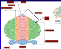
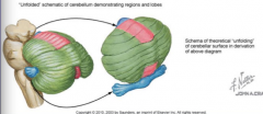
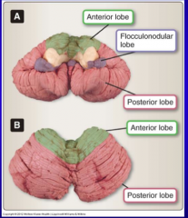
Identify: Lingua, paravermis, vermis, lateral hemisphere, anterior lobe, primary fissure, posterior lobe, flocculonodular lobe, nodule, flocculus |
|
|

|
|
|
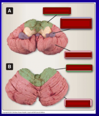
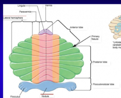
Identify: anterior lobe, flocculonodular lobe, posterior lobe |
|
|
|
What is the flocculonodular lobe heavily involved in? |
Maintaining balance |
|
Identify: fourth ventricle, fastigial nucleus, globose nucleus, emboliform nucleus, dentate nucleus
What function do these nuclei serve?
This is a section through the deep cerebellar nuclei and the medulla of the brainstem |
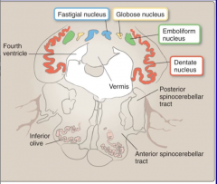
Primary relay points for efferent fibers traveling from cerebellar cortex (Purkinje cells) to other brain regions |
|
|
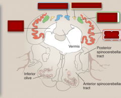
Identify where each projects:
Lateral hemispheres Paravermal zones Vermis
Use globose, emboliform, and dentate nucleus |
The lateral hemispheres project to the dentate nuclei, the paravermal zones project to the globose and emboliform, collectively known as the interpositus nuclei, and the vermis projects to the fastigial nuclei. |
|
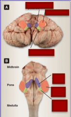
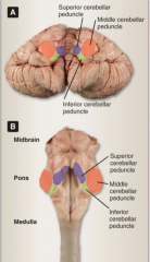
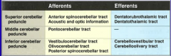
Identify superior cerebellar peduncle, middle cerebellar peduncle, and inferior cerebellar peduncle.
|
|
|
Complete |
|
|
|
What pathways carry information from he lateral portion of the anterior and posterior cerebellar cortices to the thalamus and thence onto the contralateral motor cortex to coordinate movement in the limbs ipsilateral to the cerebellar hemisphere of origin. |
Dentatothalamic tracts |
|
|
What are the five different neuron types in cerebellar gray matter? |
Basket Stellate Purkinje Golgi Granule |
|
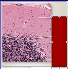
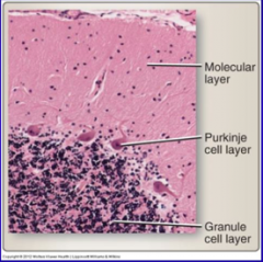
Identify molecule layer, Purkinje cell layer, and granule cell layer |
|
|
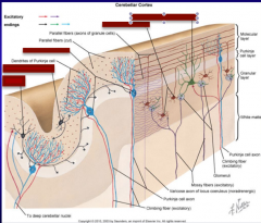
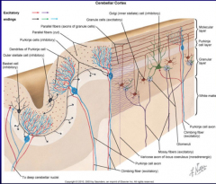
Identify the layer and whether each is inhibitory or excitatory: Basket cell, Golgi, Stellate, Granule, Purkinje |
|
|
|
Draw the wiring of the cerebellar cortex using stellate, basket, purkinje, climbing, granule, golgi, and mossy fiber.
What is the only output neurons of the cerebellum?
What is the only direct input (afferent) to the Purkinje cells from outside the cerebellum? |
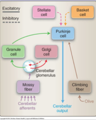
Purkinje cells
Climbing fibers (origin in olivary nuclei) |
|
|
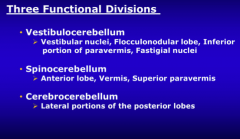
What are the three functional divisions of the cerebellum? |
|
|
|
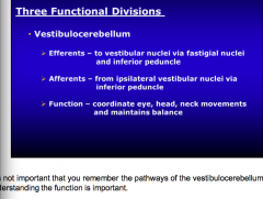
What is the function of the vestibulocerebellum? |
|
|
|
What is the function of the spinocerebellum? |
Coordinate trunk and proximal limb movements. |
|
|
What is the function of the cerebrocerebellum? |
Coordinate fine motor planning of limbs Anticipates sensory consequences of movement Cognitive memory of motor functions
Right hemisphere controls right body, arm, and leg (same for left) |
|
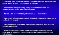
What are these all clinical signs of? |
Cerebellar dysfunction |
|
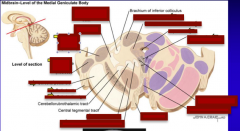
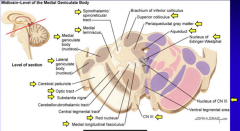
Identify: Spinothalamic/spinoreticular tract Superior colliculus Aqueduct Periaqueductal gray matter, nucleus of Edinger-Westphal, CN III, nucleus of CN III, red nucleus, medial longitudinal fasciculus, substantia nigra, optic tract, lateral geniculate body (nucleus), cerebral peduncle, medial geniculate body (nucleus), medial lemniscus |
|
|
|
functions in the control of reflex movements that orient the eyes, head, and neck in response to visual, auditory, and somatic stimuli |
Superior colliculus |
|
|
functions in the processing of autonomic and limbic activities, as well as modulation of nociception |
Periaqueductal gray matter |
|
|
passageway connecting the third and fourth ventric |
Aqueduct |
|
|
parasympathetic innervation of the eye to constrict the iris and to the ciliary muscle to alter lens shape for accommodation |
Nucleus of Edinger-Westphal |
|
|
motor control of eye muscles |
Cranial nerve III nuclei and nerve |
|
|
fiber pathway to thalamus for pain/temperature from the periphery |
Spinothalamic tract |
|
|
sensory pathway for proprioception connecting the nucleus gracilis and cuneatus with the thalamus |
Medial lemniscus |
|
|
thalamic relay nuclei for auditory information |
Medial geniculate |
|
|
thalamic relay nuclei for visual information |
Lateral geniculate |
|
|
fiber bundles of the corticospinal tract connecting the cerebral cortex to the brainstem |
Cerebral peduncle |
|
|
optic fibers from optic chiasm to the lateral geniculate |
Optic tract |
|
|
one of several nuclei involved in smooth motor control; degenerates in Parkinson’s disease |
Substantia nigra |
|
|
relay nuclei between the cerebellum to the thalamus |
Red nucleus |
|
|
fiber pathway between the vestibular nuclei and the CN nuclei III, IV, VI to coordinate head/eye movements |
Medial longitudinal fasciculus |
|
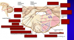
Identify: superior cerebellar peduncle, mesencephalic nucleus of CN V, middle cerebellar peduncle, locus coeruleus, medial longitudinal fasciculus, fourth ventricle, raphe nucleus (pontis), tectospinal tract, medial lemniscus, pontine nuclei, corticospinal tract, spinothalamic/spinoreticular tract, CN V, main (chief sensory nucleus of CN V), motor nucleus of CN V |
|
|
|
fiber pathway to and from the cerebellum |
Superior cerebellar peduncle |
|
|
fiber pathway to and from the cerebellum |
Middle cerebellar peduncle |
|
|
– three nuclear components of the trigeminal nerve nuclei |
Mesencephalic, Main sensory nucleus, Motor nucleus of CN V |
|
|
motor and sensory fibers of CN V |
Cranial nerve V |
|
|
collection of neurons in the pons that receive input from the neocortex and send crossing fibers through the middle cerebellar peduncle |
Pontine nuclei |
|
|
a noradrenergic brainstem nucleus involved in mood, sleep/wake cycle |
Locus coeruleus |
|
|
one of several serotonin type nuclei involved in mood, sleep/wake cycle |
Raphe nucleus pontis |
|
|
motor fibers from neocortex to spinal interneurons and lower motor neurons |
Corticospinal tract |
|
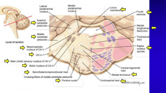
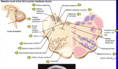
Identify the following: tracts solitarius, medial vestibular nucleus, nucleus solitarius, reticular formation, medial lemniscus, hypoglossal nucleus CN XII, inferior olivary nucleus, pyramid, spinal nucleus of CN V, spinothalamic/spinoreticular tract, spinal tract of CN V, dorsal motor nucleus of CN X, CN X, inferior cerebellar peduncle, inferior vestibular nucleus |
|
|
|
nuclei of the vestibular system that regulate balance |
Inferior and medial vestibular nuclei |
|
|
sensory nucleus for taste (from CN VII), glands, chemo/baroreceptors (CNs IX, X) |
Nucleus and tractus solitarius |
|
|
parasympathetic motor nucleus to lungs and gut |
Dorsal motor nucleus vagus (CN X) |
|
|
nuclear and tract components of CN V that extends down into the upper cervical spinal cord |
Spinal nucleus and tract of CN V |
|
|
origin of the climbing fibers to the Cerebellar Purkinje cells |
Inferior olivary nucleus |
|
|
name given to the corticospinal tract fibers in medulla |
Pyramid |
|
|
a network of neurons and axons that reside in the brain stem tegmentum involved in arousal, respiration, heart rate control |
Reticular formation |
|
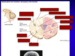
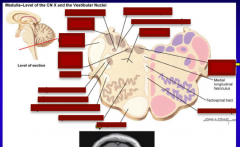
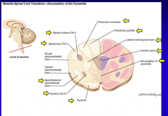
Identify the following: spinal nucleus CN V Spinothalamic/spinoreticular tract, pyramid, nucleus CN XI, fasciculus cutaenous, lateral corticospinal tract, fasciculus gracilus, central canal, decussation of pyramids |
|
|
|
glossopharyngeal nucleus with motor control over tongue and pharyngeal muscles with taste from |
Nucleus CN IX |
|

|
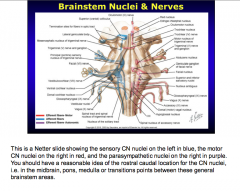
|
|
|
What is the arterial supply to the midbrain? |
Tip of the basilar artery and/or branches of the PCA |
|
|
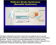
What structures would be involved with a vascular problem at each midbrain region? What would be the clinical symptoms of each?
Base Tegmentum
Base + tegmentum |
Base = CN III, cerebral peduncle => ipsilateral 3rd nerve paresis, contralateral hemiparesis Tegmentum = CN III + superior cerebellar peduncle + red nucleus => Ipsilateral 3rd nerve paresis, contralateral tremor, contralateral ataxia Base + tegmentum = same + substantial nigra => ipsilateral 3rd nerve paresis, contralateral hemiparesis, contralateral tremor, contralateral ataxia |
|
|

|
|
|
Where are PONTINE syndromes located?
|
Vascular syndromes of the medial pons |
|
|
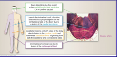
Pontine syndrome: Paramedian branches of basilar artery
What structures involved and what clinical symptoms? |
Corticospinal and corticobulbar tracts, CN VII fascicles
Contralateral hemipariesis, ipsilateral LMN, facial paralysis
Other: could also involve in addition PPRF and/or CN VI => same symptoms + ipsilateral gaze paresis |
|
What are these all symptoms of? |
Pontine syndromes |
|
|
Contralateral arm/leg weakness Contralateral decrease in position/vibration Ipsilateral tongue weakness
Identify structures involved, arterial supply, and brainstem region |
Corticospinal tract, medial lemniscus, CN XII
Vertebral arteries, paramedial branches
Medial medulla |
|
|
Ipsilateral ataxia, vertigo, nausea, ipsilateral decrease in face pain sensation, contralateral decrease in body pain sensation, ipsilateral Horner's dysphagia |
Vestibular nuclei Inferior cerebellar peduncle CN V and tract spinothalamic tract Sympathetic fibers Nucleus ambiguus Nucleus solitarius
Vertebral artery or posterior inferior cerebellar artery (PICA)
Lateral medulla |
|
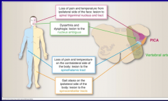
Diagnosis? |
Wallenburg syndrome |