Reading...
![]()
Play button
![]()
Play button
![]()
Use LEFT and RIGHT arrow keys to navigate between flashcards;
Use UP and DOWN arrow keys to flip the card;
H to show hint;
A reads text to speech;
65 Cards in this Set
- Front
- Back
|
The cerebellum develops from the ______.
|
The cerebellum develops from the ALAR PLATE.
|
|
|
All fiber systems except the __________ fibers are mossy fibers in the cerebellum.
|
All fiber systems except the OLIVOCEREBELLAR fibers are mossy fibers in the cerebellum.
Mossy fibers also project to the deep cerebellar nuclei. Mossy fibers chiefly synapse on granule cells. |
|
|
Define:
Vermis |
Vermis: the midline of the cerebellum which divides the two hemispheres.
|
|
|
All of the fiber systems are ____ fibers, except the olivocerebellar fibers.
|
All of the fiber systems are MOSSY fibers, except the olivocerebellar fibers.
|
|
|
What are the 3 major fissures of the cerebellar cortex and what do they separate?
|
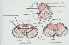
Primary - separate the anterior and posterior lobes
Horizontal: separate superior and inferior surfaces Poterolateral: separates the posterior lobe from the flocculonodular lobe or vestibulocerebellum |
|
|
Two fiber types that synapse directly on purkinje cell dendrites:
|
1.Parallel fibers
2. Climbing fibers Both synapse on purkinje cell dendrites |
|
|
3 subdivisions of the cerebellar cortex:
|
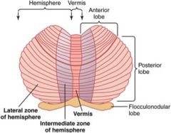
3 subdivisions of the cerebellar cortex:
1. Vestibulocerebellum - flocculonodular lobe 2. Spinocerebellum - vermis and paravermal zones of cerebellar hemispheres. 3. Pontocerebellum |
|
|
What neurotransmitter do Purkinje Cells use?
|
GABA! (inhibitory)
|
|
|
What makes Purkinje fibers of the Vestibulocerebellum special?
|

The Vestibulocerebellum is the only part of the cerebellum where purkinje cells which project DIRECTLY to location outside the cerebellum.
The project to all FOUR VESTIBULAR NUCLEI via the ICP. (Vest.Cerebellum. influences the action of vestibulospinal tracts for righting reflex and the stabilize head and neck for eye movements associated with gaze) |
|
|
Parallel fibers are the axons of ________ cells and course in the ________ layer.
|
Parallel fibers are the axons of GRANULE cells and course in the MOLECULAR layer.
|
|
|
Three layers of Cerebellum from innermost to outermost.
|
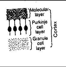
Three layers of Cerebellum from innermost to outermost:
1. Granule cell layer (innermost) 2. Purkinje cell layer 3. Molecular cell layer (outermost) |
|
|
Outer Stellate Cells inhibit ________ cells and are located in the _______ layer.
|
Outer Stellate Cells inhibit PURKINJE cells and are located in the MOLECULAR layer.
|
|
|
What is Arbor Vitae?
|
Arbor Vitae:
Cerebellar white Matter |
|
|
What are cerebellar glomeruli?
What 4 things do they contain? |

Cerebellar Glomeruli are clear areas in between clusters of granule cells.
They contain: 1. dilated axon terminals of mossy fibers 2. granule cell dendrites 3. golgi cell axons 4. glial capsule ensheathing 1-3 |
|
|
What (4) tracts travel through
the Inferior Cerebellar Peduncle to the Cerebellum? Name which are crossed or uncrossed. |
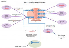
1. Olivocerebellar Tracts - Crossed
2. PSCT - Uncrossed 3. Cuneocerebellar Tracts - Uncrossed 4. Vestibulocerebellar Fibers - Uncrossed |
|
|
Olivocerebellar Tracts:
Crossed or Uncrossed? Origin: Termination: Fibers: |
Olivocerebellar Tracts:
Crossed Origin:Inferior Olivary Complex (vestibular nuclei, SC, and cortex) Termination: 1) Vestibulo cerebellum (visular vestibular info) 2) Spinocerebellum (motor feedback) 3) Pontocerebellum (motor info) FIbers: CLIMBING FIBERS!@ |
|
|
Post. Spinocerebellar Tract (PCST)
Crossed or Uncrossed? Origin: Termination: Info: Fibers: |
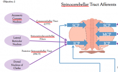
Post. Spinocerebellar Tract (PCST)
Uncrossed Origin: Dorsal Nucleus of Clark Termination:Spinocerebellum Info: Proprioception from lower limbs Fibers: MOSSY |
|
|
Cuneocerebellar Tracts
Crossed or Uncrossed? Origin: Termination: Info: Fibers: |

Cuneocerebellar Tracts
Uncrossed? Origin: Accessory Cuneate Nucleus of Medulla Termination: Spinocerebellum Info: Proprioception from upper limbs Fibers:MOSSY |
|
|
Vestibulocerebellar Fibers
Crossed or Uncrossed? Origin: Termination: Info: Fibers: |
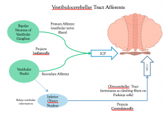
Vestibulocerebellar Fibers
Uncrossed Origin: Vestibular Nerve and Nuclei Termination: Vestibulocerebellum Info: Vestibular Fibers:MOSSY |
|
|
What tracts travel to the
Cerebellum through the Middle Cerebellar Peduncle? Crossed or Uncrossed? |

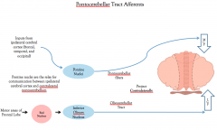
1. Pontocerebellar Fibers
Crossed |
|
|
Pontocerebellar Fibers
Crossed or Uncrossed? Origin: Terminate: Info: Fibers: |

Pontocerebellar Fibers
Crossed Origin: Pontine nuclei (with massive inputs from cerebral cortex) Terminate: Pontocerebellum and Spinocerebellum Info: Serve as a relay from the cerebral cortex to the contralateral spino-/pontocerebellum Fibers: MOSSY |
|
|
What tracts travel to the
Cerebellum through the Superior Cerebellar Peduncle Crossed or Uncrossed? |

Anterior Spinocerebellar Tract - Ipsilateral (crossed twice)
|
|
|
Anterior Spinocerebellar Tract (ASCT)
Crossed or uncrossed? Origin: Terminate: Info: Fibers: |
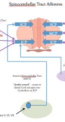
Anterior Spinocerebellar Tract
Ipsilateral - crossed twice Origin: Intermediate song of gray matter of lumbar and sacral spinal segments Terminate: Spinocerebellum Info: intergrated whole limb Golgi tendon info from lower limb Fibers: MOSSY |
|
|
What are the two types of
fibers that enter the cortex of the cerebellum, and where are they from? |
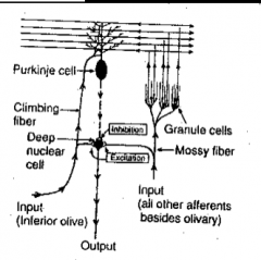
MOSSY Fibers - all afferents except for olivocereballear fibers
CLIMBING Fibers - from the inferior olive |
|
|
What three things does
the granule cell layer contain? 2 cells types 1 synaptic structure |
Granule layer:
2 cell types- Granule neurons, Golgi cells 1 synaptic structure- Mossy fibers |
|
|
Describe Granule cells:
Size: Dendrite size: Path of Axons Where they synapse: |
Describe Granule cells:
Size: small Dendrites: short dendrites Path of Axons: axons ascend to the molecular layer and bifurcate to form a PARALLEL FIBER Where they synapse: Form EXCITATORY synapses on Purkinje cell dendritic arborations |
|
|
Describe Golgi cells:
Size Type of dendrite Axon target |
Describe Golgi cells:
Size: Large neurons Type of dendrite - Radiating dendrites Axon target: INHIBITORY contacts on GRANULE cells in the synaptic glomerulus |
|
|
What 3 things make up a
cerebellar glomerulus? |
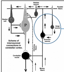
cerebellar glomerulus
1. Mossy Fiber axon terminals - excitatory synapses with Granule cells. 2. Golgi cell axon terminals - inhibitory synapses with Granule cells 2. Glial capsule |
|
|
Where do the axons of the
Purkinje cell layer go? |
Purkinje cell layer axons make INHIBITORY synapses on cells in the DEEP CEREBELLAR NUCLEI or VESTIBULAR nuclei
|
|
|
What neurotransmitter do Purkinje cells release?
|
GABA..... (Inhibitory)
|
|
|
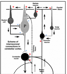
Climbing Fibers:
Origin: Function: |
Climbing Fibers:
Origin: Inf. Olivary Nucleus via Olivocerebellar Tract ONLY!! Function: Climbing Fibers powerfully EXCITE Purkinje cells as well as cerebellar interneurons. |
|
|
Parallel Fiber:
Originate: Function: |

Parallel Fibers:
Originate: Granule Cells Function: Parallel fibers make EXCITATORY synpases with dendrites of Purkinje cells and interneurons. |
|
|
Basket Cells
Function: |
Basket Cells
Function: INHIBIT Purkinje cells by synapsing on the purkinje cell stroma. |
|
|
Stellate Cells
Function: |
Stellate Cells
Outer stellate cells make INHIBITORY synapses on Purkinje cell dendrites |
|
|
How do mossy fibers influence
a large number of Purkinje cells? |

MOSSY fibers stimulate Purkinje cells INDIRECTLY via the PARALLEL Fibers of GRANULE Cells
|
|
|
What is the phenomenon of how mossy fibers influence Purkinje cells
called? |
Mossy fibers stimulate
Purkinje cells indirectly via granule cells/parallel fibers “Signal Averaging” allows Purkinje cells to process large amounts of information that is used for moment to moment coordination of motor function |
|
|
Climbing Fibers
Function: |
Climbing Fibers
DIRECTLY stimulate Purkinje (ONLY from Inf. Olive) |
|
|
Where is Cortical outflow
via Purkinje axons directed? |
1. Vestibular Nuclei
2. (4) pairs of Deep Cereballar Nuclei 4 Deep Cerebellar Nuclei: Fastigial, Globose, Emboliform, Dentate (Fat Guys Eat Donuts) |
|
|
What neurotransmitter
do the Deep cerebellar nuclei release? |
GLUTAMATE!
Excitatory! |
|
|
Why is the Vestibulocerebellum’s
output significant? |
The vestibulocerebellum is the only region whose Purkinje cells project directly to a region outside the cerebellum.
It has no projections to Deep CB Nuclei |
|
|
Purkinje cells of the
vestibulocerebellum project ipsilaterally to: |

Purkinje cells of the vestibulocerebellum project ipsilaterally to:
all (4) vestibular nuclei. Function: Extension of all extremities; Controls neck movements to stabilize the head for eye movements associated with gaze |
|
|
Purkinje cells of the
vestibulocerebellum carry out which functions? |
Vestibulocerebellum Functions:
Extension of all extremities; Controls neck movements to stabilize head for eye movements associated with gaze. |
|
|
Vermal Pathway-
Course: Function: |

Vermal pathway-
Course: From the vermis (Spinocerebellum) to the FASTIGIAL nucleus via Purkinje cells. The Fastigial nucleus projects via the ICP to the Vestibular Nuclei and Reticular Formation Funtional Region: Spinocerebellum. Function: Axial and proximal control of muscles to maintain posture and generate walking. |
|
|
Where does the fastigial
nucleus relay information? |
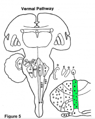
The fastigial nucleus
relays information BILATERALLY to the vestibular nuclei and reticular formation through the ICP. Function: Axial and Proximal Limb control to maintain posture. |
|
|
Paravermal Pathway:
Course: Function: |

Paravermal Pathway:
Course: Purkinje cells in the paravermal cortex (spinocerebellum) project to the GLOBOSE and EMBOLIFORM nuclei Function: Transmit- 1. Fine movemens of distal muscuature 2. Coordination and correction f ongoing movements NOTE: The paravermal pathway influences movement on the IPSILATERAL side |
|
|
From the globose and
emboliform nuclei, where does the information go? |
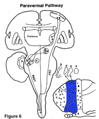
1. The info exits the SCP
2. Crosses the midline 3. Terminates in the red nucleus and thalamus which projects to the motor cortex (Rubrospinal and Corticospinal Tracts) note: the paravermal pathway influences distal limb motor control on the ipsilateral side |
|
|
Lateral Hemisphere Pathway:
Course: Function: |
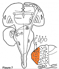
Lateral Hemisphere Pathway:
Course: Purkinje cells in the pontocerebellum project to the DENTICULATE nucleus. From the DENTICULATE Nucleus fibers course in the SCP to the Red Nucleus and Thalamus (Rubrospinal and Corticospinal tracts) Function: 1. Influence planning of complex movements 2. Facilitate initiation of distal limb movements |
|
|
Where does the dentate
nucleus project to? |

the dentate nucleus projects
via the SCP to the: -contralateral red nucleus -contralateral thalamus -a thalamic nucleus that projects to motor planning areas of the frontal cortex |
|
|
What are the two midline cerebellar lesions?
Most usual causes? |
What are the two midline cerebellar lesions?
Vestibulocerebellum - meduloblastoma Spinocerebelum: Alcoholic degeneration |
|
|
Symptoms of Vestibulocerebellum lesion
|
Vestibulocerebellum
1. Ataxic gait 2. Head Tremor 3. Nystagmus Sympotoms improve when patient is recumbent. |
|
|
Symptoms of Spinocerebellum lesion
|
Spinocerebellum
1. Truncal Ataxia (a loss of coordinated muscle movements for maintaining normal posture of the trunk) |
|
|
I which midline cerebellar lesion do
symptoms improve when the patient is recumbent? |
Vestibulocerebellum
|
|
|
What are the results of
Hemispheric lesions? |
Hemispheric lesions
result in asynergia (disordered movements) of ipsilateral limbs |
|
|
Asynergia can results in:
|
Dysmetria
-errors in distance of movements -intention errors -disdiachokinesis -speech disturbances |
|
|
Can cerebellar damage
cause paralysis? |
No, cerebellar damage
does not cause paralysis |
|
|
What are the (4) Deep Cerebellar Nuclei listed Medial to Lateral.
|
4 Deep Cerebellar Nuclei
1. Fastigial (medial) 2. Globose 3. Emboloform 4. Denticulate (Lateral) |
|
|
In genreal: The cerebrocerebellum projects primarily to the _______ nucleus.
|
In genreal: The cerebrocerebellum projects primarily to the DENTATE nucleus.
(Cerebrocerebellar pahways from dentate nucleus are mainy desined for the premotor and associaional cortices of the fronal lobe; motor planning) Cross in SCP desussation. Dentate nucleus also sends collaterals to Red Nucleus which relay to Inf.Olive (feedback loop) |
|
|
In genreal: The spinocerebellum projects primarily to the _______ nuclei.
|
In genreal: The spinocerebellum projects primarily to the INTEPOSED and FASTIGIAL nuclei.
Spinocerebellar pathways are directed towards circuits of UMN that govern motor execution. Fastigial nuclei project via he Inf.Cerebellar Pedunce to nuclei of he reticular formation and vestibular complex |
|
|
In genreal: The vestibulocerebellum projects primarily to the _______ complex.
|
In genreal:
The vestibulocerebellum projects primarily to the VESTIBULA complex. (For this reason, parts of the vestibular complex may be considred a functional and anatomical component of the deep cerebellar nuclei) Vestibulocerebellum proections course through the inferior cerebelar pedunce and terminate in nuclei of the vestibular complex that govern the movements of eyes, head, and neck, compensation for linear and rotaional accelerations of the head. |
|
|
What two Peduncles relay inputs to the cerebellum?
Which one is only afferent? Which one is afferent/efferent? |
Middle Cerebellar Peduncle - mostly AFFERENT
Inferior Cerebellar Pedunce - both efferent and afferent |
|
|
What pedunces relay info OUT of the cerebellum?
Which one is only efferent? |
Superior Cerebellar Peduncle - mostly EFFERENT
Inferior Cerebellar Pedunce - both efferent and afferent |
|
|
Vestibular axons from CN8 and axons from vestibular nuclei in the medulla project to the __________cerebellum.
|
Vestibular axons from CN8 and axons from vestibular nuclei in the medulla project to the VESTIBULOcerebellum.
|
|
|
Dorsal Nucleus of Clarke (SC) and Accessory cuneate nucles (Medulla) project to the __________cerebellum.
What information do they convey? |
Dorsal Nucleus of Clarke (SC) and Accessory cuneate nucles (Medulla) project to the SPINOcerebellum.
They convey proprioceptive information. (Proproceptive info from the face is relayed via the mesencehaic nucleus of CNV complex) |
|
|
The cerebral cortex projects (mainly) to the __________cerebellum.
|
The cerebral cortex projects (mainly) to the CEREBROcerebellum.
|
|
|
Most of the projections from the cerebellum to UMN in superior colliculus arise in the______ and ______ nuclei, which receive input from LATERAL/MEDIAL portions of cerebellar cortex.
|
Most of the projections from the cerebellum to UMN in superior colliculus arise in the DENTAE and INTERPOSED nuclei, which receive input from LATERAL/MEDIAL portions of cerebellar cortex.
|