![]()
![]()
![]()
Use LEFT and RIGHT arrow keys to navigate between flashcards;
Use UP and DOWN arrow keys to flip the card;
H to show hint;
A reads text to speech;
14 Cards in this Set
- Front
- Back
|
.. |
|
|
|
Adaptation (4) |
Exogenous stimuli induce new state, that changes cell but preserves viabilty Examples:
|
|
|
Reversible Injury |
Pathologic cell changes that can be restored to normal if stimuli is removed and if injury is mild |
|
|
Irreversible |
When stressors exceed capacity to adapt permanent pathologic changes cause cell death |
|
|
Causes (6) |
- Ischaemia
- Drugs, Alohol
- Trauma
- Viruses
- Reaction to infection
- Gene mutations
- Protein and vitamin deficiency |
|
|
Mechanisms of Cell Injury (5) |
Cell injury results from abnormalities in one or more offive cellular components:
|
|
|
Cell Death |
- Always represents the pathological process
- Represents the pathological process or severe normal physiological process |
|
|
Necrosis |
The sum of all the morphological changes that lead to cell death in living tissue/organs - denaturation of protein - enzymeatic digestion of cellular components and organelles |
|
|
Types of Necrosis (4) |
-Most common - Hypoxic death in all cells except brain - Protein denaturation with preservation of cell and tissue framework
- Common in abscessed and brain infarcts - Auto lysis/heterolysis predominate over protein denaturation - Neutrophils release toxic contents "liquefy" tissue: soft and fluid filled
- Due to trauma or release of enzymes - -
|
|
|
Apoptosis |
Programmed cell death occurs when a cell dies through regulated internal suicide program. apoptosis eliminates unwanted cells selectively - minimal disturbance surrounding cells Cell membrane remains in tact structure changes to become a target of phagocytosis - cell shrinks and chromatin degraded, blebbing and fragmentation into apoptotic bubbles which removed by phagocytosis |
|
|
Apoptosis Causes (2) |
- Programmed destruction during embryogenesis -Seletion of harmful self-reactive lymphocytes
- Cell death in certain viral infections (Hep.) - Cell death in tumours |
|
|
Dysregulated Apoptosis |
Decrease in apoptosis increase of cell survival
Increase in apoptosis and excess in cell death
|
|
|
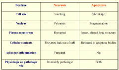
Necrosis vs. Apoptosis |
|
|
|
*Thrombosis, Ischaemia, Infarction |
|