Reading...
![]()
Play button
![]()
Play button
![]()
Use LEFT and RIGHT arrow keys to navigate between flashcards;
Use UP and DOWN arrow keys to flip the card;
H to show hint;
A reads text to speech;
52 Cards in this Set
- Front
- Back
|
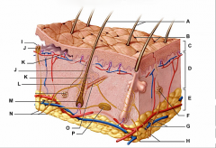
integumentary system
|
includes the skin and its derivatives such as glands, hair, feathers, claws, and scales
largest organ (15% of body weight) thickness variable, normally 1-2mm; dermis may thicken up to 6mm; stratum corneum layer increased (calluses on hands and feet) |
|
|
2 layers of the skin
|
the epidermis (outer stratified epithelium) and dermis (underlying connective tissue); separated by a non-cellular basement membrane
|
|
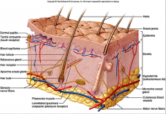
epidermis
|
identify C
stratified epithelium w/ mitotically active basal layer keratinized stratified squamous epithelium avascular, no blood vessels |
|
|
functions of the integumentary system
|
protection environment, UV rays, microbial pathogens
water and heat loss regulation, metabolic reactions excretory system vitamin D synthesis location of cutaneous sense organs |
|

hypodermis
|

(identify E)
subcutaneous tissue / superficial fascia mostly adipose tissue hypodermic injections (subQ) highly vascular |
|
|
cell types of the epidermis
|
keratinocytes
melanocytes langerhans cells merkel cells |
|
|
keratinocytes
|
most abundant
tightly connected by desmosomes produce keratin fibrils |
|
|
melanocytes
|
produce melanin pigment
melanin production increases when skin is exposed to sunlight, to protect DNA inside cells nuclei from UV induced mutations |
|
|
langerhans cells
|
play a role in immunity
|
|
|
merkel cells
|
in conjunction with sensory nerve endings, form Merkel disks (touch receptors) at epidermal juntion
|
|
|
keratin
|
fibrous protein that give epidermis its durability and protective capabilities
|
|
|
layers of the epidermis (top to bottom)
|
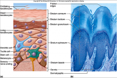
stratum corneum (horny layer)
stratum lucidum (clear layer) stratum granulosum (granular layers) - lamellated granules - keratohyaline granules stratum spinosum (spiny layer) stratum basale (basal layer) |
|

stratum corneum (horny layer)
|

(identity A)
outermost 20-30 layers of keratinized dead cells. These cells constantly rubbing of and are being replaced by division of deeper cells. consisting of dead keratinized cells. * Does not ensure water loss across the skin. |
|

stratum lucidum (clear layer)
|
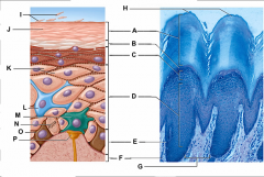
(identity B)
Flattened dead keratinocytes with indistinct boundaries. Not present in regions of thin skin. |
|
|
lamellated granules
|
located in the stratum granulosum
contain waterproofing glycolipid that is secreted into the extracellular space. |
|
|
keratohyaline granules
|
located in the stratum granulosum
Combine with the intermediate filaments in the more superficial layers to form keratin fibrils. |
|
|
Stratum germinativum
|
mitotically active basal layer to replace sheets that are constantly shed
|
|

stratum granulosum (granular layers)
|

(identity C)
where keratinization begins thin layer, cells contain abundance of granules. - lamellated granules - keratohyaline granules |
|

stratum spinosum (spiny layer)
|

(identity D
)Several layers superficial to the basal layer. Cells contain web- like bundles of intermediate filaments made of pre-keratin protein. Cells undergo rapid cell division. This is the last layer after stratum basale to receive adequate nourishment via diffusion, of nutrients from the dermis. As the daughter cells are pushed away toward the top layers, they gradually die. |
|

stratum basale (basal layer)
|

(identity E)
Single row of cells immediately adjacent to the dermis. Cells constantly undergo mitotic division to produce millions of new cells. Up to 25% of the cells are melanocytes. |
|
|
dermis
|
connective tissue layer
consists of the papillary layer and reticular layer |
|
|
papillary layer
|
- Superficial dermal region
- Areolar loose connective tissue - Dermal papillae - Abundance of capillary networks - Free nerve endings – pain receptors - Meissner’s corpuscles – touch receptors - Regions of the Dermis |
|
|
reticular layer
|
- Deepest skin layer
- Dense irregular connective tissue - Vascularized - Sudoriferous (Sweat) glands - Sebaceous (Oil) glands - Pacinian corpuscles – pressure receptors |
|
|
dermal papillae
|
uneven with fingerlike projections
*Finger prints – unique pattern of epidermal regions that remain unchanged throughout life. |
|
|
integumentary derivatives
|
claws, horns, antlers, scales, feathers, hair
glands (sebaceous, sudoriferous, eccrine, apocrine) |
|
|
claws
|
Present in all vertebrate phyla, but rare in amphibians
|
|
|
horns
|
Non-shedded, bone covered with a keratinized sheath
|
|
|
antlers
|
Shedded, bones which are covered with blood vessels (“velvet”) when first formed
|
|
|
scales
|
Derived from ancient armor-like coating known as “Ostracoderms”
|
|
|
hair
|
Originate in epidermis, but sink to dermis,
|
|
|
vibrissae
|
specialized sensory hairs (whiskers)
Whiskers, tactile hairs. Most common in nocturnal mammals. |
|
|
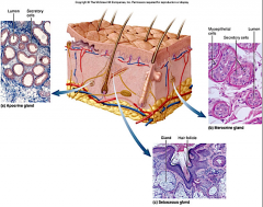
Sebaceous (oil) glands
|
found all over the skin, except for palms of the hands and soles of the feet. Ducts usually empty into the hair follicle, or directly on the skin.
(sebum, blackheads, and acne) |
|
|
sebum
|
from sebaceous (oil) glands
mixture of oil and fragmented cells, that act as a lubricant to keep skin soft and moist, and hair from becoming brittle |
|
|
blackheads
|
from sebaceous (oil) glands
accumulation of dried sebum, bacteria, and melanin in the duct |
|
|
acne
|
from sebaceous (oil) glands
active inflammation of sebaceous glands |
|
|
Sweat (sudoriferous) glands
|
widely distributed all over the skin.
Pores – epithelial openings eccrine and apocrine glands |
|
|
pores
|
epithelial openings that permit glandular excretions
|
|
|
eccrine glands
|
produce clear perspiration consisting primarily of water, salts, and urea. Under control of the nervous system, part of the bodies heat regulating apparatus
|
|
|
apocrine glands
|
axillary and genital areas. Secrete milky protein and fat-rich substance, also water, salts and urea, (nutrient source for microorganisms found on the skin)
|
|
|
integumentary system characteristics (dogfish)
|
mucous cells - produce toxins associated w/ fin spines
sensory cells - lateral line dermis - connective tissue melanophores - affect skin color placoid scales |
|
|
placoid scales (dogfish)
|
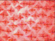
derived from the epidermis and dermis, embedded in the dermis
made of a bony base > dentin > spine > pulp cavity |
|
|
integumentary system characteristics (mudpuppy)
|
skin is a respiratory organ in amphibians (must be kept moist)
mucus gland poison gland chromatophores |
|
|
poison gland
|

common in amphibians, modified mucus gland
|
|
|
chromatophores
|
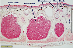
give amphibians their specific color and skin patterns
|
|
|
stratum spongiosum
|
connective tissue in the dermis
|
|
|
stratum compactum
|
heavily infiltrated with collagen fibers, in the dermis
|
|
|
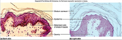
integumentary system characteristics (mammal)
|
thick skin
cat dermis is thicker than the epidermis but not well stratified connective tissue nictitating membrane vibrassea |
|
|
connective tissue
|
in the dermis, contains fibers, adipose tissue, blood vessels, nerves, glands, hair, Epidermal Derivatives
|
|
|
nictitating membrane
|
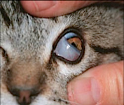
Transparent eyelid that can be drawn across the eye for protection and lubrication while maintaining visibility
|
|
|
skin pigmentation
|
hemoglobin - red pigment of red blood cells
carotene - yellow pigment (concentrates in the stratum corneum and fat) melanin - yellow, brown and black hues (pigment synthesis stimulated by UV radiation) |
|
|
cutaneous glands
|
located in the dermis
apocrine, sebaceous, merocrine glands |
|
|
structure of a hair follicle
|
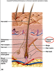
epithelial root sheath
connective tissue root sheath hair receptors entwine each follicle piloerector muscle - goose bumps, raises hair |