Reading...
![]()
Play button
![]()
Play button
![]()
Use LEFT and RIGHT arrow keys to navigate between flashcards;
Use UP and DOWN arrow keys to flip the card;
H to show hint;
A reads text to speech;
75 Cards in this Set
- Front
- Back
|
What are the main parts of the urinary system?
|
Kidneys
Urinary bladder Ureters Urethra |
|
|
Name some functions of the urinary system.
|
Filter blood
Remove waste products Recover useful metabolites Store fluid waste Transport waste to exterior Homeostasis |
|
|
Which homeostatic factors does the kidney have a role in?
|
Fluid volume regulation
Acid/base regulation Electrolyte composition regulation Blood pressure regulation Erythropoesis Vitamin D activation |
|
|
Where do blood vessels, nerves, and the ureter attach to the kidney?
|
At the hilum
|
|

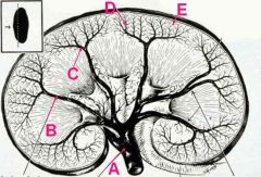
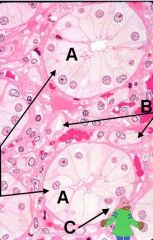
ID these regions of the kidney:
|
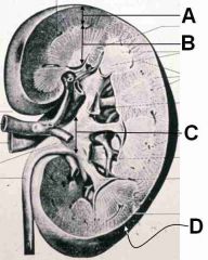
A - Cortex
B - Medulla C - Expansion of the ureter D - Capsule |
|

What type of tissue is A pointing to? B?
ID the C and D layers. What is the junction between C and D called? What are the striations called? |
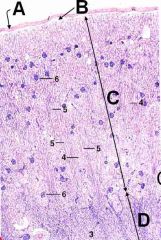
A - Dense irr CT capsule
B - Areolar CT C - Cortex D - Medulla Corticomedullary junction Medullary rays |
|
|
What are medullary rays composed of?
|
Group of nephron loop and collecting ducts
|
|
|
Which cells secrete renin?
|
Juxtoglomerular cells
|
|
|
Which cells secrete erythropoetin?
|
Endothelial cells of the peritubular capillaries
(not entirely sure about this one; I thought it was extraglomerular mesangial cells) |
|
|
What vessels run in the corticomedullary junction?
|
Arcuate vessels
|
|

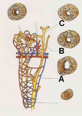
ID this!
|
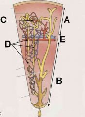
A - Cortex
B - Medulla C - Renal Corpuscle D - Nephron E - Corticomedullary junction with arcuate vessels |
|
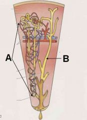
What are the vessels depicted in A? What do they surround? What is B?
|

Vessels are vasa recta surrounding nephron
B is a collecting duct |
|
|
Describe the afferent flow of arterial blood to the glomerulus.
|
Renal a.
Interlobar a. Arcuate a. Interlobular a. Intralobular a. Afferent Glomerlular arteriole |
|
|
T or F:
Little O2 is lost in the glomerular capillaries. |
True!
|
|
|
T or F:
Little O2 is lost in the vasa recta capillary bed. |
False! This is where O2 and nutritive exchange occurs.
|
|
|
What are the two capillary beds of the kidney?
|
Glomerulus
Vasa Recta |
|
ID these renal arteries
|

A - Renal a.
B - Interlobar a. C - Arcuate a. D - Interlobular a. E - Intralobular a. |
|
|
What vessel leaves the glomerulus? Where does (or can) blood go after this?
|
Leaves the efferent glomerular arteriole and can go to the peritubular capillary network OR the vasa recta
|
|
|
Describe the post-glomerular venous drainage of the renal system from the cortex.
|
Efferent glomerular arteriole
Peritubular capillary network Interlobular v. Arcuate v. Interlobar v. Renal v. |
|
|
Describe the post-glomerular venous drainage of the renal system from the medulla.
|
Efferent glomerular arteriole
Vasa Recta Arcuate v. Interlobar v. Renal v. |
|
|
What constitutes the renal corpuscle?
|
Glomerulus
Glomerular (Bowman's) capsule |
|
|
What comprises the glomerulus?
|
fenestrated capillaries
mesangial cells |
|
|
T or F:
The glomerulus is also called the capillary plume. |
False! It's the capillary TUFT! Get it right next time dumbo...
|
|
|
T or F:
There are many anastomoses between arcuate arteries. |
False! No anastomoses! I'm gonna anastomose your head to your ass if you don't start getting these right...
|
|
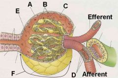
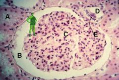
ID these regions of the Renal Corpuscle
|

A - Parietal epithelium
B - Visceral epithelium (podocytes) C - Glomerular capsule D - Juxtaglomerular cells E - Uriniferous space F - Glomerular capsule |
|
|
What is the region of the renal corpuscle called where the arteries enter? Where the proximal tubule leaves?
|
Arteries - Vascular pole
Tubule - Urinary pole |
|
|
T or F:
The capillaries in the renal corpuscle are fenestrated. |
True!
|
|
|
What is the function of the mesangial cell in the glomerulus?
|
Support, phagocytosis, and contractile action
|
|
|
What structures create the glomerular filtration barrier?
|
Podocyte pedicels, fused basal lamina, and fenestrated capillary endothelium
|
|

Hey! Isn't that Gizmo! Get that creepy mogwai outta my glomerulus!
|
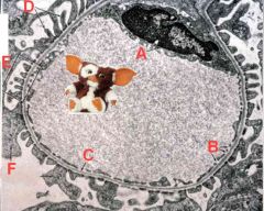
A - Endothelial cell
B - Fenestrations C - Basal lamina D - Podocyte pedicels E - Filtration barrier F - Podocyte primary process |
|
|
What syndrome is caused by the development of antibodies against the basement membrane of the glomerulus?
|
Goodpasture's syndrome
|
|
|
T or F:
Albumin cannot cross the glomerular filtration barrier. |
False! Albumin goes through slowly but it can get through.
|
|
|
What are two syndromes characterized by proteinuria?
|
Diabetes
Nephrotic syndrome |
|
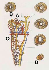
ID these regions of the urineferous tubule
|

A - Glomerular capsule
B - Proximal convoluted tubule C - Proximal straight tubule D - Thin limb (descending and ascending) |
|
|
ID
|
A - Distal straight tubule
B - Distal convoluted tubule C - Collecting duct |
|
ID
|

A - Distal straight tubule
B - Distal convoluted tubule C - Collecting duct |
|
|
Which regions of the uriniferous tubule are ONLY in the cortex?
|
Proximal convoluted tubule
Distal convoluted tubule Glomerular capsule |
|
|
Which regions of the uriniferous tubule are ONLY in the medulla? Which span the corticomedullary junction?
|
Thin descending/ascending limbs are in the medulla
Thick descending/ascending limbs (proximal/distal straight tubules) span the junction |
|
|
T or F:
The proximal convoluted tubule has less nuclei (histologically) than does the distal convoluted tubule. |
True!
|
|
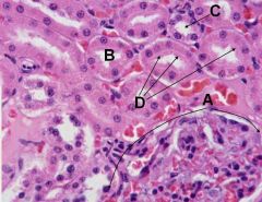
ID the structures on this cross section of the kidney.
|

A - Renal corpuscle
B - Proximal convoluted tubule C - Distal convoluted tubule D - Brush border |
|
|
What is the function of the proximal convoluted tubules?
|
Reabsorb 60 - 80% of Na, Cl, and H2O and all glucose, AAs and proteins
Eliminates organic solutes, drugs, toxins, and NH4+ |
|
|
What is the eponymous name for the nephron loop?
|
Loop of Henle
|
|

ID these structures in the renal medulla
|
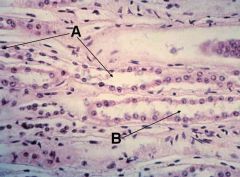
A - thin limb of nephron loop
B - thick limb of nephron loop |
|
|
Which region of the nephron loop is highly permeable to water? Which is impermeable to water?
|
Descending limb is highly permeable while ascending is impermeable.
|
|
|
Which region of the nephron loop actively pumps Cl- into the interstitial space?
|
Ascending limb
|
|
|
Which region of the nephron loop is partially permeable to urea, Na+, and Cl-?
|
Descending limb
|
|
|
T or F:
The ascending limb of the nephron loop is impermeable to water and partially permeable to Na+, Cl-, and urea. |
False!
It is impermeable to water and pumps-out Cl- |
|
|
T or F:
Both the descending and ascending limbs allow Cl- to cross. |
True!
|
|
|
Which is more prevalent? Proximal or distal convoluted tubules?
|
~7x more proximal!
|
|
|
What is the function of the distal convoluted tubules?
|
Reabsorption of Na, Cl, Ca, K, and Mg
|
|
Ho ho ho how did this dude get here? What space (B) is the jolly green giant hanging out in?
|

A - Proximal convoluted tubule
B - Uriniferous space C - Glomerulus D - Distal convoluted tubule E - Macula densa |
|
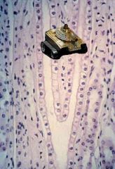
Silly Master P thinks he can drive his ghetto tank anywhere but he got lost! Tell him where he is and how you know...
|

He's in the collecting ducts! Notice the high cuboidal epithelium and less eosinophilic lateral borders.
|
|
|
What are the two cell types in the collecting ducts and what are their functions?
|
Principal (light) cells - resorb Na and Cl
Intercalated (dark) cells - release H+ for pH balance |
|
|
Where does ADH cause the insertion of aquaporins?
|
In the collecting ducts
|
|

ID these structures:
|
A - Papillary duct
B - Thin limb of nephron loop C - Dumb Donald |
|

What's Voltron doing here? Where is he and what does this region empty into?
|
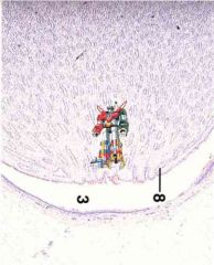
Area Cribrosa enters into the expansion of the ureter
|
|
|
What is the function of the Macula Densa? What cells does the Macula Densa work with?
|
Senses filtrate Na+ levels and volume and signals Juxtaglomerular cells when levels are too low.
|
|
|
T or F:
The Macula Densa acts when Na or filtrate volume get too high. |
False! It acts when either gets too low!
|
|
|
What do the juxtaglomerular cells secrete?
|
Renin!
|
|
|
T or F:
Filtrate osmolality in the ascending thick limb is unaffected by ADH. |
True! ADH only works on the collecting ducts!
|
|
|
Physiologically, as filtrate travels through the nephron loop, the filtrate loses __________ when descending and loses __________ when ascending.
|
Water
Na and Cl |
|
|
What are the relative levels of water and ions in the arterial and venous sides of the vasa recta.
|
Arterial side has less water but more ions while venous side has lower ions and more water. (I don't think that this is right...)
|
|
|
How many layers of tunica muscularis in the ureter? What is the orientation of each?
|
3 layers
Inner is longitudinal and the other two are circular |
|

Why is horse urine foamy?
|

Equids have goblet cells in their ureters (adds mucus).
|
|
|
What is the epithelium in the renal pelvis? In the ureter?
|
Transitional epithelium in both!
|
|
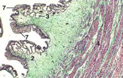
This is the urethra from which animal?
|
Horse! Check out the branched tubuloalveolar glands!
|
|
|
T or F:
The capsule of the ureter is adventitia. |
False dummy! It's half adventitia and half serosa!
|
|
|
T or F:
The capsule of the ureter is serosa. |
False dummy! It's half adventitia and half serosa!
|
|
|
Describe the orientation of the muscularis mucosa in the renal pelvis and the ureter.
|
Fooled ya! The renal pelvis and ureter have lamina propria submucosa and, thus, HAVE NO MUSCULARIS MUCOSA!!!
|
|
|
How many tunica muscularis layers in the urinary bladder? Describe their orientation.
|
3 layered:
Circular sandwiched between longitudinal |
|
|
Which layer of the tunica muscularis of the bladder forms the smooth muscle sphincter?
|
Middle circular
|
|
|
T or F:
Cats have no muscularis mucosa in the bladder while most other species do. |
True dat!
|
|
|
Describe the epithelia of the pelvic and spongy urethra.
|
Pelvic - transitional to pseudostratefied columnar epi
Spongy - stratefied columnar to str. squameous epi |
|
|
T or F:
The urethra has 3 layers of muscularis mucosa. |
False! Only two in females!
|
|
|
Where does the urethralis muscle begin?
|
Where urethra enters the perineum
|