![]()
![]()
![]()
Use LEFT and RIGHT arrow keys to navigate between flashcards;
Use UP and DOWN arrow keys to flip the card;
H to show hint;
A reads text to speech;
69 Cards in this Set
- Front
- Back
|
Concentration of ions (K/Na/Ca/Cl) |
Out:In K/ 1:20 Na/ 10:1 Ca/ 10000:1 Cl/ 12:1 |
|
|
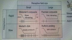
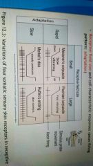
Pacinian Corpuscle |
Sense vibration |
|
|
Ruffini Endings |
Responds to skin stretch |
|
|
Free nerve endings |
Pain and temperature |
|
|
Meissner's Corpuscle |
Flutter and stroking movements |
|
|
Merkel Receptors |
Sense steady pressure and texture |
|
|
Mechanoreceptors Adaptation & Receptive field |
|
|
|
Diameter of 4 axons thickness |
group 1: Aalpha: 13-20 micrometers/ 80-120 m/s used for proprrioception group 2: Abeta: 6-12 micrometers/ 35-75 m/s used for mechanoreceptors group 3: Adelta: 1-5 micrometers/ 5-30 m/s used for pain/temperature group 4: C: 0.2-1.5 micrometers/ 0.5-2 m/s used in temperature, pain, itch |
|
|
Dorsal Column-Medial Lemniscal Pathway? |
This is the major route for touch and proprioceptive sensations ascend to the cortex |
|
|
Lemniscus? |
Means a band of white nerve fibers in the brain |
|
|
Damage to posterior parietal cortex |
(association area essential for perception of spatial perception of sensation) leads to Tactile Agnosia: can't recognized objects by feeling them |
|
|
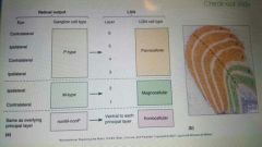
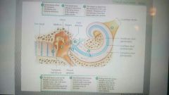
Retinal cells |
|
|
|
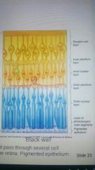
Retinal Layers |
|
|
|
Cellular pigmented epithelium |
|
|
|
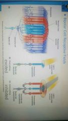
Phototransduction in Rods |
Depolarization in the dark: dark current (cGMP-gated Na+ channel) Hyperpolarization in the light Rhodopsin: Opsin + Retinal Opsins are receptors that activate by light causing a conformational change on Retinal (11-cis to all-trans) |
|
|
3 Opsins in Cones |
Red, green and blue |
|
|
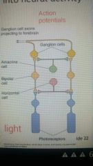
What NT is secreted in the dark |
Glutamate as a result of depolarization of cells in the dark go on to depolarize IPL of bipolar cell, then OPL of them onto ganglion cells which generate an AP |
|
|
2 kinds of Bipolar cells |
ON: respond to Glutamate by hyperpolarizing (light ON at the center) ,so depolarized when light is on OFF: respond to Glutamate by hyperpolarizing (light OFF from the center) ,so depolarized when light is off |
|
|
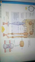
Example of direct and indirect pathways |
|
|
|
Retinal Processing picture |
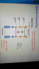
"PhBAG" Photoreceptors -> (Horizontal) -> Bipolar -> Amacrine -> Ganglion |
|
|
Purpose of on/off ganglion cells |
Detect differences in illumination in a receptive field |
|
|
Best stimuli for an on/off ganglion cell |
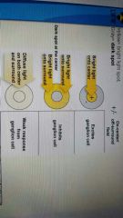
light/dark center pattern (They are sensitive and responsive to contrast!) |
|
|
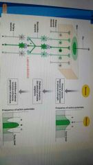
integration of presynaptic inhibition |
|
|
|
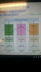
3 major types of great ape ganglion cells |
P (Parvo) - Type: 90% M (Magno) - Type: 5% nonM-nonP (K-type): 5% some P and K type cells are color-sensitive ganglion cells (color-opponent center-surround receptive field) |
|
|
Mechanoreceptors |
|
|
|
M-type cells |
-Lack of color opponency -do not send color info to brain -large receptive field -respond and adapt fast *motion detection, but low-resolution image |
|
|
P-type cells |
-color-opponency -sends color info to brain -response has no adaptation -conduction speed is slower than M-type *Object shape and color detection, with detailed images |
|
|
K-type cells |
-color-opponency -sends color info to brain -contributes to color vision *physiological properties not yet clear |
|
|
Parallel Processing |
Simultaneous input from 2 eyes for depth and distance info about light and dark (On/Off ggl. cells) different receptive fields and properties of retinal ggl cells (M/P/K type) |
|
|
Nonthalamic Targets of the optic tract (3) |
Hypothalamus: biological rhythms like sleep and wakefulness Midbrain's pretectum: size of the pupil Midbrain's Sup. Colliculus: visual reflexes, tracking, or reading left to right for example |
|
|
Superior colliculus |
receives 10% of optic fibers, and is involved in Saccadic eye movements and attention |
|
|
2 visual systems |
Primitive (eye and head movements, reaching hands and other simple behaviors) Mammalian (Speech and thinking in words: consciousness, and other complex behaviors) |
|
|
damage to each visual system |
to primitive: person is not aware of vision to mammalian: abolishes perception |
|
|
Segregation of eye input by ganglion cell types in LGN |
|
|
|
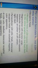
Receptive fields of LGN |
|
|
|
Retinotopy |
Map of the visual field onto a target structure: Retina -> LGN -> striate cortex perception here is related to the brains interpretation of distributions of neural activity, not a snapshot of reality |
|
|
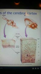
Gen features of cerebral cortex |
|
|
|
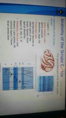
Anatomy of striate cortex |
|
|
|
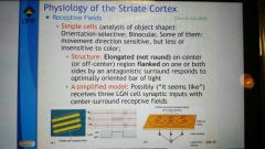
Simple Cells |
analyze shape, are orientation-selective, but not very sensitive to color |
|
|
Complex cells |
detect movement and not color regardless of position in the receptive field |
|
|
Hypercomplex cells |
Has stopping properties in larger receptive fields |
|
|
Dorsal stream |
Part of interconnected systems in the visual cortex involved in perception of spatial locations, visual disparity (depth) and movement |
|
|
Ventral stream |
anatomical pathway for perception of form and color |
|
|
Damage in the MST (of dorsal stream) |
leads to "snapshot vision" of motion perception |
|
|
Magnocellular LGN neurons project to |
layer IVC-alpha |
|
|
Parvocellular LGN neurons project to |
layer IVC-beta |
|
|
Koniocellular LGN axons bypass |
layer IV to make synapses in layers II and III |
|
|
basis of stereopsis |
-binocular disparities for depth perception -layer IVC innervates binocular neurons in superficial layers |
|
|
What are receptive fields of the striate cortex |
-input of K cells to II, III (Koniocellular in LGN) -input of M cells to IVC-alpha (Magnocellular in LGN) -input of P cells to IVC-beta (Parvocellular in LGN) |
|
|
Cortical receptive fields physiology |
Layer IVCalpha: insensitive to the wavelength Layer IVCbeta: center-surround color opponency Outside IVC: not center-surround (found upon Hubel and Wiesel's work in the 60's that won them a nobel prize in 1981) |
|
|
Orientation selectivity |
orientation-selective neurons where layers II to VI have the same orientation selectivity for vertical outlines |
|
|
What is the great impact of Hubel and Wiesel to neurobiology? |
1) ocular dominance columns 2) describing how visual signals are processed by the brain to generate edge detectors, motion detectors, stereoscopic depth detectors and color detectors: building blocks of the visual scene (Detection of lines or bars of the simple cell) |
|
|
Direction selectivity |
some neurons fire AP's more strongly to a bar moving to one specific direction |
|
|
Simple cell picture |
|
|
|
Hypercomplex cells |
exhibit end stopping properties: when a cells response increases as a stimulus expands to fill the receptive field, and then responds less when the stimulus exceeds the size of the receptive field |
|
|
3 classes of cells in V1 |
building blocks of the visual scene: simple cells (line/bar/edge detectors) complex cells (orientation detectors) hypercomplex cells (patterns/length/corners/angle detectors) |
|
|
Color blobs of the striate cortex |
are cytochrome oxidase mitochondrial enzyme in cell metabolism, which receive input from K-cells of the LGN for color as well as some P and M LGN neurons |
|
|
Blob cells |
-are color detectors (light wavelength-sensitive) -monocular, no orientation, no direction selectivity |
|
|
***3 parallel pathways to visual cortex |
|
|
|
A cortical module |
2 by 2 block in V1 of striate cortex containing ocular dominance columns, orientation columns, and color blobs to fully analyze a portion of the visual field |
|
|
Steps of hearing |
|
|
|
Dorsal Stream |
A system of interconnected regions of visual cortex involved in the perception of spatial locations, visual disparity (depth) and movement, beginning with the striate cortex (primary) and ending with the posterior parietal cortex by way of extrastriate cortex. Receiving projections mostly from M-cell pathway |
|
|
Ventral Stream |
A system of interconnected regions of the visual cortex involved in the perception of form (3D object) amd color, beginning with the striate cortex and ending with the inferior temporal cortex by way of the extrastriate cortex. Receiving projections mostly from P cell and blob pathways |
|
|
receiving projections of dorsal stream |
Mostly from M cell pathwaya |
|
|
receiving projections of ventral stream |
mostly from P cell and blob pathways |
|
|
Achromatopsia |
caused by damage to area V4 (part of Ventral stream) leading to partial or complete loss of color vision as well as shapes of objects (color cone receptors in the retina are normal) |
|
|
Area IT |
is the major output of V4 and responds to a wide variety of colors and abstract shapes, like faces and hands |
|
|
damage to ventral stream |
leads to inability to recognize objects. faces, textures or colors. although they can still perceive movement so can see form from motion, so patients with this visual agnosia can recognize their friends by the way they walk |
|
|
"Where" and "What" streams |
Where: dorsal stream (M-ovement) What: ventral stream (P-erception) |