![]()
![]()
![]()
Use LEFT and RIGHT arrow keys to navigate between flashcards;
Use UP and DOWN arrow keys to flip the card;
H to show hint;
A reads text to speech;
14 Cards in this Set
- Front
- Back
- 3rd side (hint)
|
Ossification of Pelvis |
Primary 1. Ilium -8th week IAU
2. Ischium- 4 months 3. Pubis- 4-5 months Secondary 1. 2 centres Iliac crest appears at puberty Anterior inferior iliac spine at puberty 2. 1 centre Ischial tuberosity at puberty 3. 1 centre Symphysis pubis appears at puberty |
|
|
|
Age hip bone fuses |
15-25 years |
|
|
|
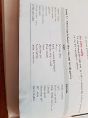
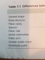
M/F pelvis characteristics |
|
|
|
|
Pelvic Girdle formed by what bones |
Sacrum and Coccyx |
|
|
|
Shoulder Blood/Nerve Supply |
Blood: branches of Axillary and Subclavian arteries Nerve: Suprascapular, Axillary and Lateral Pectoral nerves |
|
|
|
Pelvic Girdle Blood/Nerve Supply |
Femoral, obturator and gluteal nerves and arteries |
|
|
|
Perth's Diseaee |
Osteochondritis of the epiphysis of the femoral head Rad signs-widening of epiphyseal line, irregular metaphyseal outline, flattened epiphusos. Some increase in bone density within head of femur |
|
|
|
Osteoporosis (adult brittle bones) |
A deficiency in the bone matrix due to a reduction in bone formation, and therefore bones fracture easily. Causes-old age,disuse,lack of vitamin C(scurvy),post menopausal Rad signs- affected bones appear radiolucent, |
|
|
|
Osteomalacia (adult Rickets) |
An overall decrease in bone CALCIFICATION Causes-diet low in Vit D,calcium or phosphorus, malabsorption syndromes Rad signs-narrow bands of decalcification 2-3mm wide, a.k.a psuedo fractures or Looser's zones |
|
|
|
Osteomyelitis |
Bone infection, begins in medullary cavity and may spread to cortex and periosteum, bone destruction appears after 7 days when periosteum becomes elevated |
|
|
|
Osteoarthritis |
Degeneration of the articular hyaline cartilage, bone becomes thickened and spreads outwards forming SPURS round the joint margins, synovial fluid may enter bone giving it a cystic appearance |
|
|
|
Rheumatoid arthritis |
Rad appearance of bone erosion, joint deformity and a narrowed joint space due to inflammatory tissue forming over and destroying articular hyaline cartilage and eventually replacing it, joints swell and degree of movements is limited |
|
|
|
Osteochondritis Dissecans |
Caused by local bone nexfosis (death of tissue) resulting in loose fragments of bone in the joint space, most common site is knee |
|
|
|
Chondroma |
Tumour of mature cartilage, most common site is phalanges of fingers |
|