Reading...
![]()
Play button
![]()
Play button
![]()
Use LEFT and RIGHT arrow keys to navigate between flashcards;
Use UP and DOWN arrow keys to flip the card;
H to show hint;
A reads text to speech;
97 Cards in this Set
- Front
- Back
|
Perichondrium
|
dense irregular connective tissue surrounding skeletal cartilage
|
|
|
What are the three types of skeletal cartilages?
|
Hyaline cartilage
Elastic cartilage Fibrocartilage |
|
|
Hyaline cartilage
|
-the most abundant skeletal cartilage
-includes the articular, costal, respiratory, and nasal cartilages |
|
|
Elastic cartilage
|
-more flexible than hyaline
-located in the external ear, the epiglottis of the larynx, and the eustachian (auditory) tube. |
|
|
Fibrocartilage
|
-located in areas that must withstand a great deal of pressure or stretch
-ex) the knee(menisci), and the intervertebral discs |
|
|
Fibrocartilage in the menisci of the knee
|

|
|
|
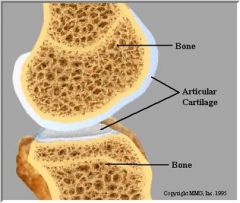
Hyaline cartilage covers the articular surfaces of bones
|
|
|
|
costal cartilages made up of hyaline
|

|
|
|
What are the two main divisions of the bones of the skeleton?
|
The axial skeleton
The appendicular skeleton |
|
|
What does the axial skeleton division consists of?
|
the skull
the vertebral column the rib cage |
|
|
What does the appendicular skeleton division consists of ?
|
-the bones of the upper and lower limbs
-the girdles that attach them to the axial skeleton |
|
|
Long Bones
(structural classification of bones) |
-longer than they are wide
-have a definite shaft and two ends -consist of all limb bones except the patellas, carpals, and tarsals |
|
|
Short Bones
(structural classification of bones) |
-somewhat cube-shaped
-include the carpals and tarsals |
|
|
Flat Bones
(structural classification of bones) |
-thin, flattened, often curved bones
-include most skull bones, the sternum, scapulae, and ribs |
|
|
Irregular Bones
(structural classification of bones) |
-complicated shapes that do not fit in any other class
-such as the vertebrae and coxae |
|
|
Sesamoid bones
(structural classification of bones) |
-"extra" bones that develop in tendons or near joints esp in feet and hands
-including patellas(the only sesamoid bones consistently present in skeleton) |
|
|
The 5 types of structural classification of bones
|
1) Long Bones
2) Short Bones 3) Flat Bones 4) Irregular Bones 5) Sesamoid Bones |
|
|
What are the 5 functions of bones?
|
1) support the body and cradle the soft organs
2) protect vital organs 3) allow movement 4) store minerals such as calcium and phosphate 5) house hematopoietic tissue in specific marrow cavities |
|
|
What are bone markings?
|
They are projections, depressions, and openings found on the surface of bones.
|
|
|
What is the function of bone markings?
|
They function as sites of muscle, ligament, and tendon attachment, as joint surfaces, and as openings for the passage of blood vessels and nerves.
|
|
|
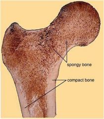
Compact Bone
|
-All bone has a dense outer layer consisting of compact bone
-appears smooth and solid |
|
|
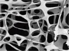
Spongy Bone
|
-internal to compact bone is spongy bone
-consists of trabeculae |
|
|
Trabeculae
|
-honeycomb, needle-like, or flat pieces of bone
|
|
|
What is the word for joint?
|
Articulation
|
|
|
What do articular cartilage prevent?
|
It prevents bones from scraping against one another.
|
|
|
Osteoarthritis
|
-degeneration of the articular cartilage
|
|
|
What makes bone tissue hard?
|
Calcium
|
|
|
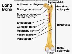
Structure of a typical long bone
(5 Key Points to Know) 1st Point |
-Long bones have a tubular bone shaft
-consists of a bony collar surrounding a hollow medullary cavity -medullary cavity is filled with yellow bone marrow in adults |
|
|
Structure of a typical long bone
(5 key points to know) 2nd Point |
-Epiphyses are at the ends of the bone.
-consist of internal spongy bone covered by an outer layer of compact bone |
|
|
Structure of a typical long bone
(5 key points to know) 3rd Point |
-The epiphyseal line (or scar) is located between the epiphyses and diaphysis
-It is a remnant of the epiphyseal plate(growth plate) |
|
|
Structure of a typical long bone
(5 key point to know) 4th Point |
-The external surface of the bone is covered by the periosteum
|
|
|
Periosteum
|
connective tissue membrane covering the external surface of the bone
|
|
|
Structure of a typical long bone
(5 key points to know) 5th Point |
The internal surface of the bone is lined by the endosteum
|
|
|
Endosteum
|
connective tissue membrane lining the internal surface of the bone
|
|
|
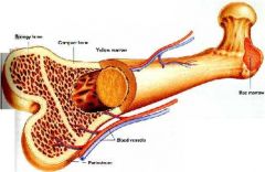
Structure of the Long Bone
|
|
|
|
What is hematopoietic tissue?
|
Red bone marrow
|
|
|
Where is hematopoietic tissue located?
|
-within the trabecular cavities of the spongy bone in flat bones
-in the epiphyses of long bones |
|
|
Location of hematopoietic tissue in adults vs infants
|
-in infants, it is found in all flat bones, epiphyses, and medullary cavities
-in adults, it is only found in flat bones and the proximal epiphyses of the humerus and femur |
|
|
Internal structure of Bone
|
|
|
|
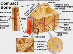
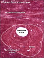
Osteon or Haversian system
(anatomy of bone) |
Structural unit of compact bone
|
|
|
What does the osteon or Haversian system consist of?
|
-concentric tubes of bone matrix (the lamellae) that surrounds a central Haversian canal
|
|
|
What is the function of the central Haversian canal.
|
It serves as a passageway for blood vessels and nerves
|
|
|
Another name for Perforating Canals
|
Volkmann's Canals
|
|
|
Function of Volkmann's canals
|
Connect the blood and nerve supply of the periosteum to that of the central canals and medullary cavity
(Anatomy of Bone) |
|
|
Where are osteocytes located?
(anatomy of bone) |
-occupy lacunae at the junctions of the lamellae
|
|
|
What is canaliculi?
|
series of hair-like channels used to connect osteocytes and the central canal
|
|
|
Anatomy of Comact Bone
|
|
|
|
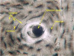
Photo of Haversian System
|
|
|
|
Locate the following:
-lamellae -canaliculi -osteocyte -haversian canal |
|
|
|
Where are circumferential lamellae located?
(anatomy of bone) |
-just beneath the periosteum
-extends around the entire circumference of the bone |
|
|
Where are interstitial lamellae located?
(anatomy of bone) |
-they lie between intact osteons, filling the spaces in between
|
|
|
Spongy bone lacks ______ but has ______ that align along lines of stress, which contain ______ lamellae.
|
osteons; trabeculae; irregular
|
|
|
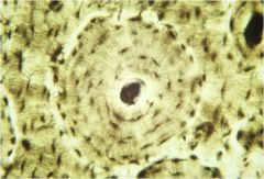
This microscopic image of bone tissue shows circumferential lamellae
|
|
|
|
Notice the trabeculae in spongy bone
|
|
|
|
What are the cells included in the organic components of bone?
|
osteoblasts
osteocytes osteoclasts |
|
|
What is osteoid?
|
ground substance and collagen fibers that are the organic components of bone
|
|
|
Organic components of bone include _____ and _____, which contribute to the flexibility and tensile strength of bone.
|
cells; osteoid
|
|
|
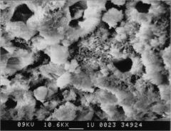
hydroxyapatite
|
-mineral salt that is largely calcium phosphate
-inorganic component of bone |
|
|
What accounts for the hardness and compression resistance of bone?
|
Inorganic components that make up 65% of bone by mass and consist of hydroxyapatite
|
|
|
Photo of hydroxyapatite
|
|
|
|
What are the two ways bone develops?
|
1) Intramembranous ossification
2) endochondral ossification |
|
|
Intramembranous ossification
|
-Bones form within fibrous connective tissue membranes.
-The cranial bones and clavicles develop in this way |
|
|
Endochondral ossification
|
-Hyaline cartilage models are laid down first
-Then they are replace by calcified bony matrix -All bones below the skull are developed this way except for the clavicles |
|
|
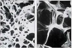
Osteoporotic Bone
(zoomed view) |

|
|
|
Normal Bone
(zoomed view) |
|
|
|
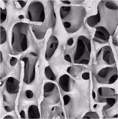
Osteoporotic Bone
|
|
|
|
Normal Bone
|
|
|
|
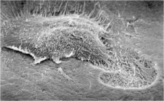
Osteoclast (photo)
|
|
|
|
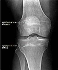
Photo of epiphyseal plates replaced with epiphyseal lines when growth in height is completed
|
|
|
|
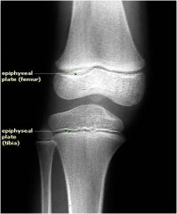
Photo of epiphyseal plates when growth is not complete
|
|
|
|
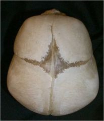
Fetal skull showing fontanel between the frontal and parietal bones
|
|
|
|
What is the function of fontanels?
|
-allows the skull to compress slightly during birth to allow the baby to fit through the birth canal
-enables the skull to grow large as the child grows |
|
|
What are the epiphyseal plates made up of?
|
Hyaline cartilage
|
|
|
What occurs at epiphyseal plates?
|
-places where long bones grow in lenght
-ex) tibia and femur |
|
|
What replaces epiphyseal plates when growth in height is complete?
|
epiphyseal lines
|
|
|
Osteogenic Zone
|
Growth in lenght of long boness occurs in this zone
|
|
|
How is the growth in lenght of long bones done at the osteogenic zone?
|
Through endochondral growth
|
|
|
What is endochondral growth?
|
1st-rapid division of the upper cells in the columns of chondrocytes
2nd-calcification and deterioration of cartilage at the bottom of the columns 3rd-subsequent replacement by bone tissue |
|
|
What is appositional growth?
|
-growth in width or thickness
-occurs due to the deposition of bone matrix by osteoblasts beneath the periosteum |
|
|
What are the 3 most common places where you are going to have fractures if you have osteoporosis?
|
-wrist
-hip -vertebra |
|
|
What is the most important stimulus of epiphyseal plate activity during infancy and childhood?
|
growth hormone
|
|
|
Where is the growth hormone from?
|
anterior pituitary
|
|
|
At puberty, what promotes growth spurt and induces the closure of the epiphyseal plate?
|
testosterone and estrogen
|
|
|
The growth hormone's effects are modulated by the _______ hormone.
|
Thyroid
|
|
|
What are osteoblasts?
|
bone forming cells
|
|
|
What are osteoclasts?
|
cells that break down bone
|
|
|
Bone remodeling is a balance of __________ and __________.
|
bone deposit; removal
|
|
|
What allows minerals of degraded bone matrix to move into blood?
|
bone resorption
|
|
|
Bone deposit occurs at a greater rate when bone is ________.
|
injured
|
|
|
Explain, what does bone do in response to mechanical stress and gravity?
|
It grows or remodels in ways that allow it to withstand the stresses it experiences.
|
|
|
What is osteoporosis?
|
a group of disorders in which the rate of bone resorption exceeds the rate of formation
|
|
|
What occurs when bone mass is reduced?
|
the bones become more porous and lighter, which increases the likelihood of fractures
|
|
|
Bones have normal bone matrix in persons with osteoporosis.
True or False |
True
Bone matrix is normal, but bone mass is reduced. |
|
|
What causes older women to be especially vulnerable to osteoporosis?
|
the decline in estrogen after menopause
|
|
|
List some factors that contribute to osteoporosis.
|
-petite body form
-insufficient exercise -immobility -diet poor in calcium and vitamin D -abnormal vitamin D receptors -smoking -certain hormone-related conditions |
|
|
What test can be performed after a certain age to help detect osteoporosis?
|
Bone mineral density test
|
|
|
Adults have ______ bone marrow and children have ______ bone marrow.
|
yellow; red
|