![]()
![]()
![]()
Use LEFT and RIGHT arrow keys to navigate between flashcards;
Use UP and DOWN arrow keys to flip the card;
H to show hint;
A reads text to speech;
88 Cards in this Set
- Front
- Back
|
Bone Tissue |
Dynamic tissue (constantly remodeling); connective tissue with matrix hardened by minerals (mostly Calcium Phosphate); made up of bone tissue, marrow, cartilage, periosteum |
|
|
Functions of Skeletal system |
Support, protection, movement, blood formation, mineral reservoir, pH balance (buffers), detoxification |
|
|
Structure of a flat bone |
Externals and internals surfaces are composed of compact bone; middle layer is spongy bone (diploe); no marrow cavity *most cranial bones, ribs, sternum, scapula, ox coxae |
|
|
Structure of long bone |
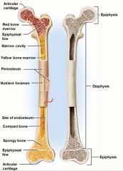
Diaphysis (shaft); Epiphysis (head); Periosteum; Articular Cartilage; compact bone; spongy bone; endosteum; yellow marrow |
|
|
Long bones of the body |
Humerus, Radius, Ulna, Metacarpals, Phalanges, Femur, Tibia, Fibula, Metatarsals |
|
|
Short Bones |
Carpals & Tarsals |
|
|
Irregular Bones |
Sphenoid and Ethmoid bones; Vertebrate |
|
|
Shaft of the bone |
*Diaphysis* - Contains compact bone, marrow cavity (medullary cavity), and endosteum |
|
|
Endosteum |
Structure inside the shaft of bones that contains cells providing bone resorption & deposition |
|
|
Enlarged ends |
*Epiphyses* - Spongy bone covered with a layer of compact bone - enlarged to strengthen joint & provide for attachment of tendons and ligaments |
|
|
Periosteum |
Outer fibrous layer continuous with tendons; inner osteogenic layer important for growth and healing; the rest of the bone is covered with this |
|
|
Ostiogenic Cells |
Reside in endosteum or periosteum; multiply continuously& differentiate into osteoblasts |
|
|
Osteoblasts |
Form organic matter of matrix & help to mineralize it in response to stress or fractures - Bone building cells |
|
|
Osteocytes |
Osteoblasts that have become trapped; reside in lacunae; signal osteoclasts/osteoblasts about mechanical stresses |
|
|
Osteoclast |
Bone-dissolving cells; develop in bone marrow by the fusion of the same stem cells that give rise to monocytes of the blood; reside in pits called resorption bays that they have eaten to the surface of the bone |
|
|
Matrix of Osseous Tissue |
Dry weight: 1/3 organic, 2/3 inorganic - Combination provides strength & resilience; minerals resists compression & collagen resists tension |
|
|
Organic matter of Osseous Tissue Matrix |
Collagen, glycosaminoclycans, proteoglycans & glycoproteins |
|
|
Inorganic matter of Osseous Tissue Matrix |
85% Hydroxypatite: crystallized calcium phosphate salt 10% Calcium Carbonate |
|
|
Osteogenesis Imperfecta |
Lack of collagen in bones |
|
|
Role of Minerals |
Keeps bones hard and fully formed |
|
|
Rickets |
Defective mineralization of bones in children; insufficient sunlight, dietary deficiency in calcium or phosphate, liver or kidney disease |
|
|
Osteomalacia |
Deffective mineralization of bones in adults |
|
|
Compact bone |
Contains osteons & perforating canals; circumferential or outer lamellae |
|
|
Osteons |
*Haversian System* Cylinders of tissue formed from layers of matrix concentrically arranged around a blood vesselo; Contains lamellae, osteocytes, haversian canal, and haversian |
|
|
Lamellae |
Individual layers of bone |
|
|
Haversian Canal |
Holds the blood vessles |
|
|
Osteocytes |
Connected to each other kl |
|
|
Perforating Canals |
Branches from nutrient arteries that run perpendicular to the haversain canals |
|
|
Spongy Bone |
Contains Trabeculae, rods and plates, and red bone marrow; trabeculae have few osteons; provides strength with little weight |
|
|
Bone Marrow |
- Soft tissue that occupies the medullary cavity of a long bone, or the spaces amid the trabeculae of spongy bone - 3 types (Red, Yellow, Gelatinous) |
|
|
Red Marrow |
looks thick like blood; mesh of reticular fibers and immature cells; Hemopoetic: produces blood cells; found in axial skeleton & girdles in adults |
|
|
Yellow Marrow |
Fatty marrow of long bones in adults |
|
|
Gelatinous Marrow |
Yellow marrow replaced with reddish jelly |
|
|
Illiac Crest |
Region of the body that is the best place for extraction of bone marrow |
|
|
Intramembranous Ossification |
Produces flat bones of skull and clavicle Steps: 1. mesenchyme condenses into a sheet of soft tissue, 2. osteoblasts gather on the trabeculae to form osteoid tissue, 3. Calcium phosphate is deposited in the matrix transforming the osteoblasts into osteocytes, 4. osteoclasts remodel the center to contain marrow spaces & osteoblasts remodel the surface to form compact bone, 5. mesenchyme at surface gives rise to periosteum |
|
|
Endochondral Ossification |
Most other bones Steps: 1. m3esenchyme differentiates into hyaline cartilage, 2. cartilage is broken down, reorganized & calcified, 3. primary ossification center forms in cartilage model (chondrocytes near center swell to form primary ossifiaction center; matrix is reduced & model becomes weak at that point) 4. primary marrow space formed by periosteal bud (osteogenic cells invade & transform into osteoblasts; osteoid tissue deposited & calcified into trabeculae at same timeosteoclasts work to enlarge the primary marrow cavity |
|
|
Secondary Ossification Center |
Begin to form in the epiphyses near the time of birth; same stages occur as in primary ossification center (result is center of epiphyseal cartilage being trasformed into spongy bone); Hyaline Cartilage forms |
|
|
Hyaline Cartilage |
Remains on joint surface as articular cartilage; remains at the Junction of diaphysis & ephiphysis (epiphyseal plate) |
|
|
Metaphysis |
- Epiphyseal Plate - The junction of the diaphysis & epiphysis - Exists only when you are growing, disappears when the person stops growing - Cartilaginous material that remains as growth plate between medullary cavity & secondary ossification centers in the epiphyses |
|
|
Bone Growth and Remodeling |
- To accommodate force applied to skeleton - Children growing bones - Bones depend on applied stress (exercising) |
|
|
Interstitial growth |
Bones increase in length atepiphyseal plate |
|
|
Appositional Growth |
Bones increase in width (mature bone only) |
|
|
Achondroplastia |
Achondroplastic Dwarfism - shortstature but normal sized head and trunk - long bones of the limbs stop growing in childhood but other bones unaffected - Result of spontaneous mutation when DNA is replicated (mutant allele is dominent) |
|
|
Mineral Deposition |
- Crystallization of ions from blood - Levels of calcium & phosphate in tissues must reach a point before crystallization can occur - most tissues have inhibitors to prevent crystallization |
|
|
Ectopic Ossification |
Abnormal calcification - may occur in lungs, brain, eyes, muscles, tendons or arteries (arteriosclerosis) |
|
|
Mineral Resorption |
Process of dissolving bone & releasing minerals into the blood - Hydrogen pumps in the cell membrane secrete hydrogen ions into the space between the osteoclast & the bone - Chloride ions follow by electrical attraction - HCl with a pH 4 dissolve bone minerals - Secrete acid phosphatase that digests the collagen |
|
|
Phosphate |
Component of DNA, RNA, ATP, phospholipids, & acid-base bufferes 85-90% of body's phosphorus is in bones |
|
|
Calcium |
Needed for communication between neurons, muscle contraction, blood clotting, exocytosis 99% of body's calciumis in bones Adult skeleton exchanges ~18% of calcium with the blood each year |
|
|
Exocytosis |
movement of substances out of a cell |
|
|
Ion Imbalance |
Changes in phosphate concentration have little effect Changes in calcium cause hypocalcemia or hypercalcemia |
|
|
Hypocalcemia |
Deficiency of blood calcium - Causes excessive excitability of nervous system leading to muscle spasms, tremors or tetanus - Caused by vitamin D deficiency, excessive diarrhea, pregnancy, lactation, underaction/removal of parathyroid glands |
|
|
Hypercalcemia |
Excessive blood calcium (rare) - Depresses nervous system-> muscle weakness, sluggish reflexes, cardiac arrest |
|
|
Calcitriol |
Activated by vitamin D Liver converts to calcidiol Kidneys convert to calcitriol - stimulates small intestine to absorb calcium & phosphate - reduces urinary secretion of calcium & phosphate - promotesosteoclast activity if calcium levels low |
|
|
Parathyroid Hormone |
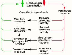
- Secreted by parathyroid glands on the posterior surface of the thyroid gland - Released when calcium blood level too low |
|
|
Functions of parathyroid hormone |
- Inhibits activity of osteoblasts - stimulates osteoclast multiplication & activity - reduces calcium secretion & increases phosphate secr3etion in the urine - stimulates production of an enzyme in kidneys that carries out the last step in calcitriol synthesis |
|
|
Calcitonin |
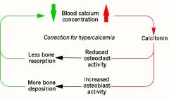
-Secreted by the thyroid gland when calcium is too high -promotes bone deposition of calcium -Important role in children |
|
|
Fractures |
Classified by their structural characteristics, or after a physician who first described it |
|
|
Stress Fracture |
A break caused by abnormal trauma to a bone |
|
|
Pathological fracture |
A break in a bone weakened by some sort of disease |
|
|
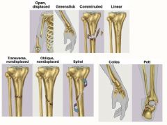
Types of fractures |
Closed/Open Complete/Incomplete Greenstick Hairline Comminuted Linear/Transverse/Oblique Spiral |
|
|
Closed Fracture |
*simple / non-displaced* skin is not broken |
|
|
Open Fracture |
*compound / displaced* bone protrudes from the skin |
|
|
Complete Fracture |
Broken in 2 or more pieces |
|
|
Incomplete Fracture |
extends only partway (pieces remain joined) |
|
|
Greenstick Fracture |
One side bent, Incomplete fracture on the other |
|
|
Hairline Fracture |
Fine crack |
|
|
Comminuted Fracture |
Broken in 3 or more pieces |
|
|
Linear Fracture |
Parallel along the long axis of the bone |
|
|
Transverse Fracture |
Perpendicular to the long axis of bone |
|
|
Oblique Fracture |
Diagonal (between linear and transverse) |
|
|
Spiral Fracture |
Resulting of a twisting stress |
|
|
Healing of fractures |
Normal healing: 8-12 weeks (longer in elderly) Stages: 1. Fracture hematoma, 2. Granulation tissue, 3. Callus formation, 4. Remodeling |
|
|
Fracture hematoma |
broken vessels form a blood clot |
|
|
granulation tissue |
fibrous tissue formed by fibroblasts & infiltrated with capillaries |
|
|
Callus formation |
soft callus of fibrocartilage replace by hard callus of bone in 6 weeks |
|
|
Remodeling |
ocurs over next 6 months as spongy bone is replaced with compact bone |
|
|
Orthopedics |
Branch of medicine that deals with the prevention & correction of injuries and disorders of the bones, joints & muscle |
|
|
Closed reduction |
treatment of fracture where fragments are aligned with manipulation & casted |
|
|
Open reduction |
treatment of fracture where there is surgical exposure & repair with plates & screws |
|
|
Traction |
Treatment of fracture: Not used in elderly due to risks of long-term confinement to bed (more susceptible to pneumonia) |
|
|
Electrical Stimulation |
Treatment of fractures that take longer than 2 months to heal *often used with athletes to help with healing* |
|
|
Osteoporosis |
Most common bone disease - Bones loose mass & become brittle due to loss of both organic matrix & minerals Post menopausal caucasian women at greatest risk (by age 70, average loss is 30% of bone mass) |
|
|
Estrogen Replacement Therapy (ETR) |
Osteoperosis Treatment: slows bone resorption, increases risk of breast cancer, stroke, and heart disease |
|
|
Osteomyelitis |
infection causes inflammation of osseous tissue and bone marrow |
|
|
Osteoma |
benign bone tumor |
|
|
Osteochondroma |
benign tumor of the bone and cartilage |
|
|
Osteosarcoma |
bone cancer |
|
|
Chondrosarcoma |
slow-growing cancer of hyaline cartilage |