Reading...
![]()
Play button
![]()
Play button
![]()
Use LEFT and RIGHT arrow keys to navigate between flashcards;
Use UP and DOWN arrow keys to flip the card;
H to show hint;
A reads text to speech;
162 Cards in this Set
- Front
- Back
- 3rd side (hint)
|
Plasma
|
liquid portion of centrifuged blood, sits atop everything, 55% of whole blood, least dense component
|
|
|
|
buffy coat
|
leukocytes and platelets, <1% of whole blood, thin white colored area when centrifuged
|
|
|
|
erythrocytes
|
45% of whole blood (hematocrit), most dense component
|
|
|
|
Functions of blood and example of each
|
Distribution: delivery of oxygen, waste products, hormones
Regulation: maintain body temperature (distributing head/absorb heat), pH 7.35 - 7.45, fluid volume (salt, ions, proteins) Protection: immunity (leukocytes), blood loss/blood clotting (platelets) |
|
|
|
What kind of tissue is blood and what is it composed of?
Is it acidic or alkaline? Accounts for what percentage of body weight? What is its temperature? |
Connective tissue which is composed of formed elements (blood cells) and plasma (fluid matrix). 55% Plasma, 45% erythrocytes, 1% buffy coat. Formed elements make it viscous.
Slightly alkaline pH Accounts for 8% of body weight. Temperature stays about 100.4 degrees. |
|
|
|
What is albumin?
|
- Makes up 60% of plasma proteins.
- It is a shuttle - binds to chemicals, ions carries them where they need to go, blood buffer, keeps water in blood to help maintain blood volume. - It maintains blood osmotic pressure. |
|
|
|
Immunoglobulins - type and what they do
|
36% of plasma proteins
Alpha, betaglobulins - most are transport proteins that bind to lipids, metal ions, and fat-soluble vitamins Gamma globulins - antibodies released by plasma cells during immune response |
|
|
|
What is fibrinogen?
|
4% of plasma proteins.
Dissolved protein, comes out of solution when blood needs to clot, forms a net to help with the clotting. |
|
|
|
Erythrocytes
|
red blood cells, lack nucleus, "bags of hemoglobin"
function in gas transport, very flexible |
|
|
|
Hematopoeisis
|
blood cell formation occurs in spongy bone; it is a general term for the maturation of erthrocytes, platelets, and leukocytes
|
|
|
|
What is Erythropoeisis? What are the three steps?
|
specific to erythrocytes; two week process to go from stem cell to mature cell, three steps: ribosome synthesis, hemoglobin accumulation, ejection of nucleus.
erythrocytes mature in the red bone marrow, synthesis hgb in massive amounts, and then excise the nucleus to become anucleate. They then enter the blood and remina for 100-120 days. |
|
|
|
Hemoglobin - What does it consist of? How much of the body's iron is present in hemoglobin? How many molecules of O2 can hgb carry? Is there a color effect?
|
consists of protein globin (4 polypeptide chains) and the heme pigment. Each heme group carries iron atom. Iron is essential for hemoglobin synthesis. 65% of body's iron supply is hemoglobin. Each hemoglobin molecule can carry four molecules of oxygen. Heme gives pinkish hue.
|
|
|
|
When and where are RBCs destroyed? Is any recycled? What is it degraded to?
|
RBCs are destroyed in the spleen after ~100 - 120 days. Iron is salvaged from heme and reused. The rest of the heme group is degraded to bilirubin.
Urobilogen gives brown color to feces, urochrome gives yellow color to urine |
urobilogen and urochrome
|
|
|
Transport of oxygen and carbon dioxide
|
Oxygen is loaded from the air sacs in the lungs into the erythrocytes where it binds to hemoglobin. When oxygen binds to to iron, hemoglobin becomes oxyhemoglobin and becomes ruby red in color. Once in the body tissues, oxygen detaches from iron, hemoglobin becomes deoxyhemoglobin, or reduced hemoglobin, and changes to dark red. The released oxygen diffuses from the blood into the tissue fluid and then into tissue cells.
|
|
|
|
What is erythropoietin? What does it do? Where is it produced? What triggers its production?
|
a glycoprotein hormone, stimulates the formation of erythrocytes; it is produced in the kidneys in response to reduced # of RBCs, reduced availability of oxygen, increased tissue demands for oxygen or heavy training
|
4 triggers
|
|
|
What controls the rate of erythropoiesis?
|
control is based on their ability to transport enough oxygen to meet tissue demands, not the number of erythrocytes in blood
|
|
|
|
Does hypoxia activate the bone marrow directly?
|
No, hypoxia stimulates the kidneys, which in turn provide the hormonal stimulus that activates bone marrow
|
|
|
|
What happens if there is to much erythropoietin?
|
Too much will make blood to viscous which will lead to stroke (polycythemia)
|
|
|
|
What is blood doping?
|
It is the use of synthetic EPO (erythropoietin) that is produced for cancer or renal dialysis patients to enhance the performance and stamina of biker racers
|
|
|
|
What dietary requirements are needed for erythropoiesis and hemoglobin synthesis?
|
Amino acids, lipids, and carbs are required for erythropoiesis.
Iron is essential for hemoglobin synthesis, which is mostly available through diet. Approx. 65% of the body's iron supply is in hemoglobin. Vitamin B12 and folic acid are necessary for normal DNA synthesis, even slight deficits jeopardize rapidly dividing cell populations, such as developing erythrocytes |
total of 6 things... 3 groups
|
|
|
Where are erythrocytes broken down? What type of cell breaks it down? How long do erythrocytes live?
|
Cell walls are broken down in spleen then macrophages engulf it, it is destroyed and then recycled. They live for 100-120 days.
|
|
|
|
What is anemia?
|
anemia is a condition in which the blood's oxygen-carrying capacity is too low to support normal metabolism
|
dictionary def...regarding the body's O2 carrying capacity
|
|
|
What are the causes of anemia?
|
blood loss - hemorrhagic anemia: an accident
not enough red blood cells produced - iron-deficiency anemia: generally a secondary result of hemorrhagic anemia, but also results from inadequate intake of iron-containing foods and impaired iron absorption; pernicious anemia: autoimmune disease that effects the elderly, lack of intrinisic factor/B12 = erythrocytes grow but cannot divide to many RBCs are destroyed - hemolytic anemias (sickle-cell) erythrocytes rupture, or lyse, prematurely, hgb abnormalities |
hemorrhagic anemia, iron-deficiency anemia, hemolytic (sickle - cell) anemia
|
|
|
What is polycythemia? How is it treated?
|
An abnormal excess of erythrocytes that increases blood viscosity, causing it to sludge, or flow sluggishly
Treated by diluting blood---removing some blood and replacing it with saline (blood letting) |
|
|
|
What are leukocytes? What is special about them in regards to formed elements? What are the two groups they are divided into?
|
White blood cells; They are the only formed element with a nucleus and organelles.
Divide into two groups: granulocytes and agranulocytes |
|
|
|
What is leukocytosis?
|
an elevated WBC count, this condition is a normal homeostatic response to an infection in the body
|
can be normal...
|
|
|
Neutrophils - %, what they do, what are they responsible for?
|
multi-lobed nuclei; the most numerous WBCs, account for 50-70% of the WBC population; MAJOR PHAGOCYTISERS; first on scene for infection; responsible for "respiratory burst" = bleach and hydrogen peroxide
|
responsibility - laundry?
|
|
|
Eosinophils - %, fight off, what do they produce, who do they balance out
|
account for 2-4% of all leukocytes; bi-lobed nuclei; parasitic worm infections; balance out basophils and produce antihistamine
|
|
|
|
What are granulocytes (nuclei, jobs...)
|
Leukocytes with lobed nuclei and cytoplasmic granules; all are phagocytes; granules are concentrated chemicals of some type that differ for each cell
|
|
|
|
Basophils - %, what do they produce, which does what in regards to vessel diameter
|
account for 0.5-1% of leukocytes; so many granules that you cannot see the nucleus; granules produce histamine (vasodilator), loose fluid, inflammation, allergies
|
|
|
|
Lymphocytes - %, divide into what two cells...which do what, can live how long
|
second most numerous leukocyte; agranulocyte; account for 25-45% of leukocytes;
divided into b and t cells (t = act directly against virus-infected cells and tumor cells; b = give rise to plasma cells which produce antibodies (immunoglobulins) that are released to the blood); smallest of the 5, purpose is specific immunity; round nucleus that has halo of cytoplasm; can live decades |
|
|
|
Monocytes - %, what do they do, what do they become, life span
|
largest leukocytes (3-8%);
PacMan; actively phagocytic; becomes either macrophage or dendritic cell in tissue, monocyte in blood; life span is 6 months |
|
|
|
What is leukopoiesis and what stimulates the process?
|
it is the production of WBC
it is stimulated by chemical messengers or hormones -interleukins: named after the stem cell population they influence; used to stimulate bone marrow transplants and treat AIDS/HIV patients -colony stimulating factors: help stimulate the growth of cells |
the stimulant is also used to stimulate bone marrow transplants and treat AIDS\HIV
|
|
|
What are platelets? What do their granules contain? What regulates their formation? How long do they survive?
|
like erythrocytes-no nucleus, cell fragments
contain granules which contain proteins for clotting (serotonin & calcium) formation is regulated by thrombopoietin survive about 10 days unless being used for clotting |
|
|
|
What is hemostasis? What are the steps?
|
Reactions set in motion to stop bleeding if a blood vessel wall breaks
Vascular spasm; Platelets plug formation; Coagulation |
|
|
|
What is vascular spasm?
|
First step in hemostasis: constriction of damaged blood vessel, triggered by injury to smooth muscle, chemicals released by endothelial cells, and platelets and reflexes initiated by local pain receptors = reduction in blood loss
|
|
|
|
What is platelet plug formation?
|
2nd step in hemostasis: temporarily seals break in blood vessel wall, platelets also help orchestrate events leading to blood clot formation
|
|
|
|
What role does nitric oxide, prostagladins, and serotonin play in hemostasis?
|
They are involved in platelet plug formation - they prevent to much platelet aggregation and are produced by injured cells
|
specifically platelet plug formation
|
|
|
What is coagulation?
|
Last step in hemostasis: blood is transformed from liquid to gel, fibrinogen converted to fibrin which creates insoluble fibers
|
|
|
|
What role does thrombin play in hemostasis?
|
thrombin is an enzyme that catalyzes fibrinogen to fibrin
|
|
|
|
What two electrolytes are important in hemostasis?
|
Potassium and Calcium: potassium is not directly involved in coagulation but is required for synthesizing four of the clotting factors; calcium binds with other factors to form prothrombin activator which is important in clot formation
|
|
|
|
What is fibrinolysis? What is the primary enzyme involved? What activates it? Why is it important? How does this help stroke patients?
|
-process that removes unneeded clots when healing has occured
-plasmin is primary enzyme -it is activated by tissue plasminogen activator (TPA) which is produced by endothelial cells -if the unneeded clots were not broken down the vessel would eventually be completely blocked -there is a genetically engineered version that is given to stroke patients |
TPA
|
|
|
What are leukemias? What are the basic types? What are symptoms? What are treatments?
|
-abnormal leukocytes which cannot function as normal leukocytes; out of control production of WBCs, no time for production of normal cells = low RBCs, anemia, and clotting issues due to decreased platelet production
-total of 4, 2 are: acute = rapid increase typically found in children and younger adults 40-85% cure rate, chronic = more prevalent in older people, can't cure it can only control, slightly slower progression than acute -symptoms = anemia, pale, fever, chills, nightsweats, headache, nausea -treatments = radiation, chemotherapy, bone marrow transplant |
|
|
|
Two hemostasis/platelet disorders
|
thrombocytopenia - decreased number of circulating platelets, petechia = small purplish spots of BV hemorrhage, spontaneous bleeding, unknown origin-radiation exposure?, treatment = platelet transfusion
hemophilias - "free bleeding", missing a clotting factor, death from internally bleeding to death, treatment is transfusion of clotting factors |
sounds like drum, think of hemostasis
sounds like a person who thinks they always have everything |
|
|
Diagnostic Blood Tests
|
-chemicals: drug, lipidemia (heart disease), HDLs and LDLs (cholesterol), glucose (diabetes), hormones
-erythrocyte shape: sickle cell, anemias -WBC w/ Diff: basophil/eosinophil counts indicate inflammation, leukemia, mononucleosis, infection, allergy -Platelet count: coagulation time of blood -CBC |
|
|
|
What is the mediastinum?
|
the medial cavity of the thorax where the heart is located
|
|
|
|
What is the pericardium?
|
a double-walled sac that encloses the heart; divided into the serous pericardium and the fibrous pericardium
|
|
|
|
What is the fibrous pericardium? What is it made of? What are its three functions?
|
it is the loosely fitting superficial part of the pericardium
it is a tough, dense CT layer (1) protects the heart, (2) anchors it to surrounding structures, and (3) prevents overfilling of the heart with blood |
|
|
|
What is the serous pericardium? What does it form? What are its' two layers?
|
a thin, slippery, two-layer membrane of the pericardium that lies deep to the fibrous pericardium
it forms a closed sac around the heart; makes a parietal layer and a visceral layer |
|
|
|
What is the parietal heart layer? What does it line? Does it attach to anything?
|
it is one of the layers of the serous pericardium
lines the internal surface of the fibrous pericardium; the parietal layer attaches to the large arteries exiting the heart, and then turns inferiorly and continues over the external heart surface as the visceral layer |
|
|
|
What is the visceral layer?
|
it is also called the epicardium and lies over the external surface of the heart; it is an integral part of the heart wall
|
|
|
|
What lies between the visceral and parietal layers of the serous pericardium? What does this structure contain and what does it do?
|
The pericardial sac
contains a film of serous fluid allows the serous membranes to glide smoothly past one another, allowing the mobile heart to work in a relatively friction-free environment |
|
|
|
What is pericarditis?
|
it is inflammation of the pericardium, roughens the serous membrane surfaces; creates a pericardial friction rub because the beating heart rubs against its pericardial sac
|
|
|
|
What, generally, causes pericarditis and how is it treated?
|
bacterial or viral infection, treated w/ antibiotics
|
|
|
|
What are the layers of the cardiac wall? What is the makeup of each?
|
epicardium: visceral layer of serous pericardium
myocardium: bulk of heart, cardiac muscle; some CT but mostly cardiac muscle endocardium: white sheet of endothelium on the CT layer, continuous with the big BVs |
|
|
|
What are the chambers of the heart? And what are their BASIC functions?
|
R) and L) atria: receiving chambers for blood
R) and L) ventricles: discharging chambers |
receiving or discharging
|
|
|
What are the heart valves?
|
bicuspid, tricuspid, aortic, and pulmonary semilunar
|
|
|
|
What is the coronary sulcus?
|
it encircles the junction of the atria and ventricles; where the coronary arteries are located
|
what does it encircle
|
|
|
What are the three types of circulation? Which is the shortest?
|
pulmonary: blood to the lungs
systemic: blood to the tissue/rest of the body coronary: blood to the heart for it to function, the shortest circulation in the body |
|
|
|
What is angina? What can cause it? Does death occur?
|
thoracic pain cause by a fleeting deficiency in blood delivery to the myocardium;
may result from stress-induced spasms of the coronary arteries or from increased physical demands on the heart; the myocardial cells are weakened by the temporary lack of oxygen but do not die |
|
|
|
What happens if you have prolonged coronary blockage? What medication can help?
|
myocardial infarction (heart attack) in which cells die; the dead cells are replaced with noncontractile scar tissue; whether or not a person survives a MI depends on the extent and location of the MI, damage to the L) ventricle is most serous
if the vessel is not completely blocked nitroglycerin tablets can be given they will dilate arteries allowing more blood to pass |
heart attack...
|
|
|
What is the function of the valves? What are the heart valves attached to? What is done with faulty heart valves?
|
They make sure that blood is going one way, Atrioventricular valves prevent backflow of blood into the atria when the ventricles are contracting;
Papillary muscles are pulled by the chordae tendinae to open; valve replacement |
|
|
|
Is the ability of the heart to beat extrinsic or intrinsic? What allows it do be this way? What system can take over?
|
the ability of the cardiac muscle to depolarize and contract is intrinsic, it does not depend on the nervous system, uses gap junctions; even if all nerve connections to the heart are severed, the heart continues to beat rhythmically (transplanted hearts)
Despite it being intrinsic, the healthy heart is amply supplied with autonomic nerve fibers that can alter its basic rhythm |
|
|
|
What are the components of the cardiac intrinsic conduction system?
|
sarcolemma, SA node, AV node, atrioventricular bundle, bundle branches, purkinje fibers
|
6 total
|
|
|
What happens in the sarcolemma? How long does total process last?
|
Calcium, instead of Sodium, rushes in during depolariztion. Calcium generates AP, which lasts 2/10 of a second from start to finish
|
|
|
|
What is the SA node? Where is it located?
|
the pacemaker of the heart, located where superior vena cava enters right atrium, it generates impulses: 68 to 72 beats per minute
|
|
|
|
What is the AV node?
|
located right above tricuspid valve in the right atrium, it delays the electrical impulse for ~ 1/10 of a second which allows the atria to contract and push blood into the ventricles
if there is an issue at the SA node, the AV node will assume responsibility as pacemaker |
|
|
|
What is the atrioventricular bundle?
|
located in the interventricular septum, it is the connection between the atria and the ventricles
|
|
|
|
What are the bundle branches?
|
located in the interventricular septum, they are the two pathways of the AV bundle, they course along the interventricular septum toward the heart apex
|
|
|
|
What are the purkinje fibers?
|
they penetrate into cardiac fibers, they complete the pathway through the interventricular septum, penetrate into the heart apex, and then turn superiorly into the ventricular walls
conduct electrical impulses into the tissues of the heart |
|
|
|
The bulk of ventricular depolarization depends on what?
|
the large fibers of the conducting network and ultimately on cell-to-cell transmission of the impulse via gap junctions between the ventricular muscle cells
|
|
|
|
What are arrhythmias? Fibrillation? Ectopic focus? PVCs? MI? What can be done?
|
- irregular heart rhythms
- Fibrillation: condition of rapid and irregular or out-of-phase contractions in which control of heart rhythm is taken away from the SA node by rapid activity in other heart regions - Ectopic focus: very common, cells that are outside the SA node start premature beat, cause by caffeine, nicotine, or genetics "abnormal pacemaker" - PVCs: rapid irregular contractions, can cause fainting - MI: damage to the SA node = heart block (no impulses get through and the ventricles beat at their intrinsic rate = to slow, insufficient circulation) - A pacemaker can be inserted |
|
|
|
What are ACE inhibitors?
|
good for people w/ high BP, inhibit renin-angiotensin system; most important mechanism for regulating BP R-AT system = kidney make renin, renin converts precursor cell angiotensinogen into angeotensin and that increases BP
example is Lisinopril |
|
|
|
What are calcium channel blockers?
|
block or inhibit calcium coming across sarcolemma; reduce BP and chest pain; alternative to nitroglycerin
example is Cardizem |
|
|
|
What are beta blockers?
|
good for managing arrhythmias if heart attack or CHF; job is to diminish effects of things like epinephrine or norepinephrine (how brain exerts its influence on heart)
example is Topril |
|
|
|
What is an ECG or EKG? What are the three distinct waves and what do they indicate?
|
An electrocardiogram - a graphic record of heart activity, recording of electrical currents of the heart
P: atrial depolarization (.08 sec), SA to AV node QRS: ventricular depolarization (.08 sec), AV to bundle of HIS to R & L bundle branches to Purkinje fibers T: ventricular repolarization (.16 sec) |
|
|
|
What is the extrinsic control of the heart? What do the sympathetic and parasym. branches do? Where are the cardioregulatory centers located?
|
cardiac centers are located in the medulla oblongata, fibers of autonomic nervous system accelerate and brake the heart (involuntary)
Sympathetic branch increase HR and force; parasym. decreases heartbeat and force Cardioaccelatory nerves - T1-T5 area, sympathetic, innervate the heart; Cardioinhibitory nerves - parasympathetic, sends message to heart off of Vagus nerve, slows things down |
|
|
|
What are the heart sounds? Why do they indicate?
|
lub-dub, S1S2
S1 - Atrioventricular valves close, indicates the beginning of systole = contraction period of heart activity S2 - Sinoatrial valves close at begininning of ventricular diastole = relaxation period of heart activity |
|
|
|
What are murmurs? What do they indicate?
|
abnormal heart sounds; swishing sound as blood backflows or regurgitates through the partially open valve after the valve has (supposedly closed)
valve problems - an insufficient or incompetent valve fails to close completely |
|
|
|
What is the cardiac cycle?
|
includes all events associated with the blood flow through the heart during one complete heartbeat---atrial systole and diastole followed by ventricular systole and diastole---mechanical events that always follow the electrical events seen on the ECG
|
|
|
|
What is ventricular filling? What is occuring? Is it passive? How is this shown on the ECG?
|
first part of the cardiac cycle - mid-late diastole; blood is passively flowing in through atrium into ventricle; semilunar valves are closed, tri & bi valves open; filling is 80% passive and 20% atrial contraction which happens at the very end to continue filling the ventricles
P wave |
|
|
|
What is ventricular systole? What is occuring during it? What is another name for it? How is the shown on an ECG?
|
ventricles begin the contraction phase - ventricular pressure rises and atrioventricular valves close----also known as the isovolumetric contraction phase----ventricular pressures rise and semilunar valves open and blood is expelled into the pulmonary trunk and aorta----the bi & tri valves have closed, aortic and semilunar valves closed for that instant also to create pressure, then open
QRS wave - isovolumetric contraction |
|
|
|
What is isovolumetric relaxation? When does it occur? What is occurring during it? What indicates it on an ECG?
|
aortic and pulmonary semilunar valves close----bi & tri valves open again----ensures blood is flowing in one direction----occurs during early diastole
T wave |
|
|
|
Two things that regulate heart rate:
|
autonomic nervous system
chemical regulation |
|
|
|
How does the autonomic nervous system regulate heart rate?
|
sympathetic - release norepinephrine at cardiac synapses, will increase cardiac activity = increase in calcium movement
parasym. - initiates nerve impulses using acetycholine, dominiate influence is inhibitory = vagal tone, causes hyperpolarization which opens potassium channels to lower CO |
|
|
|
What is involved in the chemical regulation of heart rate?
|
includes hormones (epinephrine and thyroxin) and ions (calcium, sodium, and potassium)
|
|
|
|
What roles do epinephrine and thyroxin play in heart rate?
|
epinephrine = short term, enhances heart rate and contractility
thyroxin = enhances epinephrine but slow and sustained, long term |
|
|
|
What roles do Na, Ca, and K play in heart rate?
|
hypo-Ca = decreased HR
hyper-Ca = increased/irregular HR and prolonged contraction hyper-K = heart block and arrhythmias----interferes with depolarization in Ca channels----cardiac arrest hypo-K = decreased (weak), irregular HR----cardiac arrest hypo-Na = less common hyper-Na = blocks calcium transport, decreases heart contrations----cardiac arrest |
|
|
|
What symptoms would someone with hyperthyroidism have?
|
they would be skinny, have high BP, and high HR
|
|
|
|
What are baroreceptors?
|
sensory receptors located in the large arteries near the heart, they sense BP changes and send signal to medulla which processes info and sends signals to the sym/parasym ns to act
|
|
|
|
What is cardiac output? What effects it?
|
the amount of blood pumped out by each ventricle in one minute; normal is 5 liters
it is effected by heart rate, blood volume, and activity HR x SV = CO |
|
|
|
What is cardiac reserve?
|
the difference between resting and maximal CO
|
|
|
|
What is stroke volume? What effects it?
|
the difference between end diastolic volume and end systolic volume
preload, contractility, and afterload affect SV |
|
|
|
What is preload?
|
the degree to which cardiac muscle cells are stretched just before they contract
the higher the preload the high the stroke volume |
|
|
|
What is contractility?
|
the contractile strength achieved at a given muscle length; it is independent of muscle stretch and EDV
|
|
|
|
What is afterload?
|
it is the pressure that the ventricles must overcome to eject blood; in healthy individuals, afterload is not a major determinant of stroke volume because it is relatively constant, however, in people with hypertension, afterload is important indeed because it reduces the ability of the ventricles to eject blood
|
|
|
|
What is tachy/brady-cardia?
|
tachy - rapid HR, if too rapid promotes fibrillation, HR is > than 100 bpm; occurs under stress or infection, also an indicator of heart disease
brady - slow HR, HR < 60 bpm; not necessarily bad as long as there is good CO and SV, can be caused by brain edema and head trauma |
|
|
|
What is CHF?
|
congestive heart failure - heart cannot pump enough blood to meet the needs of the body; backflow in general, fluid around lungs, heart
|
|
|
|
What is coronary atherosclerosis?
|
fatty deposits along arteries, plagues, platelets, and WBCs; lots of saturated fats, meats, and ice cream
|
|
|
|
How can high blood pressure effect the heart?
|
puts lots of stress on myocardium to pump harder, will eventually weaken the heart
|
|
|
|
What is the left coronary artery known for?
|
when this artery is blocked it is particularly serious, known as the "widow-maker"
|
|
|
|
What are the two best exercises to increase heart health? Best foods?
|
bicycling and swimming
omega fatty acids and low sodium content foods |
|
|
|
Trace blood flow through the heart
|
1. blood flows into the R) atrium from: superior vena cava, inferior vena cava, and coronary artery
2. blood flow through the tricuspid valve in the R) ventricle 3. blood flows through the pulmonary semilunar valve into the pulmonary trunk 4. blood flows to the lungs via the pulmonary arteries, where it is oxygenated 5. blood returns to the L) atrium via the pulmonary veins 6. blood flows from the L) atrium into the L) ventricle via the bicuspid valve 7. blood flows to the aorta via the aortic semilunar valve where it is then taken to the tissues |
|
|
|
What are the three major types of blood vessels and what is their BASIC function?
|
Arteries & Veins: conduits for blood
Capillaries: exchange vessels (something is actively occurring here) |
|
|
|
What are the three distinct layers of BVs?
|
Tunica intima - innermost tunic in contact with the blood
Tunica media - smooth muscle, regulated by nerve fibers (vasoconstriction or dilation) Tunica externae - loosely woven collagen fibers |
|
|
|
What are arteries? What are the three types?
|
- major type of blood vessel
- transport blood away from the heart - contain oxygenated blood - three groups include: elastic, muscular, and arterioles |
|
|
|
What are elastic arteries?
|
- conducting
- large lumen, thick-walled and near the heart (aorta and its branches) - must be elastic to expand for high pressure of blood - no constriction or dilation |
|
|
|
What are muscular arteries?
|
- distributing
- deliver blood to specific organs - contains smooth muscle similar to tunica media - dilate when more blood is needed, constrict otherwise = greater tunica media |
|
|
|
What are arterioles?
|
- far from the heart
- ultimately control blood to capillaries - well innervated (constrict or dilation) and respond to hormones - smooth muscle and endothelial lining - minute to minute controllers of blood flow |
|
|
|
Trace blood flow through the vessel system
|
elastic artery --> muscular artery --> arteriole --> capillary bed --> venule --> vein (capicitance)
|
|
|
|
What are capillaries? Name three types.
|
- smallest BVs
- very thin wall - most tissues have a rich supply, except tendons, ligaments, cartilage, and epithelia - many times are just a tunica intima - three types: continuous, fenestrated, sinusoidal |
|
|
|
What are continuous capillaries?
|
- least permeable and most common capillary
- skin, muscle - have tight junctions (cells that are fused together like glue) and intercellular clefts - allow limited diffusion |
|
|
|
What are fenestrated capillaries?
|
- large pores (fenestrations) = increased permeability
- present in areas of active absorption or filtration - abundant in kidneys and small intestine |
|
|
|
What are sinusoidal capillaries?
|
- most permeable, leaky
- abundant in special areas such as the liver, bone marrow, and spleen |
|
|
|
What are capillary beds? What are two different types?
|
interweaving networks of capillaries because capillaries do not function independently
they allow for equal distribution of nutrients within the body two types: vascular shunts (highway) and true capillaries (backroads) |
|
|
|
What are vascular shunts?
|
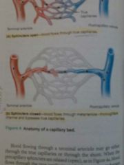
- highways
- they are a short vessel that directly connects the arteriole and venule at opposite ends of the bed - sphincters are closed: blood flows through metarteriole-thoroughfare channel and bypasses true capillaries |
|
|
|
What are true capillaries?
|
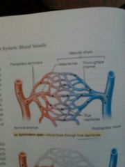
- backroads
- the sphincters are open and blood flows through true capillaries |
|
|
|
Characteristics of the venous system:
|
- carry blood from the capillary beds toward the heart
- the diameter of successive venous vessels increases, and their walls gradually thicken as they progress from venules to larger and larger veins |
|
|
|
The formation of different veins:
|
capillaries unite to form venules ---> venules join to form veins
|
|
|
|
Do veins contain all three tunics?
|
Yes, but their walls are thinner and lumens larger when compared to arteries
|
|
|
|
Is blood pressure high or low in veins?
|
blood pressure is low, they are known as blood reservoirs
|
|
|
|
What prevents the backflow of blood in veins?
|
Venous valves - they are formed from the folds of the tunica intima, resemble the semilunar valves of the heart in both structure and function
|
|
|
|
Where are venous valves most abundant?
|
in the veins of the lower limbs where gravity opposes the upward flow of blood
they are usually absent in the veins of the thoracic and abdominal body cavities |
|
|
|
What are varicose veins? How are they treated?
|
veins that have become leaky due to leaky valves due to standing for long periods of time, pregnancy, heredity, or high venous pressure
treated by surgery, salt solution injection, or laser therapy |
|
|
|
Physiology of Circulation:
|
blood pressure - force per unit area exerted on a wall of a BV by contained blood
blood flow - actual volume of blood flowing through a vessel, an organ, or an entire circulation during a period of time; it is directly proportional to difference in blood pressure between two points in the circulation resistance - opposition to flow and is a measure of the amount of friction blood encounters as it passes through the vessels |
|
|
|
What are sources of resistance?
|
blood viscosity - the more viscous (polycythemia) = decrease blood flow, the less viscous (anemia) = increased blood flow
blood vessel length - longer = less blood flow, shorter = more blood flow blood vessel diameter - smaller = less blood flow, larger = more blood flow ------- lots of resistance in the small arterioles |
|
|
|
What is pulse pressure? What increases it?
|
the difference between the systolic and diastolic pressures
atherosclerosis chronically increases pulse pressure because the elastic arteries become less stretchy |
|
|
|
What reflects arterial blood pressure?
|
- how much the elastic arteries close to the heart can stretch (their compliance or distensibility)
- the volume of blood forced into them at any time |
|
|
|
What causes a rise in arterial bp?
|
increased sv, increased hr, atherosclerosis, and increased blood volume
|
|
|
|
What is atherosclerosis?
|
changes in the walls of large arteries consisting of lipid deposits on the artery walls; one form of arteriosclerosis
|
|
|
|
Is blood pressure in the capillaries high or low? Why does it need to be this way?
|
it is low, capillaries are fragile and high pressures would rupture them and most capillaries are extremely permeable and thus even the low capillary pressure can force solute-containing fluids out of the bloodstream into the interstitial space
|
|
|
|
What regulates blood flow in tissues?
|
nervous regulation of blood flow:
- sympathetic nervous system: increases HR and stroke volume - parasympathetic n.s.: decreases HR and stroke volume |
nervous regulation of blood flow in regards to HR and SV
|
|
|
Is venous blood pressure high or low? How does blood get back to the heart?
|
it is low
respiratory and muscular pumps valves prevent backflow |
|
|
|
How do short-term neural controls manage blood pressure?
|
- maintain adequate MAP by alerting blood vessel diameter on a moment-to-moment basis
- altering blood distribution to respond to specific demands of various organs |
|
|
|
What happens under conditions of low blood volume when the body is trying to control blood pressure?
|
all vessels except those supplying the heart and brain are constricted to allow as much blood as possible to flow to those two vital organs
|
|
|
|
What is an example of altering blood distribution to respond to specific demands of various organs?
|
during exercise the blood is shunted temporarily from the digestive organs to the skeletal muscles
|
|
|
|
What are baroreceptors? Where are they located? What do they do? Carotid vs. Aortic reflexes
|
baroreceptors - clustered neurons
located: carotid sinuses, aortic arch, and large, elastic arteries of the neck and thorax they protect circulation against short term BP changes carotid - maintain blood flow to the brain; aortic - systemic blood pressure |
|
|
|
When are baroreceptors ineffective?
|
they are ineffective against long sustained increase or decrease in BP
|
|
|
|
When do chemoreceptors function? What do they do?
|
They function when oxygen content or pH of blood drops or carbon dioxide levels rise = when CO2 increases, pH decreases
Chemoreceptors in aortic arch and large arteries of neck transmit nerve impulses to vasomotor center and reflex constriction occurs. This causes a rise in blood pressure. They sense increased levels of CO2/low pH, low oxygen and constrict arteries and increase BP |
|
|
|
What are the influences of the higher brain centers on blood pressure regulation? What is an example? What is voluntary/involuntary?
|
reflexes that regulate blood pressure are integrated in the medulla oblongata of the brain stem
although the cerebral cortex and hypothalamus are not involved in routine controls of blood pressure, these higher brain centers can modify arterial pressure via relays to the medullary centers Example: fight or flight = mediated by the hypothalamus has profound effects on blood pressure voluntary - cerebral cortex; involuntary - hypothalamus |
|
|
|
Chemical controllers of blood pressure
|
- adrenal medulla hormones (fight or flight): epinephrine = increases BP and HR, vasoconstriction unless skeletal muscle (vasodilation)
- atrial natriuretic peptide: decrease BV & BP, produced in atria, cause kidneys to increase fluid output - ADH: hypothalamus, released in response to decrease BP (kidneys) = water retention = increase BP & BV - angiotensin II: increase BP, stimulates vasoconstriction and aldosterone production |
|
|
|
What is atrial natriuretic peptide?
|
chemical controller of BP
produced by atria of the heart special cells in R) atria that respond to stress, BP to high, ANP antagonizes or inhibits aldosterone production (produced outside of adrenal gland which causes body to retain sodium, no urine and BP goes up). ANP makes Na and water gets released to decrease BP Causes generalized vasodilation |
|
|
|
What is long-term regulation of blood pressure? How?
|
renal mechanisms - they regulate blood volume
an increase in blood volume causes an increase in BP Kidneys act directly, by regulating sodium and potassium levels, and indirectly, using renin-angiotensin mechanism, to maintain BP |
|
|
|
What is direct renal mechanism?
|
alters blood volume independently of hormones
when blood volume/pressure rises, the rate at which fluid filters from the bloodstream into the kidney tubules increases = increased urine production = decrease in blood volume/pressure when BP/volume is low, water is conserved and returned to the bloodstream, and BP rises |
|
|
|
How are BV and ABP related?
|
As blood volume goes, so goes the ABP
|
ABP = arterial blood pressure
|
|
|
What is indirect renal mechanism?
|
BP is low = kidney cells produce renin ---> renin converts angtiotensinogen (produced by the liver) into angiotensin ---> angiotensin II
Angiotensin: 1) stimulates aldosterone = Na retention = increased BV = increased BP 2) causes vasoconstriction = increased BP |
|
|
|
What are two ways to monitor circulatory efficiency?
|
pulse and blood pressure
|
|
|
|
What is a type of alteration in BP? What is primary and secondary?
|
Hyper or hypo-tension
Primary - there is no underlying cause identified, not "curable" just manageable Secondary: accounts for up to 10% of cases and is due to identifiable conditions, treatment focuses on correcting the problem that caused it |
high bp and low bp
|
|
|
What is tissue perfusion? And what is it involved in?
|
it is blood flow through body tissues
involved in: 1. delivering oxygen and nutrients to tissue cells and taking wastes away 2. gas exchange in the lungs 3. absorption of nutrients from the digestive system 4. urine formation by the kidneys |
|
|
|
When the body is at rest how is blood divided up? How is this changed during exercise?
|
Brain receives 13% of total blood flow
Heart 4% Kidneys 20% Abdominal organs 24% Skeletal muscles, which make up half of body mass, receive about 20% Nearly all of the increased cardiac output flushes into the skeletal muscles and blood flow to the kidneys and digestive organs declines |
|
|
|
Where is the velocity of blood flow the fastest? the slowest?
|
Fastest in aorta and large arteries - there are increases in speed in vena cavas due to skeletal and respiratory pumps, not valves
Slowest in capillaries which is necessary to allow time for gas exchange |
|
|
|
What are three important differences between systemic arteries and veins?
|
1. Arteries run deep while veins are both deep and superficial
2. Venous pathways are more interconnected making them more difficult to follow 3. The brain and digestive system have unique venous drainage systems. |
|
|
|
What is circulatory shock? What are three types/causes?
|
any condition in which blood vessels are inadequately filled and blood cannot normally flow through; blood flow is inadequate to meet tissue needs; if it persists cells die and organ damage follows
hypovolemic, vascular, cardiogenic |
|
|
|
What is hypovolemic shock?
|
- results from a large-scale blood or fluid loss
- BP decreases, HR increases to account for it - vasocontriction: trying to move blood into mian circulatory areas (vena cavas, arteries) to get blood to and through the heart - need to replace fluid volumes |
|
|
|
What is vascular shock?
|
- blood volume is fine
- circulation is poor due to extreme vasodilation - example is anaphylactic shock - histamines are rapidly produced |
|
|
|
What is cardiogenic shock?
|
- pump failure
- occurs when heart cannot sustain adequate circulation - usually caused by myocardial damage (as may follow numerous MIs) |
|
|
|
What is autoregulation? How is this controlled?
|
the automatic adjustment of blood flow to each tissue in proportion to the tissue's requirements at any instant
local conditions regulate this process independent of control by nerves or hormones by modifying the diameter of local arterioles feeding the capillaries |
|
|
|
metabolic autoregulation?
|
when blood flow is too low to meet a tissue's metabolic needs, oxygen levels decline and metabolic products accumulate - these changes serve as autoregulation stimuli that lead to automatic increases in tissue blood flow
declining levels of nutrients are strongest stimuli for autoregulation exercising, need glucose, protein, oxygen = all can be strong stimuli |
|
|
|
myogenic autoregulation
|
keeps tissue perfusion fairly constant in spite of changes in systemic pressure
stretching of smooth muscle to keep things going normally despite little changes in BP |
|
|
|
What is long-term autoregulation? When is it common?
|
involves increasing number of BVs and enlargement of existing BVs
common in the heart when a coronary vessel is partially occluded and occurs throughout the body in people who live in high-altitude areas, where the air contains less oxygen |
|
|
|
What is angiogenesis?
|
phenomenon in which the number of blood vessels in the region increases, and existing vessels enlarge
common when coronary vessels become occluded and when a person moves to an area of higher altitude |
occurs in long-term regulation
|
|
|
What is collateral circulation?
|
dependent upon age, a body will develop new BVs to go around vessels that are occluded
|
|