![]()
![]()
![]()
Use LEFT and RIGHT arrow keys to navigate between flashcards;
Use UP and DOWN arrow keys to flip the card;
H to show hint;
A reads text to speech;
26 Cards in this Set
- Front
- Back
|
The average mass of Hb in a red cell is shown by what test? Is it sex dependent?
|
MCH (Mean corpuscular hemoglobin)
NOT sex dependent |
|
|
What is red cell distribution width?
|
Since MCV represents the average red cell vol., it is less reliable in identifying marked variation in cell size (anisocytosis)
RDW measures the variation in size |
|
|
What do high and low red cell distribution width's mean?
|
High RDW: Increase in the heterogeneity of RBC size
Low RDW: A more uniform RBC population, little or no variability in size. |
|
|
what is a high RDW associated with? What about small?
|
high: Anemia
Small: Hereditary hemoglobinopathy |
|
|
What are the 2 types of macrocytosis? is macrocytosis normochromic or hypochromic?
|
Oval or Round
Normochromic; no polychromasia remember: macrocytosis is >8 um diameter; MCV>100 |
|
|
What type of macrocytes predominate in:
Vitamin B12 and folate deficiencies Aplastic anemia Myelodysplastic Syndromes Chemotherapy effect |
Oval macrocytes
|
|
|
Microcytes are associated as a normochromic or hypochromic process?
|
Hypochromic
|
|
|
What is an Acanthocytes? What do you normally see?
|
Smaller than normal red cell
3 to 12 irregular membrane spikes, unevenly distributed. No Central Pallor |
|

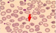
What does this show? what are they associated with?
|
Acanthocytes
Seen with liver disease |
|
|
spikes evenly distributed on a RBC membrane, maintain central pallor...this describes?
|
Echinocyte
Remember: Acanthocytes have NO central pallor and the spikes are more random |
|
|
Other than improperly prepared smears, what are some of the causes of echniocytes (2 major)
|
Uremia and chronic renal failure
Immediately post-transfusion with aged or metabolically depleted blood. |
|
|
What are schistocytes?
|
Fragments of RBCs...
Irregularity of cell size and shape, . Small fragments lack any significant central pallor. |
|
|
what can be very indicative of very serious hemolytic conditions?
|
Schistocytes
|
|
|
Resembles a pear: round with a single elongated or pointed extremity. Unipolar tapered end with a blunt tip
Maybe artifactual in improperly made smears. (tails line up in same direction) this describes? |
teardrop cell
|
|
|
Rod shaped with nearly parallel sides
If >10% suspect hereditary ovalocytosis (25% is diagnostic), an abnormality of skeletal membrane proteins. |
Ovalocyte (Elliptocyte)
|
|
|
Schistocytes, acanthocytes, and sphereocytes are the ones to know ...
|
for the test!!
|
|
|
Central pallor appears slit like, straight, or fish mouth.
put in here just for completion, feel free to breeze on through |
Stomatocyte
|
|
|
Increased surface membrane to volume ratio results in a central darker region within the area of central pallor. “Bulls eye” cell.
What kind of cell is this? |
Target Cell
|
|
|
iron deficiency anemia, Thalassemia, Hemoglobinopathies, and Liver disease can be diseases associated with what cells?
|
Target Cells
|
|
|
What is a reticulocyte?
|
an immature RBC
|
|
|
this cell is larger than an RBC and has a blue color, which is referred to polychromatophillia...
|
Reticulocyte
|
|
|
Numerous, evenly distributed blue-gray granules within the cytoplasm. Maybe fine or coarse
this describes what? |
Basophilic Stippling
|
|
|
A DNA fragment produced when nuclear extrusion from the RBC takes place
not seen in normal state because the spleen will eat these bad boys up |
Howell-Jolly Body
|
|
|
Iron containing particles visible on WG stain.
Abnormal sidersomes composed of both iron and protein matrix. this describes what? |
Pappenheimer Body
|
|
|
under a microscope you see blue granules, with irregular sharp edges. You ask for an iron stain and it is positive. This confirms that you are looking at a
|
Pappenheimer Body
|
|
|
RBCs stick together and are in a "stack of coins" formation... this describes? where can you find it on a smear?
|
Rouleaux formation
found in the feathers edge |

