![]()
![]()
![]()
Use LEFT and RIGHT arrow keys to navigate between flashcards;
Use UP and DOWN arrow keys to flip the card;
H to show hint;
A reads text to speech;
54 Cards in this Set
- Front
- Back
|
DNA size is expressed in what 3 ways? |
1. Number of base pairs 2. Molecular eight ~660 is molecular weight of 1 base pair 3. Length ~34angstrom per helical turn of 10.4 base pairs |
|
|
What methods are used to measure DNA size? |
1. Electron Microscopy 2. Gel electrophoresis |
|
|
What is the most popular way to express DNA size? |
Number of base pairs |
|
|
What is the formula used to determine molecular weight? |
Molecular weight = # of Base Pairs * 660 |
|
|
What is the formula used to determine Length |
Length = # of base pairs * H |
|
|
What is the formula used to determine H? |
Rise/Residue |
|
|
What is H traditionally? |
3.4 angstroms OR 3.4x10^-4nanometers |
|
|
Given that SV40 virus has 5226 base pairs. What is it's molecular weight? |
Molecular weight = #bp * 660 --> MW= 5226*660 --> MW= 3.5x10^6 |
|
|
Given that SV40 virus has 5226 base pairs. what is the length? |
Length = #bp * H --> Length = 5226 * 3.4x10^-4 Length = 1.7 nanometers |
|
|
Explain how the same DNA molecule could assume two to be different sizes? |
The same DNA molecule could assume two different conformation (Relaxed and Supercoiled) |
|
|
Between "relaxed" and "supercoiled" DNA. which moves faster through a CsCl gradient and there for has greater density? |
Supercoiled DNA |
|
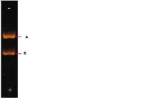
Based on the image, which band represents a relaxed DNA? |
A |
|

Based on the image, which band represents supercoiled DNA? |
B |
|
|
What reasons would supercoiled DNA have to have a higher density than relaxed DNA? |
1. Supercoiled DNA binds to more CsCl 2. It has a smaller molecular volume, hence more molecules packed in a given volume, leading to a higher density. |
|
|
The number of supercoils in a DNA molecule |
Writhe |
|
|
The positive # indicating the total number of turns and supercoils regardless of whether the supercoils are negative |
Twist |
|
|
The total number of turns and supercoils taking into account positive and negative integers |
Linking number |
|
|
Given the following information. What would happen if a DNA molecule were nicked and unwound 2x. Twist= 25 writhe= 0 Linking number = 25 |
If DNA were nicked and unwound two times then it will spontaneously negatively supercoil right-handedly. This will cause the following Twist=25 Writhe= -2 Linking number=23 |
|
|
Given the following information. What would happen if a DNA molecule were nicked, unwound 2x, and the molecule doesn't spontaneously supercoil Twist= 25 writhe= 0 Linking number = 25 |
If the DNA were left as is and not allowed to supercoil then the following would occur. Twist= 23 Writhe= 0 Linking number 23 |
|
|
What would happen to the Writhe if DNA was overwound |
If DNA was overwound then it would result in a left-handed turn and a positive supercoil resulting in positive writhe number |
|
|
What would happen to the Writhe if DNA was underwound |
If DNA was underwound then it would result in a right-handed turn and a negative supercoil resulting in negative writhe number |
|
|
Which of the following must be integers? 1. Writh 2. Linking number 3. Twist |
Linking number |
|
|
The free energy is minimal when about ______ of the change in L is expressed in T and ______ in W |
30% ; 70% |
|
|
Given that Linking Number change is 2 what is the most stable (geometrical) isomer if T0=25 and W=0. |
25-2*0.3 = 24.4 T=24.4 0-2*0.7 = -1.4 W=-1.4 |
|
|
What is the formula to determine Supercoiling Density? |
(L-L0)/L0 |
|
|
What happens to the DNA molecule when histones are removed |
30-nm fibers and nucleosomes disappear |
|
|
Can nicks be used to relax supercoiled DNA? |
yes |
|
|
The lagging strand is also known as the ______________ and the leading strand is also known as the ________________ |
Discontinuous strand ; continuous strand |
|
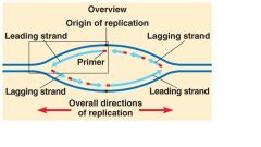
Label the 5' and 3' end of each DNA fragment shown |

|
|

How many replication forks are shown here? |
2 |
|
|
Describe the experiment performed by Matthew Meselson and Franklin Stahl perform to detemine if DNA replicates conservitively, semiconservatively, or through random replicates |
Meselson and Stahl ran a DNA molecule amongst N15 heavy isotopes for 3 generations. This ensured that all of the DNA in the last generation had the N15 then They moved these DNA molecules in an environment of light N14 isotopes. They then allowed the DNA to replicate for 2 generations. After each generation they measured the DNA through a CsCl gradient and photographed their results under UV light. If the DNA replicated conservitively then after the first generation of DNA molecules would show one two distinct bands one with heavy and the other with light. If the they replicated dispersively then after the first and second generation there would only be one band indicating that the DNA molecules contained both the heavy and light isotopes in the CsCl gradient. If they replicated semiconservatively then after the first generation there would be one band and then after the second generation there would be 2 bands. They found that the DNA molecules replicate semiconservatively. |
|
|
Matthew Mesleson and Franklin Stahl determined that DNA replicates semiconservatively. What isotopes did they use and why? Were they radioactive? |
N15 (heavy) and N14 (light) were used in order to be used in a CsCl gradient which separates molecules based on density. These are not radioactive isotopes. |
|
|
Why did Meselson and Stahl wait 3 generations before introducing the DNA molecules to the N14 solution? |
By waiting 3 generations, Meselson and Stahl ensured that all of the DNA molecule were N15 labeled. |
|

Using the image, which of generation shows that the DNA is H/L |
1.0 |
|

Based on the image at what generation does DNA show a L/L and H/L band |
Begining from 1.9-2.5 |
|

Using the image explain why H/H are no longer seen in later generations? |
H/H is no longer seen because the DNA is not exposed to the N15 solution (heavy) and is only in the N14 (light) solution. Thus any replication that goes on will only progress with N14. |
|
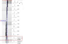
T/F: The following image taken by Meselson and Stahl was taken by a radiograph |
False. The isotopes used were not radioactive and therefore no radiograph was used. The image shown was taken using a camera under UV light. |
|
|
T/F: For many years "labeled" has been synonymous with "radioactive" |
True |
|
|
Why are radioactive tracers used as opposed to nonradioactive tracers? |
Radioactive tracers allow vanishingly small quantities of substances to be detected |
|
|
T/F: All labels are radioactive |
False. Such examples of unradioactive tracers include N15 |
|
|
Radioactive emissions from a sample create photons of visible light detected by a photomultiplier tube
|
Liquid Scinitllation Counting |
|
|
Describe the process of Liquid Scintillation Counting |
1. Radioactive material is put into a vial containing scintillation fluid. 2. Fluid containing fluor fluoresces when hit with radioactive emissions. 3. The Liquid Scintillation Counter acts to convert invisible radioactivity into visible light when radioactive emissions hit the Fluor 4. The unit is counted per minute or CPM |
|
|
If Liquid Scintilation Counting material uses Fluor (a fluorescent material) and should then be visible to be counted, why would radioactive emissions be required? |
The radioactive emissions hit the Fluor which emit fluorescence. This process converts radioactivity into visible light which is then counted. |
|
|
Knowing that DNA replication is semi-conservative. The next question is to determine if DNA proceeds through replication Continuously, Discontinuously, or Semi-Discontinuously. What experiment was performed to determine if DNA replication proceeded Continuously?
|
In order to determine if DNA replication proceeded continuously. |
|
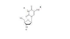
Using the image, which of the following (A, B, or C) would not be a place where to label thymidine with tritium? |
C. Tritium can't label this -OH because this H+ is lost when it is built on the DNA strand. |
|
|
What organism was used when demonstrating the semidiscontinuous mode of DNA replication? |
T4 |
|
|
How were thymidine labeled in order to demonstrate the semidiscontinuous mode of DNA replication? |
It was labeled with Tritium. |
|
|
Explain how Okazaki fragments were used to demonstrate the semidiscontinuous mode of DNA replication |
Okazaki fragments are used to demonstrate that DNA replication does not occur continuously. |
|
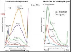
Describe the experiment performed based on the image |
The image is the results of an experiment used to demosntrate the semidiscontiuous mode of DNA replication. T4 bacteriophage was the organism used. Thymidine were labeled with tritium. As thymidine is added to the growing DNA strands, they will label the strand. The label allowed experimenters to determine if DNA replication produced okazaki fragments. If okazaki fragments are formed and then later ligated during DNA replication then it will be shown that DNA replication proceeds through a continuous mode. In order to determine which is true, experimenters eliminated ligating enzymes responsible for the joining of okazaki fragments. The graph onthe left shows normal DNA replication. The longer the Thymidine labels were present, the longer the strands formed. The graph on the right showed demonstrated disabled ligase. The results of the graph on the right show that a great majority of replicated DNA were actually short pieces. These implied that DNA replicates in small chunks and are then ligated later. This showed that DNA does not replicate continuously. However at thi point it is not known whether it proceeds discontinuous or semi-discontinously. |
|
|
What does the dut gene do? |
Dut gene codes for the dUTPase which converts dUTP to dUMP, hence preventing dUTP (as dUMP) from being incorporated into the DNA. |
|
|
What is the ung gene responsible for? |
Ung gene codes for Uracil N-glycosylase. It removes uraicl molecule that have been incorporated into DNA. |
|
|
What two mechanisms keep DNA "dUMP-free"? |
1. The dUTPase (encoded by dut gene) keeps the ratio of dUTP/dTTP low in the cell so that few dUTP molecules can be incorporated into DNA 2. The uracil-N-glycosylase (encoded by the ung gene) removes the uracil group from DNA if any dUMP has been incorporated into DNA |
|
|
How did Okazaki determine that DNA replicates semidiscontinuously? |
Okazaki developed a dut+ung- mutant which minimized the incorporation of dUMP (presence of dUTPase) and formation of abasic sites (no uracil-N-glycosylase). He found that 50% o newly labeled DNA in these cells was still in short pieces. Hence, the conclusion is that DNA replication is semidiscontinuous |
|
|
Why did Okazaki develop mutants to determine if DNA replicated semidiscontinuously |
Ung produceds Uracil-N-glycosylase which is responsible for removing uracil from growing DNA strands which would create abasic sites and nicks. WIthout ung, abasic sites won't form and neither will nicks. With that he found that 50% of newly formed DNA were still in short pieces while the other 50% were in long continuous strands.
|