Reading...
![]()
Play button
![]()
Play button
![]()
Use LEFT and RIGHT arrow keys to navigate between flashcards;
Use UP and DOWN arrow keys to flip the card;
H to show hint;
A reads text to speech;
166 Cards in this Set
- Front
- Back
|
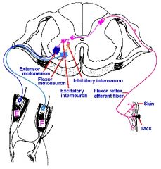
why is flexation an ex of an ipsilateral reflex
|
b/c sensory input and motor output are on same side of spinal chord
|
|
|
what is contra lateral reflex arc
|
when sensory input comes in one side of the spinal chord and motor output to extend the hand occurres on the other side
|
|
|
in relfex arc where do u have a vague feeling? where does it go after ?
|
thalmus . goes to the cortex to cause further physical action and emotion
|
|
|
why is the middle "butterfly" in the spinal chord grey?
|
it lacks myelinated sheaths
|
|
|
what is the basic unit of structure ofthe nervous system
|
the neuron
|
|
|
what are the support cells of the neurons?
|
Neuroglia cells
|
|
|
what are the four neuroglia cells
|
oligodendrocytes, microglial , astrocytes, and ependymal
|
|
|
what do oligodendracytes do
|
form the myelination inthe brain and spinal chord (CNS)
|
|
|
what to microglia do
|
develope form wht blood cells and wander thru CNS to destroy dead nervous tissue , microorganisms and other foreign matter,
|
|
|
what do astrocytes do?
|
constitute over 90% of tissue in some area of the brain, they cover the entire brain surface and most non synaptic regions of the neurons in the gray matter of the CNS, supportive frame work, make food, assist blood brain barrier, ans form scar tissue
|
|
|
what do ependymal cells do?
|
lint hte ventricles of the brain and secrete and circulate cerebrospinal fluid
|
|
|
what is gray matter of the spinal chord
|
made up of huge number of axon ends, dendrites, bodies, interneurons, no mylenation therefor its grey. it is the site of all synaptic contact btx neurons, it is the site of all synaptic integrations (information proscessing ) in the CNS
|
|
|
divisions of gray matter in the spinal chord?
|
Horns, Lateral dorsal(posterior) and Ventral(anterior)
|
|
|
gray matter of spinal cord is refred to as what?
|
wal mart computer, it can receive , process , and send information. ie. simple things like reflexes. anythign beyond simple computer must be sent up to the cortex (good computer) ex, the reflex action is form the spinal chord. but if u know its is hot then its form the cortex. the brain of the nervous system is the grey matter.
|
|
|
myelinated axons are what for the nervous system
|
they are a highway system brings info in and out of the brain
|
|
|
what are the component parts of a reflex arc
|
receptor, insonimg sensory neuron, processing center, outgoing neuron, and effector
|
|
|
what is monosynaptic relfex arc
|
means only one synapse beteween aferent and efferent neuron (very little delay and prompt response)
|
|
|
waht is a soma of a cell also known as
|
the body
|
|
|
what type of mater surrounds the gray matter. why is it that color
|
white, its made of axons of neurons bundled together, tey ARE myelinated
|
|
|
the white bundles in spinal cord are called
|
1) dorsal, lateral and ventral funiculi, they provide communication between different levels of the CNS
|
|
|
a group of grey matter outside the CNS is called?
|
a ganglion, it is the swelling of the cell bodies outside the CNS
|
|
|
Posterior branch , meningeal branch and anterior branch takes care of the needs of the what
|
back, the chord , and everything else
|
|
|
reflex arc refers to what
|
receive amessage, take it to a processing center, send out message to effect a change, there has to be a sensory receptor (dendrite), a sensor neuron, grey matter to process, outgoing motor neuron to an effector (skeletal muscles)
|
|
|
what are the two types of protiens on the cell membrane
|
transmembrane(integral) protiens,which pass all the way thruthe membrane, and prephreial protiens - stuck on the outside of inside of the membrane (not all the way thru)
|
|
|
what are the three types of integral protiens
|
1) channels or pores, pumper molecules(driven by ATP), and enzymes
|
|
|
two types of channels or pores are
|
open, and gated
|
|
|
gated channels are regulated by wht 3 ways
|
1)ligand regulated gates (chemicle), voltage regulated ( changes in electrical potential in membrane, and , mechanicalls regulates (physical stress on cell)
|
|
|
waht is the resting membrane potential
|
-70mv (avg) (just inside cell membrane)
|
|
|
where is sodium found reference cell membrane?
|
outside (its positive charged), more guraded and more difficult to move in and out of the cell
|
|
|
where is potassium found reference cell membrane
|
inside the cell also positive chg, able to move in and out with greater ease than sodium, varitey of channels and pored to move
|
|
|
what makes the inside of the cell membrane negatively charged
|
lg amounts of phosphate ions, sulfate ions, and protiens that are negatively charged. a protien molecule nh2-c-cooh that looses its hydrogen to become nh2-c-coo- it is now neg charge, the hydrogen is used as a buffer. better allowing us to maintain PH
|
|
|
what is the stimulis in thehand on the hot plate scenerio?
|
hot.
|
|
|
hand on the hotplate, what has to happen in order to transfer that signal to the CNS how is it done
|
the "hot" needs to be changed to an electrochemicle signal that out body uses to transport messages. It is done when the heat stimuli, causes the perephreal protien out of the way for sodium to begin to enter cell.
|
|
|
when a receptor has the ability to transform one type of energy stimuli to another what is it called
|
receptor potential
|
|
|
when the perephrial protien moves out fo the way why does soduim rush into the cell.
|
1) its following th econcentration gradient. 2) opposite charges attract (sodium + and the - protiens and suck inside the cell
|
|
|
when sodium enters the cell and begins to move the resting potential away from -70 how far will it go to start the nex step
|
it will go to +30 mv, at that time the soduim channels will close and the potassium chanels will open.
|
|
|
what is the threshold for action in a cell (Mv)
|
it is -50 mv, this is when the body decides it will respond to the stuimuli if it only reaches -55 or so it is faciliatead not necerraly going to respond
|
|
|
when potassium channels open and soduim channels close in order to balance the cell charge, when till it stop
|
it will over shot adn become hyperpolarized at -90mv, at this point the cell can not respond to new stimuli
|
|
|
what maked the process "move to the next cell" to start eh sodium channel opening on that one?
|
voltage disruption, encourages future reactions towards the axon.
|
|
|
waht is the receptor potential?
|
it is a graded potential (it can die out before reaching axon), however its specific to the receptor that changes heat to electro chemical
|
|
|
a free end dendrite is aslo known as a what?
|
receptor
|
|
|
facilitated response. is what
|
when one stimulis has changed the cell to -55 it wont fire off the reaction, but if a second stimuli comes in it can easly trigger it cause it was already half way there ,,,, it has been facilitated
|
|
|
what is the refractory period?
|
this is the time immediatly after and during an action potential, difficult or impossible to stimulate that regin of a neuron to fire again.
|
|
|
what is absolute refraction?
|
it is when aqbsolutely no stimuli of any strength can cause it to fire again. occurres during repolarization
|
|
|
what is the electrical event called when the action potential gets to the axon knob?
|
voltage disruption
|
|
|
how does the voltage disruption get the ACH to release?
|
it causes calcium (which came from ur extra cellular fluid) makes small vesicles merge to larger which then make way to the membrane and ach will ooze out into the synapse.
|
|
|
what controls the amouht of ACH released in the synapse?
|
the number of action potentials recieved that the axon knob
|
|
|
what happens to ACH once its job is finished?
|
achtylchinesterase chops it into aceytle and choline where it is brought back in once inside an enzyme merges both halves together again
|
|
|
the release of ACH is a graded potential determined by what...
|
neurotransmitter chemicles
|
|
|
what the difference btx graded potentials and action potentials?
|
graded potentials can die out, action potential dont
|
|
|
which (graded or action) are controlled by chemicles from neurotransmitters and which by voltage disruption?
|
graded potentials are caused by chem. controlled neurotransmitters, action potentials casued by voltage disruption
|
|
|
portions of a myelinated axon that have no myelination are called what?
|
Nodes of Ranvier
|
|
|
what cells meylinate the axons in the PNS
|
Schwann cells
|
|
|
how do schwannsells act.
|
they wrap tightly around the axon so that no electiracl impulses will flow cause of the phosphlipid layer. causing the signal to hop over it.
|
|
|
what is the movement where signals hop over the myelination areas called
|
saltatory transmission, doenst take less time for each action potential , there are just less of them
|
|
|
what two thing make action potentials get form poitn a to b quicker
|
1) myelination, 2) diameter
|
|
|
waht cell makes myelination in the CNS
|
olgiodendracytes
|
|
|
why would u rather cut a nerve in ur leg (PNS) rather than one in ur spine (CNS)
|
Schwan cells (PNS) can reapir them selves, oligodendracytes(CNS) dont repair they scar.
|
|
|
MOnosynaptic?
|
no neurons inbetween, just in and out in CNS only one synaptic space
|
|
|
poly synaptic??
|
another neuron inbetween (interneuron)
|
|
|
what is reciprocal inhibition?
|
ex flexing arm... one muscle to flex is activated (the agonist) while a second muscle is told not to do anything (the antagonist ) this is why u need two neurons involved in sending message to flex
|
|
|
what is the agonist?
|
the musscle causing the action u see .... bicep in flexing arm
|
|
|
what is antagonist?
|
the muscle that oposes the agonist... the tricep relaxing or not contraction so the bicep can flex the arm
|
|
|
why is flexing ur arm ipsilatteral?
|
becuse the stimulis enters one side and exits that same side causing action on the same side
|
|
|
what is cross extensor what is cross flexation?
|
when an interneuron exists to bring the message from one side to the other side to cause extension there.
cross flexation is the same as above but the action carried out is flexation |
|
|
what is contralateral
|
the term for the crossing over of the signal
|
|
|
draw a cross section of the spinal cord and the spinal nerves place in the picture neurons the represent a polysynaptic cross extension
|
|
|
|
what is it called when an axon attatches to a individual muscle cell?
|
neuromuscular junction
|
|
|
what is a motor unit
|
it is the neuron and all the muscle cells to which it is associated with
|
|
|
how many muscle cells are ususally associated with a motor unit
|
5- 200 the fewer u have associated with it the finer control u have
|
|
|
why is the threshold important for muscle cells
|
important for amount of contraction
|
|
|
unit of structure and function for muscles is?
|
sarcomere
|
|
|
the cell membrane of a muscle cell is?
|
sarcolema
|
|
|
the cytoplasm of a musscle cell is?
|
sacroplasm
|
|
|
what are in the t-tubules and why?
|
extracellular fluid, they are outside the cell membrane. this is usefull for ca ions to cause contraction
|
|
|
waht is a triad..
|
it consisitsof the t-tubule, and the stackes sacrolasmic reticulium called cisternae.
|
|
|
the boundaries on eaither end of a sarcomere is called what
|
z- line
|
|
|
the section on either end consisting of the the thin myofibrils form a line called
|
the I band (i for thin)
|
|
|
the regoin in the center of just thick myofribrils is what
|
the h-zone (the cross bar of the H)
|
|
|
waht is the main molecule of thin myofibrils
|
actin
|
|
|
what is embedded in the actin (thin myofribils?
|
ADP active sites (mini velcro)
|
|
|
what what covers the ADP active sites (velcro)?
|
complex of molecules called the troponin/tryptomyosin complex
|
|
|
what makes up the thick myofibrils
|
myosin. have a main axis and globulels (oars) on top of the globules is the source of energy AtP and ATPase. when activated these two produce ADP (energy)
|
|
|
what from the brain causes a muscle to contract
|
the signal sent dwon the neurons along the axons in form of action potentials , this is refered to as the wave of depolarization
|
|
|
the wave of depolarizatoin once it reaches the axon knob does what
|
it causes a voltage disruption in the knob which releases CA++ and causes the ACH to be released
|
|
|
once the ACH acts on the muscle cell what happenes.
|
a wave of depolarization begins and travels down the cell, this goes into the t tubules causing CA++ to be released, this activates the atpase on the thick myofirbrils , and binds to the troponin of the troponin.tropomyosin complex causes the complex to move out of the way
|
|
|
how do u bring CA++ back into the sarcoplasmic retic.?
|
this is done bye a transpot carrier molecule, which uses energy, we get from the mitochindria (cellular respiratoin)
|
|
|
what are the 4 types of neruoglial cells
|
olgiodendrocytes, microglial, astrocytes, and ependymal
|
|
|
what are astrocytes
|
supportive framework for the nervouse tissue, nourishment( convert glucose to lactate (food for neurons), assist in forming th eblood brain barrier, release protiens (nerve growth factors) synapse formation and nerver growth, regulate neurotransmitters and potassium by absorbing them, scar tissue formation when neuron dies
|
|
|
path of the CSF in brain?
|
twolateral ventricles--interventricular foramen--3rd vent.--cerebral aquaduct--4th vent.
|
|
|
what is common name for the medulla oblongatta
|
the foreman, has pairs of neucli to control,,, breathing (medulary rhythmic center), vomiting, coughing, sneezing, hiccups, vasomotor center(blood vessel diameter, regulated BP) , the cardiac center-- controls rate and force of heart beat. doesnt do the things it makes the body do it "cracking the whip" (nyquil???)
|
|
|
another name for the pons
|
the bridge, it has the ponti nucli (pneumotaxic and apneutactic nucli) respiration, its main job is the bridge between the medulla and the higher brain
|
|
|
what is the recticular formation
|
circuit board traveling between all parts of the brainstem, comunicates with other parts of the brain. also controls alertness or consciousness, maintain tone and posture(circuit board, stiff straight. ie posture tone alert like a robot)
|
|
|
mesonencephalon (midbrain) does? and made up of?
|
corpora quadrigemina (lt rt infer. superior colliculi) dorsal surface of the mesencephalon. the superior contorl visual reflexes, inferior control auditory reflex, also in midbrain is the cerebral peduncle,,, inside is the substancia nigra relays inhibitory signals to thalmusand basal nuceli
|
|
|
what does the limbic system do
|
allows expression of emotion
|
|
|
where are the four basil nuceli located
|
white mater of the cerebrum ( the four circles under the cortex in the drawing
|
|
|
most improtant basial nuceli is
|
amigydeloid.. works with the libmic system for emotions
|
|
|
3rd ventircle surrounds what
|
the thalmus
|
|
|
what does thalmus do
|
regulates (gate keeper) to the cerebrum and cortex 99% of stimulus form outside the body outside or inside is not allowed thru .....
|
|
|
what does the hyopthalmus do
|
it is the homeostasis capital, many pairs of neucli .. rage, thirst, hunger, waterlevels (surpaoptic), making oxytocin (paraventricular), controls the pituitary (neurosecretory neuclius)
|
|
|
mamillary bodies do what
|
eating.. (mamaries breasts food)
|
|
|
cerebellum what does it do how attatched
|
its attatched by the superior ( connects to mesencephalon) middle (connects to the pons) and inferior (connects to the medulla, these are pathways of neurons to the speciffic parts. tree of life ( white is th eaxons that enter ) the grey are the nuclei. one major nuclei is the dente nuclei. this is sensory information, place in space
|
|
|
what does the denti nuclei do where is it.
|
it is in the cerebellum, this nuclei is responsible for the place inspace info and if it should go up to the cortex for the primary motor of the sensory cortex.
|
|
|
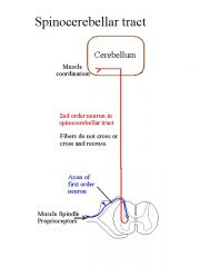
what does the spino cerebellar assending tract deal with and where does it start and end
|
starts in the spinal cord , ends in the cerebellum. axons are found in the inferior peduncle then to the dente nuclius, deals with where ur parts are in ur space.
|
|
|
teasing needle from the anterior to the posterior side at the mesencehpahlon....
|
pia mater-- spinothalmic ascending tract,cerebral penducle,substantia nigra, ependymalcells of the cerbral aquaduct, csf, ependymal cells, inferior coliculi, superior colliculi, pia matter adn sub arachniod space
|
|
|
wht is the cortex referred to as:
|
the expensive main computer
|
|
|
the central sulcus divides brain into
|
the motor cortex (front) and sensory (back) cortex
|
|
|
pre frontal ,, what does it do #1 on pic
|
higher intellectual, conscious, simple math simple problem solving (works with limbic system
|
|
|
pre motor what does it do #2 on pic
|
sends messages for repetative motion (typing , piano), involved in planning muscle motion, close with sending messages to and from primary motor
|
|
|
primary motor what does it do #3 on pic
|
region sends msg directly to control motor skills mesages are carried on the corticospinal decending tract
|
|
|
primary sensor what does it do #4 on pic
|
ANY SENSATION (Pain, toucch , heat) mesages come in on the fasiculis gracilis and cuneatus
|
|
|
sensory association #5 on pic waht does it do
|
called somasthetic, called memory bank wehre sensations are stored
|
|
|
visual association #6 on pic what does it do
|
visual memroy bank goes here after primary visual
|
|
|
primary visual #7 on pic what does it do
|
first place visual image goes then sent to visual assosiation for later use
|
|
|
brocas area #8 in the premotor area #2 waht does it do
|
only onleft side of brain, sends motor messages to use mouth tophysically speek
|
|
|
primary auditory #9 on pic inthe midbrain what does it do
|
where regular sounds come
|
|
|
short term memory #10 n pic in midbrain
|
where alzheimers pts have problems
|
|
|
wernicke's area #11 on pic midbrain waht does it do
|
only on left side of brain, makes sure what u say is appropriate and reasonable at that time, in temporal lobe
|
|
|
genreal interpretive #12 on pic midbrain what it do
|
wher eocipital, parietal, and temporal lobes come together, believed geniusus ahve this area well developed, original knowlege
|
|
|
what do ependymal cells lack
|
basement membranes
|
|
|
how much CSF is made per day
|
500 ml
|
|
|
how much CSF is in our bodies
|
100to 160 ml
|
|
|
where does CSF leave the 4th ventricle and where does it go after
|
it goes thru the russian holes (or medial and lateral appetures, into the sub arachniod space, the arachniod villias will allow it into the blood sinus where it will then mix with the blood
|
|
|
what tissue do the ependymal cells attatch to
|
white matter of the brain
|
|
|
where are choriod plexus mostly found
|
lateral ventricles
|
|
|
what are the roles of the CSF
|
1- bouyancy, 2-protection 3) chemical stability
|
|
|
what layers are the menengies made of
|
threelayers -- dura mater, arachnoid mater and pia mater
|
|
|
meninges of the spinal cord describe
|
epidural space btx vertebra and dural sheath that surrounds it, arachniod mater adheres to it from we have: vertebra--epidural space, duramater, arachniod mater, subarachniod space, pia mater, then spinal chord
|
|
|
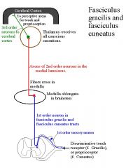
fasciculus gracilis and cuneatus ascending tracts carry what sensory impoulses
|
sensory impulses from skin muscle, tendons and joints.. touch pressure and body movement
|
|
|
describe the fasciluis gracilic and cuneatus tract for elbow
|
from elbow dendrite-- up axon of the 1st order neruon ---into gray matter--- continues into white matter and up the spinal cord. (stops in the medulla) in a nucleus (gracilis or cuneatus here if it continues will go to the 2nd order neuron it crosses over (decussation) to its corresponding nuceli and travel up to the thalmus there it stoppes again, if continues it enters the 3rd order neuron and travels up to the cerebrum (specifically the primary sensory cortex,
|
|
|
draw the fasciculus gracilis of cuneatus ascending tract
|
|
|
|
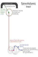
draw the spinothalmic ascending tract
|
|
|
|
draw the spinocerebellar ascending tract
|
|
|
|
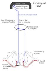
draw teh corticospinal descending tract
|
|
|
|
draw the reticulospinal descending tract
|
|
|
|
what infor mation does the spinothalmic tract convey
|
it carries messages of pain, temp, and some specific types of touch (light and crude) and pressure,
|
|
|
describe the pahtyway of the spinothalmic ascending tract
|
enters in the right side afferent unipolar sensory 1st order neuron synapses with an interneuron, goes below the central axis crosses over to other side of the gray matter into the white matter, there it forms the spinothalmic ascending tract at the point of synapse beginning the 2nd order neuron, travels up the spinal cord to the thalmus travels along ventral surface of brain, it can synapse into tthe 3rd order neuron if it needs to go to cerebrum
|
|
|
where does the spino cerebeller ascending tract begin
|
in the white matter dorsal and ventral right next to where the spinal nerves enter and exit
|
|
|
describe the pathway of the spinocerebeller ascending tract
|
1st order neurons originate in muscle and tendons, tothe spinal cord, synapses with 2order takes it to the dorsal spinocerebeller tract and travels up thru the medula enter the cerebellem thru the inferior peduncle to the dente nucleus
|
|
|
what woudl u find in the inferior peduncle?
|
neurons from the spinocerebeller tract
|
|
|
waht information does the spino cerebeller tract carry
|
"place in space" where ur body parts are in relation the the rest
|
|
|
what is the beginning of the corticospinal descending tract?
|
the pyramid cells in the primary motor cortex
|
|
|
describe the corticospinal descending tract
|
starts in primary motor cortex, there is a lateral and dorsal tract, they travel downit stops in the spinal cord in the white matter in the anterior area wher it then enters the gray area thru the dorsal horn, there is synapses with a motor neuron, (no interneuron) This is the lateral branch and controls the efector adn recipricol inhibition of the antagonistic response
the anteriro branch travels donw to the spine where it does have an interneuron and corsses over. anterior branch gives off signals to the cerebellum tolet it konw waht its doing to ur body parts this is thru the middle peduncle |
|
|
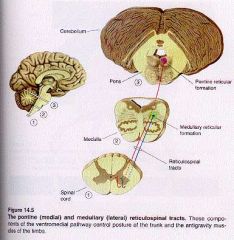
describe the reticulospinal descending tract
|
the circuitboard in the brainstem goes down to spinal cord synapses with motor neuron to go to effector, also sends msg to cerebellem
|
|
|
what does tectospinal tract do where it from
|
superior coliculi to neck coordinates eye movement with head (techno , light visual)
|
|
|
what does vestibulo tract do ,where it from
|
vestibular nucleis inthe brain stem works on ballance from impluses in ner ear
|
|
|
reticular formation sends signals to?
|
cardiac and vaso motor in medulla, thalmus and cortex --alertness, cerebellum visual auditory and motion coordination, and to ponds to regulate sleep patterns (RAS) epinephrine and seritonin
|
|
|
what does the limbic system do
|
gives u ur emotions,proper emotional responses, corpus colosum, olfactory (cerebrum), hypothalmus, mammilary bodies, ant thalmus,fornix, basal nuclei
|
|
|
waht are the two parts of the nervous system that send information
|
sympathetic (adrenergic , flight or fright) and parasympathetic (cholernergic)
|
|
|
sympathetic is for what?
|
prparing body for energy expending situations,
|
|
|
parasympathetic for what
|
pepare for conserving enregy
|
|
|
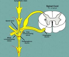
how does sympathetic work?
|
motor neuron comes form the spinal cord, goes to a ganglion and synapses, wil lhave a long post ganglionic fiber, and release epinephrine at teh effector this makes it adrenergic
|
|
|
how does parasymapthetic work
|
comes of the cranial nerves synapse in chain ganglion adn neurotransmoitter is ACH, has a short post ganglionic fiber, then release ACH in both places therfore its cholinergic
|
|
|
how many neurons in the sympathetic system go to an effector
|
two
|
|
|
in the sympathetic nerve system where to the preganglionic fibers arise?
|
lateral grey mater of the spinal cord associated with t1 thru l2, comes out the anterior branch goes to chain paravertrebral ganglion and stop for the post ganglion fiber to synapse
|
|
|
what path does signals follow to use theanterior branch
|
from lateral gray horn to the white ramus loops down synapses inthe paravertebral ganglion, up the gray ramis then to its efector
|
|
|
wht path does signal follow to go up or down
|
out form gry lateral horn to the white ramis down to the parevertibral ganglion then up or down the chain to its intended place
|
|
|
what path does it follow to use collateral ganglion
|
out lateral gry horn to the white ramis, then past the paravertebral ganglion the the cloatteral ganglion wherer it synapses to the psot ganglion neuron then to the appropriate organ
|
|
|
whats the path followed for the adrenal medula ascending
|
out from gry lateral horn to the white ramis past paravertebral ganglia to the cieliac col.l ganglia then to adrenal medulla ( its its own efector)
|
|
|
where does celiac ganglion go to
|
stomach pancreas and liver
|
|
|
what does the sympathetic collateral superior ganglion go to
|
small intestine
|
|
|
what does the inferior ganglion go to
|
lg intestine
|
|
|
what does the hypogastric ganglion go to
|
urindary bladder and genitles
|
|
|
waht part of body can be dangeroulsy effected if lg amounts of epinhphrine were to introduce to body?
|
kidneys, can burst the capillary beds due to increas in bp
|
|
|
waht makes the autonomic nervouse system fire?
|
brainstem, cortex, limbic system, adn incoming sensory visceral info. hypothal and recct. form. help too
|
|
|
draw how the impulses travel for for the sympathetic nerve sys, to use ant. branch adn also travel up an ddown chain
|
|