![]()
![]()
![]()
Use LEFT and RIGHT arrow keys to navigate between flashcards;
Use UP and DOWN arrow keys to flip the card;
H to show hint;
A reads text to speech;
42 Cards in this Set
- Front
- Back
|
GABA and Glutamate are in 99% of neurons. What do agonists and antagonists of each do? |
GABA: agonists= anesthesia, muscle relaxants etc antagonists= seizures Glutamate: agonists= seizures antagonists= anesthesia (specifically dissociative anesthesia) |
|
|
what are neuromodulators? Where are they? How do they function? |
- Acetylcholine (attention memory), dopamine (motivation motor movements), serotonin (mood depression), noradrenaline (attention cognition)
- not in most neurons, localized mostly to midbrain, hindbrain
- most use G-protein coupled receptors and they modulate the activity of neurons |
|
|
When you have a cigarette___ is activated. Parkinsons is treated with a ____. |
ACh; dopamine agonist |
|
|
What does a neuron need to use a neurotransmitter?
|
- always: vescicle transporter to concentrate the transmitter into a vesicle
- sometimes: biosynthetic enzymes to synthesize the neurotrans. (glutamate and glycine not specifically synthesized by the cell) - uptake transporters in plasma membrane - mechanism to degrade the transmitter |
|
|
What are neuropeptides? where are they? |
WHAT: Neuropeptides are cotransmitters (as opposed to classical transmitters like GABA) synthesized by neurons which use g-protein liked receptors ex. vassporessin or oxytocin
WHERE: the hypothalamus and pituitary use them as primary transmitters. You can have multiple neuropeptides in one neuron. They have connections all over the brain
|
|
|
two types of receptors |
1) ionotrophic- fast receptors, make ions move, neurotrans binds and it opens, like glutamate 2) metabotrophic- use metabolic pathways, Gprotein, slower (>100 sec) |
|
|
Glutamate can bind to.... (4) |
AMPA NMDA (needs glutamate and needs to remove a magnesium blockade to fire) Kainate (mostly extrasynaptc) ^ ionotropic
mGLuR1-8 - metamorphic |
|
|
Dopamine receptors. which are inhibitory? |
D1, D2 (D2s and D2L), D3, D4
(D2, D3, D4 inhibitory) |
|
|
What are G Protein Ligands? What determines the effects of binding? |
- neuromodulators, transmitters and neuropeptides can be ligands for G proteins - the effects of ligand binding to any particular G protein-liked receptors is determined by the specific sub-types of the G protein it binds to and the effectors of those G proteins
(for ex beta adrenergic receptors bind to Gs and activate adenylate cyclase while alpha adrenergic receptors bind to Gi and inhibit adenylate cyclase.)
|
|
|
What differentiates neuropeptides from conventional neurotransmitters? (4) |
- peptides are secreted from dense core vesicles while transmitters are in small secretory vesicles - peptides vesicles dock far from the calcium entry on the presynaptic terminal , while transmitters dock very close - peptides are synthesized in soma and released once- no reuptake. Transmitters are replenished by local synthesis and also typically reused - peptides dont have direct synapse to synapse communication like transmitters but diffuse throughout the brain |
|
|
Oxytocin- what is it, where is it produced? |
the "bonding hormone" it is both a hormone (when in the blood) and a neuropeptide (when binds in brain)
- produced by hypothalamus and released in brain and also into the bloodstream via the pituitary - found after orgasm and labour and stimulation of nipples |
|
|
Prairie Voles and pair bonding |
-injection of oxytocin into the brain of monogamous female prairie voles accelerates pair bonding. - oxytocin receptor antagonists delivcvered specifically to the nucleus accumbens prevented pair bonding - compared to parry voles, other rodents have very few oxytocin receptors in NA this the question is this general role across species or is it specific to parry voles? |
|
|
Conditioned Place Preference Assay, Social preference version+ results |
- used to measure the motivation effects or reinforcing properties of objects or experiences - put mouse in 2 room chamber. Then lock them in one room containing either aversive or rewarding stimulus then remove barrier see which room they choose. - in social version, each room has dif bedding. they put all mice together with a certain type of bedding, and the mouse alone with another type. train them to associate one bedding w friends, then see which room the mouse goes to - normal mice prefer friends, but if you block oxytocin all over body, they no longer showed this preference - then the injected oxy antagonist into NAc by microdilalysis. Just blocking oxy in NAc blocked place preference, only in social context.
|
|
|
How does viral-mediated gene delivery (gene therapy) work? |
- remove the DNA of virus so it is replication deficient (cant reproduce inside cell) - we can add foreign DNA such as that which codes for fluorescent proteins like GFP - when you inject it, it infects cells it comes into contact with - either cell bodies (AAV) or terminals (rabies) - used to see projections
|
|
|
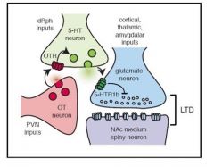
Do oxytocin neurons project to the NAc? |
- they found using RbV that neurons from the PVN of the hypothalaus project to the NAc - using immunohistochemistry, they showed that there are indeed PVN cells that both project to NAc and produce Oxytocin |
|
|
What are two ways to see if certain neurons contain a certain protein? |
1) Receptor reporter mice: Inject DNA of the fluorescent protein into animal when it is in womb so that the DNA is in its cells. Now any cell that will want to make that protein (ie oxytocin) receptor will also produce this fluorescent protein and will glow
2) Immunohistochemistry: synthesize the protein you want to label and then attach a carrier protein to it which is known to illicit a large immune response in the animal (usually rabbit). See which cells are attacked by the antibodies |
|
|
Which neurons in the NAc have oxytocin receptors? |
- used antibodies to identify different cell types, which express different proteins - PARV is the one that has receptors mostly- it is an interneuron |
|
|
Are the oxytocin receptors on the neurons in the NAc required for social reward? How did the authors conclude this? |
- No, they found that when you delete oxytocin receptors from all the cells in the NAc there is no behavioural change on place preference, so the PARV interneurons arent mediating the effect
- they did this by using mice with DNA with loxP injected. This loxP site goes on either side of the oxytocin receptor gene - when you inject Cre (cre-recombinase) to the cells you want to not have receptors (using a virus containing its DNA), it makes cuts at the loxP sites, effectively deleting the oxy receptor from the DNA |
|
|
Do any neurons that project to the NAc have oxytocin receptors (on their axon terminals)? Why might this be important? is this required for social reward?
|
- Yes PVN neurons do, so do thalamus, amygdala, hippo, VTA and dorsal raphe
- transmitter receptors on axon terminals determines the probability that a vesicle will be released in response to an action potential by affecting other things such as: calcium levels, number of vesicles docked, and number of reserve vesicles - yes receptors located on other axons going into the NAc from other brain areas ARE important for the behavioural response - we think the dorsal raphe nuclei are the critical pathway |
|
|
How can the probabilty of a vesicle release be increased? |
by a g-protein receptor signaling cascades in the axon terminal |
|
|
Oxytocin receptors on dorsal raphe neurons and social reward |
- AAV encoding the DNA for Cre and GFP was injected into the dorsal raphe nucleus in conditional oxytocin receptor knockout mice - eliminating the receptors on this pathway eliminated the behavioural effect |
|
|
The dorsal raphe neurons are required for social reward and they contain serotonin. Are serotonin receptors in NAc required for social reward? What did the authors find in terms of the neurochemistry? |
injected serotonin antagonist and found blocking serotonin (as well as oxy) in NAc eliminated social behaviour - they found that if you do electrophys and take a slice of NAc, stimulating around cells (which activates neurons nearby), adding serotonin weakens the glutaminergic synapse strength (antagonizing it does nothing). - Adding oxytocin alone also has this effect, but adding it to the serotonin has no effect, so they concluded that oxytocin is probably causing release of serotonin and then serotonin blocking glutamate
|
|
|
Look at this picture summary of how oxytocin influences social reward in mice |
|
|
|
What is a disadvantage of the paper presented on oxytocin and social reward? |
it doesn't assess when oxytocin neurons actually fire- during the interaction or in avoidance of an interaction it is hard to draw strong conclusions about the behavioural significance of oxytocin signalling without this |
|
|
What is calcium imaging? |
- calcium regulates many cell processes so its concentrations inside cell is very regulated and LOW - calcium enters cells during action potentials and a bit during synaptic activity - thus increased internal calcium levels correlate well with electrical recordings of firing rates, although its not absolutely precise - we do this by sticking a fibreoptic cable into the area of interest (here the VTA) and having a rewardng activity like sucrose in a bowl for ex. When the mouse drinks the sugar, we see increases in the calcium fluctuations in the neurons |
|
|
How can we tell when dopamine neurons are firing - specifically how can we see if they are active during social interaction? |
- calcium imaging - GCaMP is a genetically encoded calcium indicator which has been created to glow whenever calcium binds to its subunit - this allows us to detect single action potentials by monitoring changes in GCaMP fluorescence - then you create transgenic mice that express cre only in dopamine neurons and use cre-dependent viruses (Will only express its DNA if the cell also expresses cre) to ensure that only dopamine neurons will make GCaMP protein |
|
|
what is GCaMP created from? GCaMP glows for around how long after each action potential? |
designed by fusing proteins: GFP, Calmodulin and M13 peptide from the myosin chain kinase; 100ms |
|
|
How do the authors of the dopamine article go about creating a cre-dependent GCaMP virus? |
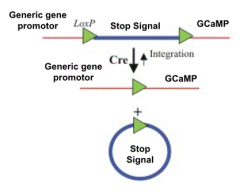
The GCaMP protein has a stop signal on it after the promotor. Only neurons that express cre (They have already made it so the dopamine neurons express cre) will have this stop signal bypassed and GCaMP will be created. Non- cre expressing neurons (so non dopamine neurons in the midbrain for ex) will be unaffected by having this viral DNA in them
|
|
|
So, are dopamine neurons active during social interactions?
|
- yes, they found that there appears to be a lot of calcium fluctuation in midbrain dopamine neurons during social interactions
- however they found that this is also seen when the mouse interacts with a novel object |
|
|
Is the dopamine neurons' activity different between social interactions & novel object interactions? |
- they found that the peak fluctuations - the levels of activity- is not different but there is a temporal difference - timing of firing for social interactions is during the interaction, whereas firing for novel object happens mostly after withdrawl form this object |
|
|
Does dopamine neuron activity during an interaction predict how fast the animal will return to the stimulus (either a social interaction or novel object)? |
Yes, the higher the firing, the less time before the animal returns to the object, but only for a social interaction, not a novel object |
|
|
Ok so we know that dopamine neurons in the midbrain are more active in social and novel object interaction... but does stimulation of the dopamine neurons promote social interactions and/ or object interactions? |
Yes, mice with artificial dopamine stimulation causes longer social interactions, but NOT novel object interactions opposite is also true- using optical inhibition shortens social interactions, but it does not effect novel object interaction |
|
|
How did the authors stimulate dopamine neurons to see if this promoted social interactions? what are two other less cool ways they couldve done this (and their issues)? |
- the authors used optogenetics. they injected cre-dependent viral DNA that encodes for channelrhodopsin in cell bodies into dopamine-cre dependent (TH:Cre VTA) mice
Other ways: - put metal wires in midbrain ad deliver electrical pulses, however this could stimulate all the other cell types too - give a dopamine receptor agonist- but this way both extrasynaptic and synaptic receptors would be activated and also activating receptors with drugs can be very different than natural activation |
|
|
What is optogenetics? How does it work? |
- the use of light to control neuronal firing in cells which have been made sensitive to light through the introduction of foreign DNA - the foreign DNA (from algae) encodes light-sensitive proteins called opsins - you insert the DNA into specific neurons in the brain and then shine a light - the light causes ion channels to open and the cell to depolarize and begin firing
|
|
|
excitatory vs inhibitory opsins |
in optogenetics you can either use excitatory opsins like ChR2 and C1V1 which drive action or inhibitory ones like Halo, NpHR or Arch which inhibit action potential activity - for the excitatory ones you can pulse light (allows you to very precisely drive potentials) or leave it on, but for inhibitory ones you typically use a long light pulse |
|
|
Dopamine neurons project to many different places. Which pathway is driving this social interaction promoting effect? how did they find this? |
- they stimulated dopamine neurons, killed animal and placed brain slice in with antibodies for IEGs (specifically cFOS) to measure neuronal activity - they also used an antibody for dopamine just to make sure that these cFOS active cells actually produced dopamine -Two regions they found was the NAc and the prefrontal cortex that had a lot of neurall activity in response to DA stimulaiton - then they injected a cre-dependent, opsin-encoding virus into the midbrain of cre-dopamine mice and shined the light this time into the projection target, either the NAc or the PFC (shining into midbrain would activate all dopamine neurons). - NAc neuron stimulation caused increase in social reaction, PFC didnt |
|
|
Immediate Early Genes (IEGs) aka activity dependent genes |
- genes transcribed in response to any elevations in normal firing activity - people measure IEG expression as an index of recent neural activity - cFOS and Arc are the most commonly measured IEGs - in the study they waited 90 mins for these genes to be transcribed |
|
|
Some dopamine axons just pass through the NAc. how can we be sure we are just measuring the neurons that actually end in the NAc? |
- they inject virus into the NAc which has a red protein (cherry) and WGA- Cre (WGA is a protein that can jump across synapses) - the WGA- cre protein transports cre to all the cells that project to the NAc - now al the axons that project to the accumbens have cre - then they go to the VTA and inject cre dependent opsin DNA so that VTA neurons will be sensitive to light but only if they project to the NAc |
|
|
What happens when you optically stimulate dopamine neurons in the PFC?
|
causes conditioned place aversion
- one chamber they stimulate dopamine neurons in the PFC - the mice dont like this and avoid this chamber - also causes anxiety like effects: When you activate DA axons in the PFC mice rarely go to the open arms of elevated plus maze, they stay in the closed arms of the maze - no promotion of social interaction! |
|
|
When are the neurons that project specifically to the NAc active? How did they find this? |
*The activity in the NAc is selectively active during social interactions and not novel objects, whereas dopamine neurons in the VTA may fire to both of these -injected cre-dependent GCaMP into VTA and then put optical fibre into NAc neurons coming from VTA and imaged calcium fluctuations in NAc
|
|
|
If dopamine release in the NAc promotes social behaviour, what receptors there are important? Describe how they showed this. |
Half of the neurons in NAc are D1 half are D2. - they found that blocking the D1 receptors in the NAc blocks the increase in social interaction caused by dopamine neuron stimulation - they also wanted to be super sure it was only D1 receptor since dopamine neurons can also release GABA and glutamate. - they took a Cre- dependent virus encoding for an artificial dopamine receptor protein made from extracellular rhodopsin and intracellular D1 receptor fused. It is light-sensitive and produces dopamine upon stimulation. Called opt-D1 - they found that stimulating only the D1 receptor was enough to cause social interction changes. When you activate this opt-D1 receptor the cell fires more |
|
|
List 3 ways to stimulate social interactionin mice. |
1) stimulating the midbrain VTA dopamine neurons 2) stimulating the dopamine neurons that project to the NAc- these neurons are active in social interactions 3) activating dopamine D1 receptors in the NAc as well as direct activation of dopamine receptor-containing NAc projection neurons
|