![]()
![]()
![]()
Use LEFT and RIGHT arrow keys to navigate between flashcards;
Use UP and DOWN arrow keys to flip the card;
H to show hint;
A reads text to speech;
45 Cards in this Set
- Front
- Back
|
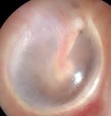
Eardrum is pinkish gray; see the malleus lying behind upper drum; from umbo, bright cone of light fans anteriorly and downward. Small blood vessels along handle of the malleus are normal.
|
Normal Eardrum
|
|
|
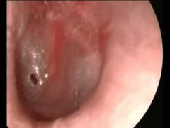
Holes in the eardrum; usually from purulent infections. Eardrum scarred; no landmarks are visible.
|
Perforation of the Drum
Q: What is the difference between central and marginal perforation? |
|
|
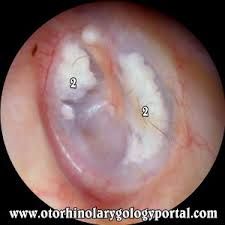
Large, chalky white patch with irregular margins; deposition of hyaline material within the layers of the tympanic membrane that sometimes follows severe episodes of otitis media.
Does not usually impair hearing and is rarely clinically significant. |
Tympanosclerosis
|
|
|
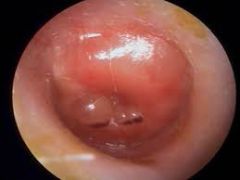
Air is absorbed from middle ear into the bloodstream and causes serous fluid accumulation.
Usually caused by viral upper respiratory infections. |
Serous Effusion
|
|
|
The eustachian tube cannot equalize the air pressure in the middle ear and outside air.
S/S: Fullness and popping sensations in the ear, mild conduction hearing loss and sometimes pain. |
Serous Effusion
|
|
|
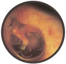
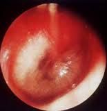
Commonly caused by bacterial infection from S. pneumoniae or H. influenzae.
S/S: Earache, fever and hearing loss |
Acute Otitis Media with Purulent Effusion
|
|
|
painful hemorrhagic vesicles appear on the tympanic membrane, the ear canal, or both.
S/S: Earache, blood-tinged discharge, conductive hearing loss. |
Bullous Myringitits
|
|
|
Normal Eardrum
|
|
|
Perforated Eardrum
|
|
|
Tympanosclerosis
|
|
|
Serous Effusion
|
|
|
Acute Otitis Media with Purulent Effusion
|
|
|
Bullous Myringitis
|
|
|
What is the otoscope used for? |
to visualize the ear canal and drum. note discharge, foreign bodies, redness. |
|
|
What is the Weber test? What results do you look for? |
Test for lateralization (hearing out of one ear only or better than the other) |
|
|
What is the Rinne test? What results do you look for? |
air conduction vs. bone conduction of sound ( can they still hear the sound waves after they stop feeling the movement of the fork?) |
|
|
What is the difference between the Weber and the Rinne tests? |
Weber: tuning fork is placed on top of head; do they hear equally on both sides? Rinne: tuning fork placed on mastoid bone until they stop feeling it, then placed near ear ( cant hey still hear the fork movements?/ AC>BC) |
|
|
What is a PE tube? |
A PE tube (AKA tympanostomy tube) is a pressure equalization tube used to allow air into the middle ear and to drain fluid from the ear; used to decrease infections. |
|
|
Name the Lymph nodes (10). |
Preauricular, Posterior auricular, occipital, tonsillar, submandibular, submental, superficial cervical, posterior cervical, deep cervical chain, supraclavicular. |
|
|
This node is located in front of the ear. |
preauricular |
|
|
This node rests in the area superficial to the mastoid process. |
posterior auricular |
|
|
This node is located at the base of the skull posteriorly |
Occipital |
|
|
This node is located at the angle of the mandible |
Tonsillar |
|
|
This node is midway between the angle and the tip of the mandible. |
Submandibular |
|
|
This node lies in the midline a few centimeters behind the tip of the mandible |
Submental |
|
|
This node is superficial to the sternomastoid |
Superficial cervical |
|
|
This node lies along the anterior edge of the trapezius |
Posterior cervical |
|
|
This node is located in deep to the sternomastoid; often unpalpable |
Deep Cervical Chain |
|
|
This node is located deep in the angle formed by the clavicle and the sternomastoid |
Supraclavicular |
|
|
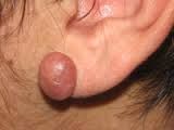
This lesion may develop in any scarred area but is most common on the shoulders and upper chest and are most common in dark–skinned people.
|
Keloid (def)
|
|
|
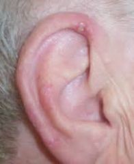
Chronic inflammatory lesion; starts as painful papule on the helix or antihelix and turns into crusty ulcer. Reddening may occur.
|
Chondrodermatitis
|
|
|
You should biopsy this lesion to rule out carcinoma.
|
Chondrodermatitis (recommendation)
|
|
|
Firm, nodular, hypertrophic mass of scar tissue extending beyond the area of injury.
|
Keloid (Pyhsical manifestation)
|
|
|
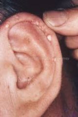
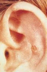
Deposit of uric acid crystals characteristic of chronic gout (or chronic sustained levels of uric acid); appears as hard nodules in helix & antihelix.
|
Tophi (def)
|
|
|
May discharge chalky white crystals through skin. Appear on joints, hands, feet & other areas.
|
Tophi (physical manifestation)
|
|
|
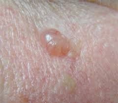
Raised nodule; has lustrous surface with small vessels; growth and ulceration may occur.
|
Basal Cell Carcinoma (physical manifestation)
|
|
|
Common slow–growing malignancy that rarely metastasizes. Common in light–skinned people.
|
Basal Cell Carcinoma (Def)
|
|
|
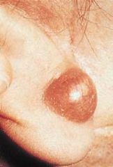
Dome shaped lump in the dermis forms benign, firm sac attached to the epidermis. Blackhead may be visible on surface.
|
Cutaneous Cyst– AKA– Sebaceous Cyst
|
|
|
Small lumps on the helix or antihelix; ulceration may result from repeated injuries. Additional nodules may appear on hands, arms, knees or heels.
|
Rheumatoid Nodules
|
|
|
Keloid (PIC)
|
|
|
Tophi (PIC)
|
|
|
Cutaneous Cyst (PIC)
|
|
|
Chondrodermatitis Helicis (PIC)
|
|
|
Basal Cell Carcinoma (PIC)
|
|
|
Rheumatoid Nodules (PIC)
|