![]()
![]()
![]()
Use LEFT and RIGHT arrow keys to navigate between flashcards;
Use UP and DOWN arrow keys to flip the card;
H to show hint;
A reads text to speech;
50 Cards in this Set
- Front
- Back
|
The skin provides a physical barrier that regulates ___________ loss and protection against ________, ___________, and ___________ insults from the external environment.
Dysfunction of the skin leads to __________, ___________, _________, and ____________. |
Water, mechanical, chemical, and microbial
Injury, dehydration, infection, and inflammation |
|
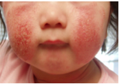
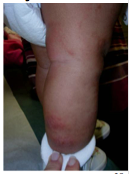
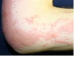
What does this child have? |
Atopic dermatitis = chronic skin condition associated with barrier dysfunction |
|
|
As an immunologic barrier, the skin both _______ and ________ to pathogens.
Dysfunction of the immunologic barrier leads to _________, __________, ___________, and ___________. |
Senses and responds
Infection, skin cancer, inflammatory skin conditions, and allergy |
|
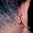
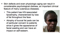
What does this HIV man have? What is it caused by? |
Molluscum contagiosum = skin infection caused by a virus |
|
|
How does the skin help maintain constant body temperature? |
Insulating properties of fat and hair Accelerating heat loss with sweat production and a dense superficial microvasculature |
|
|
What does dysfunction of temperature regulation lead to? What is the term for chronic episodic attacks of digital ischemia provoked by exposure to cold? |
Hyper- or hypothermia
Raynaud phenomenon |
|
|
What pigment protects against UV? Where is it located? What could dysfunction lead to? |
Melanin = epidermis More susceptible to skin cancer |
|
|
__________ receptors allow the skin to constantly monitor the environment. __________ receptors are important for the body's interactions with physical objects.
Dysfunction leads to _________ (itch), _________ (abnormal sensation), and ______, |
Sensory receptors Mechanoreceptors
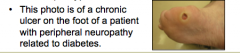
Pruritus, dysesthesia, insensitivity to injury (diabetes leprosy)
|
|
|
What are the four phases of cutaneous wound repair? What does loss of ability to repair injury (such as post-radiation treatment) lead to? |
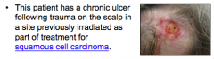
1. Coagulation 2. Inflammatory phase 3. Proliferative-migratoy phase (tissue formation) 4. Remodeling phase
Delayed wound healing |
|
Appearance and quality of life |

|
|
|
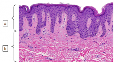
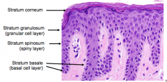
What are the three layers of the skin? |
1. Epidermis 2. Dermis 3. Subcutis |
|
|
Top = epidermis Bottom = dermis |
|
|
What is the primary component of the epidermis? |
Epidermis = keratinocytes Dermis = fibroblasts, collage, and elastic fibers |
|
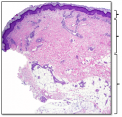
Identify the three layers? What lies below the dermis? Other names? |
Epidermis Dermis Fat (subcutis, panniculus, hypodermis) |
|
|
|
|
|
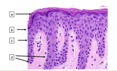
What is the length of the epidermal cell maturation? When are they shed after reaching the stratum corneum (how long)?
The lowest epidermal layer is what? What is it the source of? Does cell division occur here? |
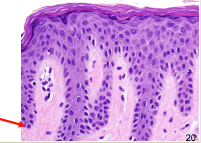
Two weeks = maturation Shed two weeks after reaching stratum corneum (28-day cycle)
Basal layer = source of epidermal cells where CELL DIVISION occurs |
|
|
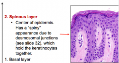
What is the layer above the basal layer? How is its appearance described and why? What do desmosomal junctions do? |
|
|
|
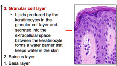
What is the layer above the spinous layer? What is produced here and why? |
|
|
|
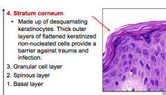
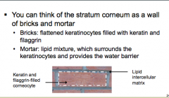
What is the top layer called? What is it made up of? Are keratinized cells nucleated? What does it provide a barrier for? |
|
|
|

|
|
|
What is the protein found in the granular cell layers of the epidermis? What does it do within keratinocytes? What do mutations causes? |
Filaggrin = retains water within keratinocytes Mutations = atopic dermatitis and asthma (atopic diseases) |
|
|
Bullous pemphigoid: Disease type? Age of patients? Disease process? Clinical presentation? |
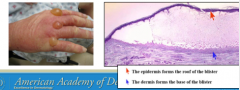
Type = autoimmune blistering disease Age = older patients Disease process = autoantibodies form to antigens directly beneath the basal layer of the epidermis Clinical presentation = tense bullae on an erythematous base on the skin (mucous membranes may also be affected) |
|
|
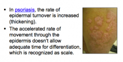
What is the main dysfunction of psoriasis? What does this cause? |
|
|
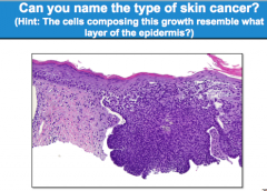
What type of skin cancer? |
Basal cell carcinoma |
|
|
What is the most common form of skin cancer? It is composed of cells that resemble what? How does it most commonly present? |
Basal cell carcinoma Cells that resemble basal keratinocytes Presentation = Pearly, erythematous papules or plaques with rolled borders and telangectasias in sun-exposed areas |
|
|
What are the tree main cell types that make up the epidermis? |
1. Keratinocytes 2. Melanocytes 3. Langerhans cells |
|
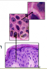
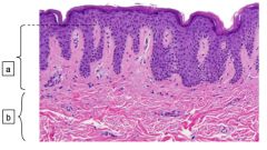
What cell types is shown here? what makes of the majority of cells? What are they held together by? In which layer are these primarily visible? |
Keratinocytes = majority of cells Desmosomes = macromolecular structures that look like stripes (or spines) between cells. Visible in SPINOUS layer |
|
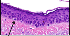
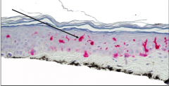
What type of cell is this? Where are they located? What do they do? |
Melanocyte
Staggered along basal layer one in every 10 keratinocytes
Produce pigment (melanin) and transfer it to keratinocytes in the basal cell layer |
|
|
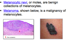
What are benign collections of melanocytes called? What is a malignancy of melanocytes? |
|
|
|
What type of cells are Langerhans cells? Where are they located? What is their main function? |
Dendritic cells in mid-epidermis
Afferent limb of immune response: recognition, uptake, processing, and presentation of antigens to sensitized T-lymphocytes
Important in induction of delayed type hypersensitivity |
|
What is a common disease in which Langerhans cells play a prominent role? |
Contact dermatitis = poison oak |
|
What is shown here? |
Langerhans cells |
|
Identify the two layers of the dermis. |
Top = papillary dermis Bottom = reticular dermis |
|
|
What type of support structure does the dermis provide? How thick is it? Is it thicker than epidermis?
What does the dermis contain? |
Flexible but tough Thickness = 1-4 mm Much thicker than epidermis
Blood and lymphatic vessels Nerves which supply skin Sweat glands and hair follicles |
|
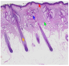
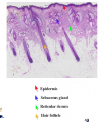
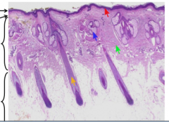
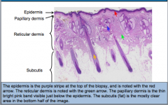
Scalp biopsy: Identify the arrows |
Yellow = hair follicles running through dermis Blue = sebaceous or oil glands Red = epidermis Green = reticular dermis |
|
|
Where do fibroblasts and mast cells reside? What are fibroblasts responsible for?
What can result from uncontrolled synthesis and excessive deposition of collagen at sites of prior dermal injury and wound repair? |

Dermis
Synthesis and degradation of connective tissue proteins --> wound healing and scarring
Keloids! |
|
|
What are mast cells responsible for?
What is a vascular reaction of the skin characterized by wheals surrounded by a red halo or flare? |
Immediate-type hypersensitivity reactions in the skin
Urticaria |
|
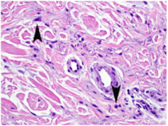
What type of cells are shown here? |
Mast cells |
|
|
What is the fat layer that separates the dermis from deeper underlying structures such as fascia and muscles? |
Subcutis |
|
|
What are the four functions of the subcutis? |
Insulates the body Serves as an energy supply Cushions and protect the skin Allows for mobility over underlying structures |
|
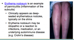
What is shown here? What is it an example of? |
|
|
|
What are the two "adnexal" structures? |
Pilosebaceous unit Eccrine gland |
|
|
What does a pilosebaceous unit consist of? |
1. Hair follicle 2. Sebaceous (oil) gland 3. Apocrine sweat gland 4. Arrector pili muscle (when these contract you get goosebumps) |
|
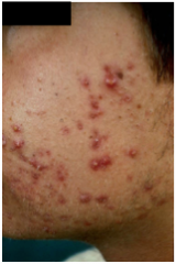
What is a disoder of the pilosebaceous unit? What four factors cause it? |
Acne vulgaris
1. Plugging of hair follicle as a result of abnormal keratinization of the upper portion (--> comedones)
2. P. acnes bacteria in the hair follicle (lives on oil --> breaks down to free FA --> inflammation)
3. Hormones (androgens)
4. Sebaceous gland activity (increased in presence of androgens) |
|
|
In contrast to apocrine glands, eccrine sweat glands to not involve ______________. They open directly onto the skin surface and are present where?
How do eccrine glands help regulate body temperature?
What will a patient by predisposed to if eccrine glands are genetically absent? |
Do not involve hair follicle (present throughout the body)
Excreting sweat onto the skin surface --> cooling and evaporation take place
Hyperthermia
|
|
Identify layers and arrows again. |
|
|
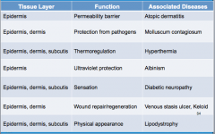
Give the function and associated disease of each tissue layer. |
|
|
|
What are the six important functions of the skin? |
Barrier Immune recognition and surveillance Damage repair Thermoregulation Photoprotection Communication |
|
|
Which immune processes are in skin? Innate, adaptive, both? |
Both |
|
|
What is the disease if you see "palpable purpura?" |
Vasculitis!
|