![]()
![]()
![]()
Use LEFT and RIGHT arrow keys to navigate between flashcards;
Use UP and DOWN arrow keys to flip the card;
H to show hint;
A reads text to speech;
115 Cards in this Set
- Front
- Back
|
Human Movement System |
The combination and interrelation of the nervous, muscular, and skeletal systems |
|
|
Kinetic Chain |
Human movement is accomplished through the integration of the nervous, skeletal, and muscular systems. The nerves, muscles, and joints must work together in a chain to produce motion(kinetic). These three systems are also referred to as the kinetic chain.
|
|
|
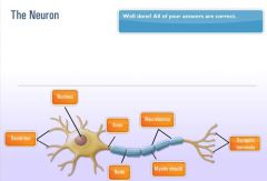
The Neuron |
|
|
|
Neuron |
Functional unit of the nervous system. Billions of neurons make up nervous system, provides it with ability to communicate internally with itself as well as externally with environment. Transmits impulses through both electrical and chemical signals. Forms the core of the nervous system which includes the brain, spinal cord, and peripheral ganglia
|
|
|
What are neurons composed of |
Neurons are composed of cell body, axon, and dendrites. |
|
|
Cell body of Neuron contains: |
The cell body of Neuron contains a nucleus, lysosomes, mitochondria, and a Golgi complex. |
|
|
Axon – |
cylendrical projection from the cell body that transmits nervous impulses to other neurons or effector sites(muscles, organs). Provides communication from brain and spinal cord to other parts of the body. |
|
|
Dendrites |
– gather information from other structures and transmit it back into the neuron. |
|
|
Sensory (afferent) neurons |
– Respond to touch, sound, light, and other stimuli and transmit nerve impulses from effector sites(muscles, organs) to the brain and spinal cord. |
|
|
Interneurons |
– Transmit nerve impulses from one neuron to another. Hence INTER neuron. Between neurons |
|
|
Motor (efferent) neurons |
– transmit nerve impulses from the brain and spinal cord to the effector sites such as muscles or glands. |
|
|
role of different neurons |
So sensory neurons transmit from muscles and organs to the CNS. Motor neurons transmit nerve impulses from CNS to muscles and organs. Interneurons transmit impulses between neurons. |
|
|
2 divisions of nervous system |
The nervous system is composed of two interdependent divisions, the CNS and the PNS |
|
|
Central Nervous System |
– Consists of the brain and the spinal cord, and its primary function is to coordinate the activity of all parts of the body. |
|
|
Peripheral Nervous System |
– Nerves that connect the CNS to the rest of the body and the external environment. Nerves of PNS are how CNS receives sensory input(from sensory afferent neurons) and initiates responses(through motor efferent neurons). |
|
|
The PNS serve two main functions. |
They provide a connection for the nervous system to activate different effector(organ, muscle) sites. Second they relay information from effector(organ, muscle) sites back to the brain via sensory receptors, providing constant update to the relation of the body and the environment. |
|
|
The PNS consists of . |
the somatic and autonomic nervous system |
|
|
The somatic nervous system consists of |
the nerves that serve the outer areas of the body and skeletal muscle and are responsible for the voluntary control of movement. So somatic is what controls your biceps and legs and whatnot. |
|
|
The autonomic nervious system |
supplies neural input to the involuntary systems of the body, like your heart. Autonomic, auto, think autopilot, you don’t have conscious control over the autonomic process. |
|
|
The autonomic is divided into |
sympathetic and parasympathetic nervous systems |
|
|
The sympathetic system |
increases the activation level of neurons in preparation for activity(ramps you up). |
|
|
The parasympathetic |
ramps your system down, decreases levels of activation. |
|
|
What are Sensory receptors |
Sensory receptors are specialized structures that convert environmental stimuli(heat, sound, taste, etc) into sensory information for the brain. These receptors are divided into four categories, mechanoreceptors, nociceptors, chemoreceptors, and photoreceptors. |
|
|
Mechanoreceptors –
|
specialized structures that respond to mechanical pressure within tissues and then transmit signals through sensory nerves. Respond to outside forces such as touch, pressure, stretching, sound waves, and motion. Senses distortions in body tissues.
|
|
|
Muscle Spindles –
|
Sensory receptors, run parallel to muscle fibers. Are sensitive to changes in muscle length and rate of length change. Help regulate the contraction of muscles via the stretch reflex mechanism. This mechanism is a normal response to the body to a stretch stimulus in the muscle, it is designed to protect and prevent overstretching and muscle damage.
|
|
|
Gogli Tendon Organs(GTOs) –
|
Specialized sensory receptors located where the skeletal muscle fibers attach to the tendons. Sensitive to changes in muscular tension and rate of tension change. Activiating the Gogli tendon organ causes the muscle to relax which prevents the muscle from excessive stress or injury.
|
|
|
Joint receptors –
|
located around joint capsule, respond to pressure, acceleration, and deceleration of the joint. Signals extreme joint positions and thus helps prevent injury.
|
|
|
movement/performance and CNS |
Performance increases in early stages of training result from changes in the way the CNS controls and coordinates movement. Unsuccesful performances can be cross referenced with other sensory input and new movement strategys found. Regular training causes adapations int he CNS, allowing greater control of movements, thus causing movements to be more smooth and more accurate – improving performance.
|
|
|
Skeletal System |
– Body’s framework, composed of bones and joints. Provides shape and focus for bodies. Produces blood for the body and stores minerals. Growth, maturation, and functionality of skeletal system are greatly affected by posture, physical activity, and nutrition |
|
|
Bones – |
Provide a resting ground for muscles and protection of vital organs. |
|
|
Joints – |
Junctions of bones, muscles, and connective tissues at which movement occurs. Also known as articulation. |
|
|
how many divisions of skeletal system |
The skeletal system is divided into two divisions. |
|
|
Axial Skeleton |
– Portion of skeletal system that consists of skull, rib cage, and vertebral column. Think torso and head. 80 bones. |
|
|
Appendicular Skeleton
|
– Portion of skeletal system that includes the upper and lower extremeties. Arms, legs. Think appendage, appendicular, arms, legs. 126 bones.
|
|
|
bones in body total |
206 bones in the skeletal system, 177 used in voluntary movement, more than 300 joints in the body.
|
|
|
2 vital functions of bones |
Bones serve two vital functions – leverage and support.
|
|
|
Remodeling – |
Process of resorption and formation of bone. Old bone is broken down and removed by osteoclasts, new bone is laid down by cells called osteoblasts.
|
|
|
Osteoclasts – |
Bone cell that removes bone tissue. Clast. Clap. You want to get rid of the clap. Osteoclasts get rid of bone. |
|
|
Osteoblasts – |
Bone cell that forms bone. Blast. You like having a blast. You like building bone. Osteoblasts build bone.
|
|
|
posture/exercise affect on bone growth |
Remodeling follows lines of stress placed on bone. Exercise and habitual posture fundamentally influences the health of the skeletal system. Incorrect exercise and posture will lead to remodeling process that reinforces predominating bad posture.
|
|
|
how many types of bones |
Five major types of bones. Shape, size, and proportion determine their classification.
|
|
|
Long bones
|
– long cylindrical body, irregular or widended ends. Shaped like a beam and have slight curvature. Predominantly composed of compact bone tissue for strength and stiffness. Has considerable amount of spongy tissue for shock absorption.
|
|
|
Epiphysis – |
End of long bones, mainly composed of cancellous bone and house much of the red marrow involve din red blood cell production. One of primary sites for bone growth. End of long bones, red marrow which produces red blood cells. Knobby end looking parts of the bone.
|
|
|
Diaphysis |
– Shaft portion of long bone. The shaft. Long part. Compact bone(strong).
|
|
|
Epiphyseal Plate |
– Region of long bone connecting the diaphysis to the epiphysis. A layer of subdividing cartilaginous cells in which growth in length of the diaphysis occurs. |
|
|
Periosteum |
– Dense membrane composed of fibrous connective tissue that closely wraps (invests) all bone, except that of the articulating surfaces in joints, which are covered by a synovial membrane. Inner surface provides materials for nutrition repair and facilitates growth in the diameter of the bone. |
|
|
Medullary cavity – |
Central cavity of bone shafts where marrow is stored. Contains fatty yellow marrow, predominantly fat and serves as energy reserve, center of diaphysis. |
|
|
Articular (hyaline) cartilage – |
Covers the articular surfaces of bones.“articular surface” means the parts of the bone that moves in joints. Hard, white, shiny tissue that along with synovial fluid helps reduce friction in freely moving synovial joints. Fundamental to smooth joint action. |
|
|
Short bones |
– Similar in length and width. Somewhat cubical in shape. Consist predominantly of spongy bone tissue to maximize shock absorption. Carpals of hands and tarsals of feet
|
|
|
Flat bones – |
Thin bones, two layers of compact bone tissue surrounding a layer of spongy bone tissue. Involved in protection of internal structures and also provide broad attachment sites for muscles. Sternum, scapulae, ribs. |
|
|
Irregular bones |
– Unique shape and function. Veterbrae, pelvic bones, facial bones. |
|
|
Sesamoid Bones |
– Small bones embedded in a joint capsule or found in locations where tendon passes over a joint. Develop within particular tendons at a site of considerable friction or tension. Serve to improve leverage and protect the joint from damage. |
|
|
bone surface markings |
Bones have specific distinguishing structures called surface markings. They increase stability in joints as well as provide attachment sites for muscles. Divided into depressions and processes. |
|
|
Depressions – |
Flattened or indented portions of bone, which can be muscle attachment sites. Indents. Grooves.
|
|
|
Processes – |
Projections protruding from the bone where muscles, tendons, and ligaments can attach. Part that sticks out on bones. Where there is a depression on both sides will generally be processes. |
|
|
Vertebral Column – |
Backbone, spinal column, series of irregularly shaped bones called vertebrae that houses spinal cord. |
|
|
1st seven vertebrae |
First seven vertebrae starting from top are cervical vertebrae, flexible framework and provide support and motion for the head(your neck, basically). |
|
|
middle 12 vertebrae |
Next 12 are upper and middle back, called thoracic vertebrae, move with the ribs to form rear anchor of rib cage. Larger than cervical vertebrae and increase in size from top to bottom |
|
|
lower 5 vertebrae |
Next five are lumbar vertebrae. Largest in spinal column, support most of the body’s weight and attached to back muscles, often location of pain because they are subject to largest forces and stresses.
|
|
|
bone below lumbar vertebrae |
The sacrum is a triangular bone located below lumbar vertebrae, four or five sacral vertebrae in a child which become fused into a single bone during adulthood.
|
|
|
bottom of spine |
Bottom of spinal column is coccyx or tailbone, 3 to 5 bones fused together |
|
|
Intervertebral discs |
Intervertebral discs are fibrous cartilage that act as shock absorbers and allow the back to move |
|
|
neutral spine |
Optimal arrangement of curves is referred to as a neutral spine. Vertebrae and associated structures under the least amount of load.
|
|
|
joints |
Formed by one bone that articulates with another bone. Categorized by structure and function. |
|
|
Arthrokinematics –
|
Joint motion. Rolled, slide, and spin. Motions rarely occur in isolation. Rolling movement – bicycle roll on street. Sliding – tire skidding on street. Spinning movement – twisting lid off a jar.
|
|
|
Synovial joints
|
– Held together by a joint capsule and ligaments and are most associated with movement in the body. 80% of all joints in the body, have greatest capacity for motion. Produce synovial fluid, resembles egg whites and works like engine oil.
|
|
|
Nonsynovial joints – |
do not have a joint cavity, connective tissue, or cartilage. Exhibit little to no movement, seen in skull, distal joint of tibia and fibula.
|
|
|
Ligaments – |
Primary connective tissue that connects bones together and provides stability, input to the nervous system, guidance, and the limitation of improper joint movement. Fibrous connective tissues, bone to bone, provide static and dynamic stability as well as input to nervous system (proprioception). Made up of collagen. Ligaments have poor vascularity, blood flow, thus do not heal or repair well. |
|
|
Muscular system |
– series of muscles that move the skeleton. Muscles generate internal tension, under control of nervous system, manipulates bones to produce movements. Movers and stabilizers. |
|
|
muscle types |
Skeletal muscle one of three major muscle types, others are cardiac and smooth. Made up of individual muscle fibers. Bundles of muscle fiber can be broken down into layers. First layer is fascia, connective tissue. |
|
|
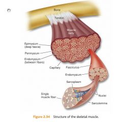
Epimysium –
|
layer of connective tissue that is underneath the fascia and surrounds the muscle. Inner layer immediately surrounding the muscle. Fascia and epimysium are connected to bone to help form muscle’s tendon.
|
|
|
perimysium.
|
The next bundle of muscle fiber is called fascicle. Each fascicle is wrapped by connective tissue called perimysium.
|
|
|
Perimysium
|
– connective tissue that surrounds fascicle.
|
|
|
Endomysium –
|
deepest layer of connective tissue that surrounds individual muscle fibers.
Each fasicle is made up of many individual muscle fibers which are wrapped in a connective tissue called endomysium. |
|
|
role of connective tissue |
Connective tissues within muscle play vital role in movement, they allow forces generated by muscle to be transmitted from contractile components of muscle to bones, each layer of connective tissue extends the length of the muscle helping form the tendon.
|
|
|
Tendons – |
Connective tissues that attach muscle to bone and provide an anchor for muscles to produce force.
|
|
|
structure of skeletal muscle |
|
|
|
sarcolemma
|
Fibers are encased in a plasma membrane known as sarcolemma.
|
|
|
Sarcomere |
– functional unit of muscle that produces muscular contraction and consists of repeating sections of actin and myosin
|
|
|
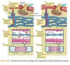
Neural Activation |
– Contraction of a muscle generated by neural stimulation. |
|
|
Motor Unit – |
Motor neuron and all of the muscle fibers it innervates. |
|
|
Neurotransmitters |
Chemical messengers that cross the neuromuscular junction (synpase) to transmit electrical impulses from nerve to the muscle |
|
|
Acetylcholine (ACh) |
Acetylcholine (ACh) is what is used by neuromuscular system. Once attached ACh stimulates fibers to go through a series of steps that initiates muscle contractions. |
|
|
motor units/movement |
Muscles are divided into motor units. Single motor unit consists of one motor neuron(nerve) and the muscle fibers it innervates. If stimulus is strong enough it will spread through whole length of muscle fiber, all of the muscle fibers supplied by a single nerve. If the stimulus is not strong enough then there will be no action potential and no muscle contraction. Motor units cannot vary the amount of force they generate. They either contract maximally or not at all. |
|
|
all or nothing |
Because of all or nothing law the overall strength of skeletal muscle contraction depends on size of the motor unit recruited(how many muscle fibers are contained within the unit) and number of motor units activated. |
|
|
muscle fibers based on movement |
Muscles that control fine movements are made up of many small motor units. Large muscles are made up of larger motor units. 10-20 fibers in each eye motor unit. 2,000 to 3,000 fibers in intestinal motor units. |
|
|
excitation-contraction diagram |
|
|
|
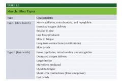
two muscle fiber types |
Fiber types vary in chemical and mechanical properties. Two main types, type I and type II. |
|
|
Type I (slow twitch) |
-contain large number of capillaries, mitochondria(transforms energy from food into ATP), myoglobin(increased delivery of oxygen). Red fibers -smaller in diameter, slow to produce maximal tension, more resistant to fatigue. Produce long term contractions. Think marathons. Maintaining posture against gravity. |
|
|
Type II (fast-twitch) |
-subdivided into Type IIa and Type IIx. -Contain fewer capillaries, mitochondria, and myoglobin. White fibers. - larger in size, quick to produce maximal tension, fatigue more quickly. Sprint muscles. -Type IIx have low oxidative capacity and fatigue quickly. -Type IIa higher oxidative capacity and fatigue more slowly than IIx. IIa are known as intermediate fast-twitch fibers. |
|
|
do muscles have combo of 1 and 2 or only one type of twitch? |
All muscles have combination of slow and fast twitch. Ex. shin has 735 slow twitch type I whereas calf muscle has 49% slow twitch. |
|
|
muscle fiber type chart |
|
|
|
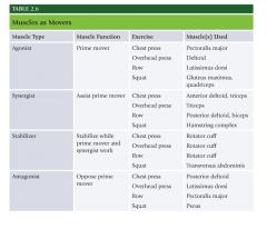
Agonist muscles |
Agonist muscles act as prime movers. They are most responsible for a particular movement.
|
|
|
Synergist muscles |
Synergist muscles assist prime movers. Synergist, think synergy. Assists with. |
|
|
Stabilizer |
Stabilizer support and stabilize the body. |
|
|
Antagonist muscles |
Antagonist muscles perform opposite action of prime mover. |
|
|
Muscles as Movers |
|
|
|
Endocrine system |
System of glands that secrete hormones into bloodstream to regulate variety of bodily functions. Mood, growth, development, tissue function, and metabolism.
|
|
|
Primary endocrine glands |
Primary endocrine glands are hypothalamus, pituitary, thyroid, and adrenal glands. |
|
|
which is the master gland |
Pituitary, “master” gland. Controls functions of other endocrine glands. |
|
|
thyroid |
Thyroid produces hormones that regulate metabolism and affect growth. |
|
|
adrenal glands |
Adrenal glands secret hormones – corticosteroids, catecholamines, cortisol, adrenaline in response to stress. |
|
|
hormonal activity |
Hormonal activity control rests with hypothalamus and pituitary gland. |
|
|
primary energy source
|
Glucose is primary energy source during vigorous exercise. Glucose principal fuel for the brain. Too much glucose can damage vascular system. Control of glucose regulated by pancreas – producing insulin and glucagon.
|
|
|
Insulin |
– Regulate energy and glucose metabolism. Glucose rich blood is circulated through pancreas, elevated levels of glucose trigger release of insulin. Circulating insulin binds with receptors in skeletal muscle and liver cells and cell membranes become more permeable to glucose. Glucose then diffuses from bloodstream into cell resulting in drop in blood glucose. Thus insulin causes fat, liver, muscle cells to take up glucose from the blood and store it as glycogen in liver and muscle. |
|
|
Glucagon |
– Opposite effect of insulin, functions to raise blood glucose by triggering release of glycogen stores from liver. Drop in circulating blood glucose triggers release of glucagon from pancreas. |
|
|
levels of glucose |
As activity levels increase the glucose uptake by cells increases. Increases insulin sensitivity in cells. Glucagon also increases helping m aintain steady supply of glucose. |
|
|
Catecholamines |
Catecholamines – two, epinephrine(adrenaline) and norepinephrine. Produced by adrenal glands(on top of each kidney). Help prepare body for activity. Fight or flight. Hypothalamus triggers adrenals to secrete epinephrine for fight. |
|
|
Epinephrine
|
Epinephrine – increases heart rate and stroke volume, elevates blood glucose levels, redistributes blood to working tissues, opens up airways. |
|
|
Testosterone |
Testosterone – produced in testes in males, ovaries and adrenal glands in females. Males produce up to 10x more. Fundamental role in growth and repair of tissue. Estrogen produced in ovaries in female and small amounts in adrenals in males. |
|
|
Cortisol |
Cortisol – Catabolic hormone. Secreted by adrenals, serves to maintain high energy supply. Chronic cortisol can lead to significant breakdown of muscle tissue. |
|
|
Growth Hormone |
Growth Hormone – Released from pituitary, regulated by hypothalamus. Stimulated by several factors: estrogen, testosterone, deep sleep, vigorous exercise. Primary anabolic hormone responsible for most of growth and development during childhood until puberty when primary sex hormones take over. Increases development of bone, muscle tissue, and protein synthesis. Increases fat burning and strengthens immune systme. |
|
|
where are thyroid glands located |
Thyroid gland located at base of the neck below thyroid cartilage(adams apple). Releases hormones responsible for metabolism regulation. |
|
|
Testosterone and growth hormone levels increase after |
Testosterone and growth hormone levels increase after strength training and moderate to vigorous aerobic exercise. |
|
|
what lowers testosterone and raises cortisol |
Prolonged bouts of endurance training or extremely intense training lowers testosterone levels while raising cortisol levels. |