![]()
![]()
![]()
Use LEFT and RIGHT arrow keys to navigate between flashcards;
Use UP and DOWN arrow keys to flip the card;
H to show hint;
A reads text to speech;
42 Cards in this Set
- Front
- Back
|
What is the basic function of the basal ganglia? |
Movement modulation |
|
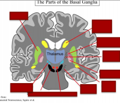
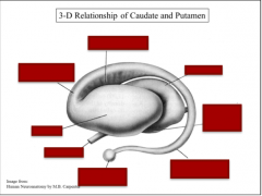
Identify putamen, caudate nucleus (body), globus palidus (ext. segment), internal capsule, globus palidus (int. segment), caudate nucleus (taile), red nucleus, cerebral peduncle, substantia nigra, subthalamic nucleus. |
|
|
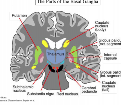
Identify caudate, corpus callosum, internal capsule, GPi, GPe, putamen, thalamus. |
|
|
|
What is the other name for the somatic basal ganglia? What is it involved in? What are the three parts? |
Dorsal basal ganglia Movement control Caudate & putamen, globus pallidus |
|
|
What is another name for the limbic basal ganglia? What is it involved in? What are the three parts? |
Ventral basal ganglia Motivation, reward, effect Nu. accumbens & olfactory tubercle, ventral pallidum |
|
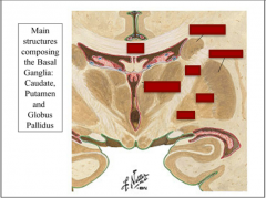
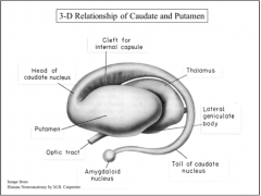
Identify: putamen, optic tract, head of caudate nucleus, amygdaloid nucleus, cleft of internal capsule, tail of caudate nucleus, lateral geniculate body, thalamus |
|
|
|
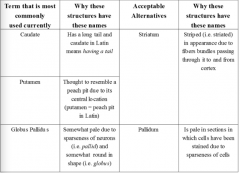
What is another name for the caudate and putamen?
What is another name for globus pallidus? |
Review this. |
|

Review this |
Review this |
|
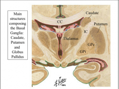
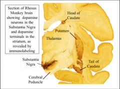
Identify head of caudate nucleus, putamen, thalamus, tail of caudate, substantia nigra, cerebral peduncle |
|
|
|
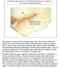
What is the region of the substantia nigra where the dopaminergic neurons are located?
What is the region below this in the substantia nigra that contains a net-like, meshwork of fibers (few dopaminergic neurons). |
|
|
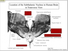
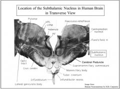
Identify the sub thalamic nucleus, cerebral peduncle, infundibulum, and VPL/VPM |
|
|
|
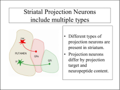
What are the two main types of neurons in the striatum? What type makes up 95% of the neurons? |
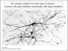
A = aspiny (95%) SN = spiny (5%) |
|
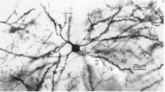
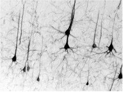
What is this a close-up of? What is the purpose of these? |
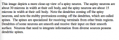
Neurons that need to integrate information from diverse sources possess dendritic spines. |
|
|
What is another name for spiny neurons in the striatum? Why? Another name for aspiny neurons? Why? |
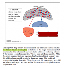
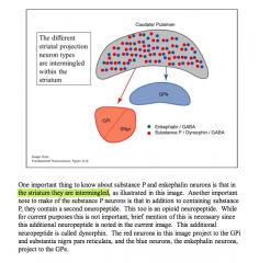
One important point about the spiny neurons, which this image shows, is that the spiny neurons have a long axon that leaves the striatum. By contrast, aspiny neurons have axons that are short and do not leave the striatum. Because of this, the spiny neurons in the striatum are called projection neurons, while the aspiny neurons are called the local circuit neurons, or interneurons. Thus, whatever the striatum decides based on the pattern of input it receives, it is the spiny neurons that transmit that decision to other brain areas. |
|
|
What neurotransmitter do all spiny neurons use? Nearly all ENK neurons in the striatum project to what location? Nearly all substance P neurons project to what location? |
GABA |
|
|
Striatal projection neurons projecting to GPi contain ______. Striatal projection neurons projection to GPe contain _____. |
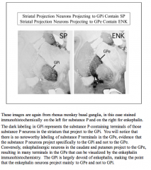
SP ENK |
|
|
Striatal projection neurons projecting to the substantia nigra predominantly contain ____. |
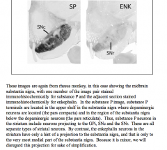
SP |
|
Identify which is enkephalin, which is substance P/dynorphin. |
|
|
|
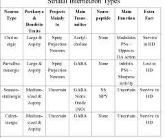
What are the four types of interneurons in the striatum?
Which type has been the target of therapies directed at the basal ganglia? |
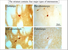
Cholinergic |
|
|
What type of interneuron uses calcium binding protein parvalbumin and uses GABA. Larger than projection neurons but smaller than the large cholinergic interneurons. |
Parvalbuminergic |
|
|
The third type of interneuron in the striatum is the somatostatin-containing interneuron, somatostatin being a neuropeptide. This type of interneuron can be identified by immunostaining for somatostatin,butthemaintransmitteritusesisGABA. Theseneuronstendtohavecellbodiesthat are the same size as the cell bodies of the spiny neurons. |
Somatostatinergic |
|
|
Calretinin is also a calcium binding protein. These neurons have a cell body that is the same size as the cell body of the spiny neurons of the striatum. These neurons too use GABA as their primary neurotransmitter. |
Calretinergic |
|
|
True or false: Somatostatin interneurons are impervious to Hunting's Disease (affects basal ganglia).
What neuropeptide do somatostatin neurons make? |
True
NPY |
|
|
As Huntington’s disease progresses, _____ neurons progressively die. By grade 3 (very advanced), about 60% of _____ neurons are lost, and the striatum, GPe, and GPi are all greatly shrunken as a result. Nonetheless, _____ neuron abundance is preserved in the grade 3 case illustrated, and remains around 600. |
As Huntington’s disease progresses, spiny neurons progressively die. By grade 3 (very advanced), about 60% of spiny neurons are lost, and the striatum, GPe, and GPi are all greatly shrunken as a result. Nonetheless, NPY neuron abundance is preserved in the grade 3 case illustrated, and remains around 600. |
|
|
By grade 4 Huntington’s disease, about 90% of _____ neurons are lost, and the striatum, GPe and GPi are yet further shrunken. Nonetheless, there are still around 600 _____ neurons in the putamen. Note that there are even more than 600 neurons, because of the compression of these neurons into a narrower space as the striatum shrinks. Thus, _____ neurons are impervious to Huntington’s disease. One approach for understanding how Huntington disease kills striatal spiny neurons involves focusing on the NPY neurons in comparison to spiny neurons. What do NPY neurons have that spinyneuronsdonot? Thus,whilethe functional role of the NPY interneuron type is not well understood, its behavior in Huntington’s disease makes it interesting and important. |
By grade 4 Huntington’s disease, about 90% of spiny neurons are lost, and the striatum, GPe and GPi are yet further shrunken. Nonetheless, there are still around 600 NPY neurons in the putamen. Note that there are even more than 600 neurons, because of the compression of these neurons into a narrower space as the striatum shrinks. Thus, NPY neurons are impervious to Huntington’s disease. One approach for understanding how Huntington disease kills striatal spiny neurons involves focusing on the NPY neurons in comparison to spiny neurons. What do NPY neurons have that spinyneuronsdonot? WhatdoNPYneuronsnothavethatspinyneuronsdohave? Thus,whilethe functional role of the NPY interneuron type is not well understood, its behavior in Huntington’s disease makes it interesting and important. |
|

|

|
|

Review |
|
|
|
What is the major neuron type in the globes pallidus?
What is the neurotransmitter involved? |
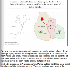
Aspiny GABA |
|
|
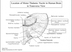
The GPe projects to the _____ nucleus, whose location is shown in this image. This is an important fact in understanding how the basal ganglia works. The GPi projects to what is called the _____ thalamus. There are two nuclei making up the motor thalamus. One is the ventral _____ nucleus (VA) and the other is the ventral _____ nucleus (VL). The ventral lateral nucleus has two parts, a __ part (caudalis) and an __ part (oralis), as shown in this illustration. |
The GPe projects to the subthalamic nucleus, whose location is shown in this image. This is an important fact in understanding how the basal ganglia works. The GPi projects to what is called the motor thalamus. There are two nuclei making up the motor thalamus. One is the ventral anterior nucleus (VA) and the other is the ventral lateral nucleus (VL). The ventral lateral nucleus has two parts, a C part (caudalis) and an O part (oralis), as shown in this illustration. |
|
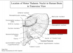
Identify VLo and VLc |
|
|
|
The VA is located at the _____ of the motor thalamus, with the VL region _____ the VA, and with the VL having an oral part toward the _____ and a caudal part toward the _____. |
The VA is located at the front of the motor thalamus, with the VL region behind the VA, and with the VL having an oral part toward the front and a caudal part toward the back. |
|
|
The GPi projects to the V_ and the V_. Note the substantia nigra pars reticulata also projects to the _____ thalamus. |
The GPi projects to the VA and the VL |
|
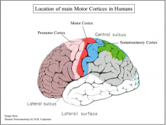
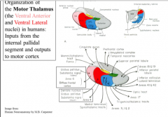
The VA projects to area _, which is pre-motor cortex, and the VL projects to area _, which is motor cortex. The basal ganglia sends much of its output to _____-related cortex. |

The VA projects to area 6, which is pre-motor cortex, and the VL projects to area 4, which is motor cortex. the basal ganglia sends much of its output to motor-related cortex. |
|
|
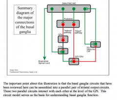
What are the three major sources of input to the striatum? |
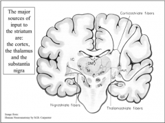
Cortex, thalamus, and the substantia nigra. |
|
|
The ______ thalamus is the source of one of the major inputs to the striatum. Because the function of this input is not well defined, however, we will devote no further attention to it. The major input to striatum that is clearly central to the function of the basal ganglia is the input from the cerebral cortex. Almost all of the cerebral cortex projects to the _____. The third major input to the striatal part of the basal ganglia is the _____ input from the substantia nigra pars compacta. |
The intralaminar thalamus is the source of one of the major inputs to the striatum. Because the function of this input is not well defined, however, we will devote no further attention to it. The major input to striatum that is clearly central to the function of the basal ganglia is the input from the cerebral cortex. Almost all of the cerebral cortex projects to the striatum. The third major input to the striatal part of the basal ganglia is the dopaminergic input from the substantia nigra pars compacta. |
|
The cortical neurons that project to the striatum are the layer __ pyramidal neurons. Many of the neurons in layer __ that project to striatum also send motor commands |
The cortical neurons that project to the striatum are the layer 5 pyramidal neurons. Many of the neurons in layer 5 that project to striatum also send motor commands |
|
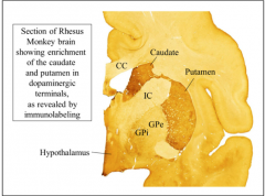
Note the dopaminergic terminals are located in the caudate and putamen, but not the ___ and ___. |
Note the dopaminergic terminals are located in the caudate and putamen, but not the GPe and GPi. |
|
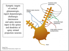
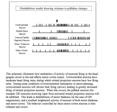
This illustration summarizes the inputs to striatal spiny neurons, and shows where on these neurons the dopaminergic and cortical inputs end. The cerebral cortex input ends on the tips of the spines of the spiny neurons. The cortical input uses _____ as its neurotransmitter. The substantia nigra dopaminergic input ends on the sides of the spines. Thus, the _____ input can modulate the cortical input to spines. |
This illustration summarizes the inputs to striatal spiny neurons, and shows where on these neurons the dopaminergic and cortical inputs end. The cerebral cortex input ends on the tips of the spines of the spiny neurons. The cortical input uses glutamate as its neurotransmitter. The substantia nigra dopaminergic input ends on the sides of the spines. Thus, the dopaminergic input can modulate the cortical input to spines. |
|
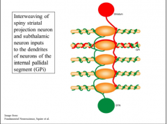
An important point about globus pallidus is that while the GPe projects to the _____ nucleus, the subthalamic nucleus mainly projects back to GP_. The subthalamic inputs wrap around the smooth dendrites of the GPi neurons, which also receive terminals from striatal substance P-containing neurons. This illustration schematizes those two inputs for GPi pallidal neurons. |
An important point about globus pallidus is that while the GPe projects to the subthalamic nucleus, the subthalamic nucleus mainly projects back to GPi. The subthalamic inputs wrap around the smooth dendrites of the GPi neurons, which also receive terminals from striatal substance P-containing neurons. This illustration schematizes those two inputs for GPi pallidal neurons. |
|
Draw this out! |

|
|
|
PUT IN INFORMATION FROM IN CLASS SESSION! |
IN CLASS SESSION! |
|

Review |
|