![]()
![]()
![]()
Use LEFT and RIGHT arrow keys to navigate between flashcards;
Use UP and DOWN arrow keys to flip the card;
H to show hint;
A reads text to speech;
26 Cards in this Set
- Front
- Back
|
- Recurrent arterial and/or venous thromboembolism - Recurrent spontaneous abortions OR - Thrombocytopenia in the presence of antiphos-pholipid antibodies but without features of SLE. Livedo reticularis, skin ulcers and neuropsychiatric disturbances have also been noted Dx? Rx? |
Dx: Anti-phospholipid antibody syndrome Rx: If suspected, commence aspirin 150–300 mg (o) daily and refer to a consultant. |
|
|
Diagnositic triad: Polyarthritis + fatigue + skin lesions Dx? |
SLE (systemic lupus erythematosus) |
|
|
What features are needed for a diagnosis of SLE? Name at least 4 |
Classification criteria(SLE = four or more of these 11 criteria) Malar (butterfly) rash Discoid rash Photosensitivity Arthritis (non-erosive arthritis in ≥ 2 peripheral joints) Oral ulcers (usually painless) Serositis (pleurisy or pericarditis) Kidney features (proteinuria or cellular casts) Neurological features (intractable headache, seizures or psychosis) Haematological features (haemolytic anaemia, leucopenia, lymphopenia or thrombocytopenia) Immunological features (positive anti-DNA, antiphospholipid antibodies or anti-Sm tests and false positive syphilis serology) Positive antinuclear antibody (ANA) test |
|
|
What are the initial screening tests you should do in a patient with suspected SLE? |
ESR/CRP—elevated in proportion to disease activity ANA test—positive in 95% (perform first) (key test) |
|
|
What are the more specific tests for a patient with SLE that you should conduct if initial screening tests come back positive? |
dsDNA antibodies—90% specific for SLE but present in only 60% (key test) ENA antibodies, especially Sm—highly specific NOTE: Rheumatoid factor—positive in 50% |
|
|
What should your management approach to a patient with newly diagnosed SLE be? |
Appropriate explanation, support and reassurance, use of sunscreens Refer to consultant rheumatologist for shared care in a multidisciplinary team |
|
|
What treatment options may a rheumatologist consider in a patient with SLE? |
Based on severity and organ involved. Mild: NSAIDs (for arthralgia) Moderate (especially skin, joint serosa involved): low-dose antimalarials (e.g. hydroxychloroquine up to 6 mg/kg once daily) (e.g. 400 mg (o) daily for 3 months, then 200 mg daily long term) Consider: fish body oil 0.2 mg/kg (o) daily Severe: corticosteroids are the mainstay (e.g. prednisolone 7.5–15 mg (o) daily) immunosuppressive drugs (e.g. azathioprine, methotrexate with folic acid, rituximab) may be used for severe arthralgia Avoid drugs in those in clinical remission and with normal complement levels. Other treatments, such as plasma exchange and immunosuppressive regimens, are available for severe disease. Keep in mind antiphospholipid antibody syndrome, especially with recurrent fetal loss and thrombotic episodes. |
|
|
Diagnostic triad: finger discomfort + arthralgia + GORD (± skin tightness) Dx? Other features? |
Scleroderma Clinical features Female to male ratio = 3:1 A progressive disease of multiple organs (if does then CREST) Raynaud phenomenon Stiffness of fingers and other skin areas (sausage fingers) ‘Bird-like’ facies (mouth puckered) Dysphagia and diarrhoea (malabsorption) Oesophageal dysmotility Respiratory symptoms: pulmonary fibrosis Cardiac symptoms: pericarditis, etc. Look for tight skin on chest (Roman breastplate) |
|
|
What investigations should you consider for diagnosis of scleroderma? |
ESR may be raised Normocytic normochromic anaemia may be present ANA test—up to 90% positive (relatively specific) Rheumatoid factor—positive in 30% Anticentromere antibodies—specific (positive in 90% with limited disease and 5% with diffuse) Antitopoisomerase I (anti-Scl-70) antibody is specific but only positive in 20–40% Skin biopsy—increase in dermal collagen |
|
|
What are the clinical features of CREST syndrome?
|
Calcinosis Raynaud phenomenon Oesophageal dysmotility Sclerodactyly (tight skin over fingers/ toes) TelangiectasiaAnticentromere antibody (invariably positive) |
|
weakness + joint and muscle pain + violaceous facial rash Dx? Ix? Rx? |
Dermatomyositis Differential diagnosis: statin-induced necrotising myositis (↑ CK levels) Diagnosis Muscle enzyme studies (serum creatine kinase and aldolase) Biopsies—skin and muscle EMG studies—show characteristic pattern Treatment includes corticosteroids and cytotoxic drugs. Early referral is appropriate. |
|
|
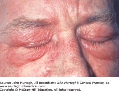
dry eyes + dry mouth + arthritis Dx? Other features? |
Sjögren syndrome Clinical features Fatigue Sicca (xerostomia, dry eyes, dry vagina) Difficulty swallowing food Increased dental caries; denture dysfunction Salivary gland enlargement Xerotrachea → chronic dry cough; hoarseness Dyspareunia Arthralgia ± non-erosive arthritis |
|
|
What do patients with Sjögren syndrome have an increased risk of? |
Although considered benign can transform into non-Hodgkin lymphoma (44 times risk). |
|
|
How do you diagnose Sjögren syndrome? |
Autoantibody tests—positive ANA (ENA), Ro (SSA), La (SS-B) Schirmer's tear test |
|
|
What management options are there for Sjögren syndrome? |
Referral to rheumatologist Treatment is symptomatic for dry eyes, mouth and vagina; arthralgia NSAIDs, hydroxychloroquine or steroids for arthritis |
|
|
arthralgia + weight loss + fever (± skin lesions) Dx? |
Polyarteritis nodosa Other clinical features: Young to middle-aged men Constitutional symptoms: fever, malaise, myalgia, weight loss Migratory arthralgia or polyarthritis Subcutaneous nodules along arterial lines Livedo reticularis and skin ulcers Kidney impairment and hypertension Cardiac disorders: arrhythmia, failure, infarction Diagnosis confirmed by biopsy or angiogramESR raisedTreatment with corticosteroids and immunosuppressantsDeath is usually from kidney disease |
|
|
Diagnosis and treatment for polyarteritis nodosa? |
Diagnosis confirmed by biopsy or angiogram ESR raised Treatment with corticosteroids and immunosuppressants Death is usually from kidney disease |
|
|
malaise + painful shoulder girdle + morning stiffness (>50 years) Dx? Other clinical features? |
polymyalgia rheumatica Pain and stiffness in proximal muscles of shoulder and pelvic girdle, cervical spine Symmetrical distribution Typical ages 60–70 years (rare <50) Both sexes: more common in women Early morning stiffness May be systemic symptoms: weight loss, malaise, anorexia Painful restriction of movement of shoulders and hips Signs may be absent later in day |
|
|
What is important to consider in a patient with polymyalgia rheumatica that presents with headaches? |
Temporal arteritis The clinical manifestations of polymyalgia rheumatica invariably precede those of temporal arteritis, of which there is about a 20% association. |
|
|
fatigue + headache + jaw claudication Dx? |
Temporal arteritis |
|
|
What investigations would you consider for PMR and temporal arteritis? |
Investigation No specific test for polymyalgia rheumatica ESR—extremely high, around 100 C-reactive protein—elevated Mild anaemia (normochromic, normocytic) GCA - above plus biopsy |
|
|
What treatment options are there for PMR and temporal arteritis? |
Treatment—for uncomplicated disease PREDNISOLONE Starting dose temporal (giant cell) arteritis: 40–60 mg (o) daily initially for 2–4 weeks (+ aspirin 100 mg/day) then gradual reduction according to ESR/CRP Polymyalgia rheumatica: 15 mg (o) daily for 2–4 weeks, then taper Taper down gradually to the minimum effective dose (often <5 mg daily) according to the clinical response and the ESR and CRP. Aim for treatment for 2 years. Relapses are common. If complicated (e.g. evolving visual loss) give IV methylprednisolone for 3 days prior to oral agents. OTHER DRUGS Azathioprine or methotrexate can be used as steroid-sparing agents. |
|
|
Why is it important to diagnose giant cell arteritis? |
In giant cell arteritis, a delay in diagnosis after presenting with amaurosis fugax and non-specific symptoms can have tragic consequences, in the form of ischaemic events such as blindness and strokes. |
|
|
Male to female ratio = 2:1 Recurrent oral and/or genital ulceration Arthritis (usually knees) Ocular symptoms—pain, reduced vision, floaters (ocular inflammation) Dx? Ix? Rx? |
Behçet syndrome There is no specific diagnostic test. Associated problems/complications: repeated uveitis and retinitis → blindness, colitis, venous thrombosis, meningoencephalitis. Treatment: high-dose steroids and specific ulcer treatment. DMARDs may be required. Patients with Behçet eye disease should be referred promptly for an ophthalmological opinion, which may be sight-saving.2 |
|
|
malaise + URTs (e.g. rhinitis, sinusitis) + LRTs (e.g. wheeze, cough) Dx? Ix? Rx? |
Granulomatosis with polyangiitis (Wegener granulomatosis) = upper respiratory tract (URT) granuloma, fleeting pulmonary shadows (nodules) and glomerulonephritis. Chest X-ray points to diagnosis—multiple nodes, cavitations Antineutrophil antibodies (c-ANCA) are a useful diagnostic marker (not specific) Diagnosis confirmed by biopsy, usually an open lung biopsy Better prognosis with early diagnosis and treatment with cyclophosphamide |
|
|
asthma + rhinitis + vasculitis + hypereosinophilia Dx? |
Churg–Strauss vasculitis |