![]()
![]()
![]()
Use LEFT and RIGHT arrow keys to navigate between flashcards;
Use UP and DOWN arrow keys to flip the card;
H to show hint;
A reads text to speech;
37 Cards in this Set
- Front
- Back
|
True (or indirect) hernia:
|
protrusion though a normal aperture in the abdomen and contains a complete peritoneal sac
|
|
|
False (or direct) hernia:
|
protrusion through an aperture that is not normal and do not contain complete peritoneal sac
|
|
|
Parietal (or Richter or Littre) hernia:
|
involves only a portion of the intestinal wall
|
|
|
When does risk of strangulation increase?
|
contraction of the hernial ring scar, inflammation of the hernial sac, faster peristaltic motion of intestine into the sac than out of the sac, and increased secretion or pooling of peritoneal fluid in the sac causing distention of the sac
|
|
|
How can umbilical hernias form?
|
foaling trauma, excessive straining, and umbilical cord infection
|
|
|
How are umbilical hernias managed?
|
Hernias less than 5 cm usually spontaneously regress and should be treated with daily digital reduction. Hernias that are greater than 10 cm or do not regress after 4 months should be surgically repaired
|
|
|
What is the disadvantage of the hernia clamp?
|
risk of incarcerating bowel and the formation of enterocutaneous fistula
|
|
|
Types of umbilical hernia surgical repair:
|
closed, by leaving the hernia sac intact and inverting it prior to linea closure or repair can be open, removing the hernia sac prior to linea closure
|
|
|
How can risk of ventral midline hernias be reduced?
|
closing initially with proper suture placement, suture selection, soft tissue handling, and aseptic technique
|
|
|
Optimal bite size for linea closure:
|
15mm from incision edge
|
|
|
Strongest linea closure suture:
|
6 polyglactin 910 and 5 polyglycolic acid
|
|
|
What suture is recommended for hernia repair in adult horses?
|
7 PDS and 6 polyglactin 910
|
|
|
Where does linea suture fail?
|
93% of suture fails at the knot and suture loops fail before fascial disruption
|
|
|
Where do most incisional hernias occur?
|
Because the linea is thinner cranially, more incisional hernias occur in the cranial aspect of the incision
|
|
|
Classes of incisional hernias:
|
acute total dehiscence or chronic incisional hernias
|
|
|
When does acute total dehiscence usually occur?
|
first 4-7 days post-operatively
|
|
|
How is acute total dehiscence repaired?
|
performed with monofilament stainless steel wire with rubber stent tubing in a through and through interrupted vertical mattress pattern. The wire suture is placed 5cm (2 inches) and 2.5 cm (1 inch) from the incision edge
|
|
|
What is the benefit of rubber stents in repairing acute total dehiscence of a ventral midline incision?
|
prevents wire cutting through the skin
|
|
|
What post-operative care is recommended for acute total dehiscence herniorrhapy?
|
Bandaging is changed daily for a month. At 14 days from the repair, alternate wire sutures are removed, with complete removal by day 21
|
|
|
How are chronic incisional hernias managed?
|
Conservatively, or arge defects should be reconstructed
|
|
|
Conservative management for chronic incision hernias:
|
Infection should be treated with antimicrobials, drainage, flushing only if good drainage exisits, and bandaging for a prolonged period of time
|
|
|
When is chronic incision herniorrhapy performed?
|
at least 8-10 weeks after complete resolution of infection
|
|
|
Mesh implant options for chronic incisional herniorrhaphy:
|
knit polypropylene mesh (Marlex), coated polyester (Mersilene), and polyglactin 910
|
|
|
Advantages of polyglactin 910 mesh implant:
|
absorbable and may not need to be removed if it becomes infected
|
|
|
Options for chronic incisional mesh hernioplasty:
|
subperitoneal mesh placement with fascial overlay, subperitoneal mesh placement with hernial ring apposition, subcutaneous placement with hernial ring apposition, and laparoscopic intraperitoneal mesh overlay
|
|
|
Describe subperitoneal mesh placement with fascial overlay:
|
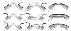
incision follows the contour of the hernia, the skin & SQ tissue are reflected, and the fascia is dissected from the peritoneum (if possible). The mesh is placed retroperitoneal and subfascially. Sutures are preplaced and when tightened, the mesh lays flat. The hernia flap is trimmed and the free edge is sutured to the edge of the hernia ring. The linea is not apposed
|
|
|
Describe subperitoneal mesh placement with hernia ring apposition:
|
linear skin incision is made and the skin and SQ tissues are reflected. The fascia is dissected from the peritoneum (if possible.) The mesh is placed between the body wall and the peritoneum and sutured to one side. Sutures are preplaced in the other side of the mesh, the linea is apposed, the mesh sutures are tightened, and the linea is closed.
|
|
|
Describe subcutaneous placement of mesh:
|
linear incision is made and the skin & SQ are reflected. The hernia sac is inverted and the ring is closed with tension relieving suture. Mesh is placed over the ring closure and sutured to the abdominal tunic
|
|
|
Describe laparoscopic mesh hernioplasty approach:
|
mesh is positioned subperitoneally, overlaying the inner surface of the internal rectus sheath. Mesh sutures are placed transfascially, securing the mesh to the peritoneum, and tied extrafascially
|
|
|
Complications of chronic incisional hernia repair:
|
greater in horses weighing more than 450 kg, incisional drainage and tearing of the internal abdominal oblique muscle
|
|
|
Predisposing factors for prepublic tendon rupture:
|
hydrop allantois, hydrops amnion, trauma, twins, fetal giants, draft breeds, and age
|
|
|
How do congenital diaphragmatic hernias develop?
|
failure fusion of 1 of 4 embryonic components of the diaphragm
|
|
|
Hoe do acquired diaphragmatic hernias develop?
|
Trauma
|
|
|
Types of diaphragmatic hernias:
|
peritoneal-pericardial or peritoneal-pleural
|
|
|
How are diaphragmatic hernias repaired?
|
Small defects are repaired with primary apposition, with sutures placed in the scared hernial ring. Large defects can be reconstructed with mesh placed on the abdominal side of the defect
|
|
|
What approach can be taken for very large traumatic diaphragmatic hernias?
|
thoracotomy may be required, in addition to VMC, for access. Alternatively, thoroscopy can be used in conjunction with thoracotomy and rib resection to repair diaphragmatic hernias and mesh placement
|
|

|

|

