![]()
![]()
![]()
Use LEFT and RIGHT arrow keys to navigate between flashcards;
Use UP and DOWN arrow keys to flip the card;
H to show hint;
A reads text to speech;
10 Cards in this Set
- Front
- Back
|
Key information for AST and ALT in general: |
- Generally associated with Hepatocellular damage - Generally not associated with cholestasis - Ratio of AST and ALT can be useful in differential - ALT is more specific for liver damage than AST |
|
|
What do different AST:ALT ratios indicate? |
AST: ALT =1: - Associated with ischaemia (CCF and ischaemic necrosis and hepatitis) AST: ALT >2.5: - Associated with Alcoholic hepatitis - Alcohol induced deficiency of pyridoxal phosphate AST: ALT <1 (usually a/w high rise in ALT, with slower rise in AST) - High rise in ALT specific for Hepatocellular damage - Paracetamol OD with hepatocellular necrosis - Viral hepatitis, ischaemic necrosis, severe toxic hepatitis |
|
|
When is ALP elevated? Where is ALP secreted? |
1) primarily associated with cholestasis and malignant hepatic infiltration 2) Marker of rapid bone turnover and extensive bony metastasis 3) Pregnancy ALP is secreted in bile cannalicular membrane of hepatocytes, bone, placenta, small intestines. ***Be cautious in isolated in ALP, may not be a liver problem. |
|
|
When is GGT elevated? |
1) Sensitive to alcohol ingestion 2) Marker of Hepatocellular damage but non-specific 3) Sharpest/rapid rise associated with biliary and hepatic obstruction |
|
|
For AST, the levels correlates to degree of liver damage. It can be monitored to predict the differential diagnosis. What are the levels and differentials? |
Serum level >20 x normal (in thousands) - Severe skeletal muscle trauma - Acute viral hepatitis - Toxic hepatitis (Drug induced hepatic injury) - Ischaemic hepatitis (Severe passive liver congestion (CCF)) Serum level 10-20 times normal (high hundreds) - CVS (Severe myocardial infarction) - Infection (Infectious mononucleosis) - Liver (Alcoholic cirrhosis) Serum level 5-10 times normal (mid hundreds) - Liver (Chronic hepatitis) - Skeletal muscle: Duchenne muscular dystrophy, Dermatomyositis, Influenza B calf myositis in children Serum levels 2-5 times normal (below hundred to low hundreds) - Blood (Haemolytic anaemia, haemolysis) - Liver (Fatty liver, Metastatic hepatic tumour) - Other: Pulmonary embolus, Alcoholic delirium tremens, Acute pancreatitis, IM injection, Strenuous physical exercise - Drugs: Opiates, Erythromycin, Sulphonamides, anti-tubercular, large doses of paracetamol/ aspirin/ vitamin A |
|
|
Where is LDH produced? |
In most tissues, and includes 5 different isozymes. LD1,2: CVS, Hematological LD3: Respiratory LD5: Hepatobiliary |
|
|
What are the differentials of raised LDH? |
CVS (LD 1 and 2) – AMI +/- hepatic congestion: - Rheumatic carditis - Myocarditis, CCF, Shock Respiratory (LD3) - Pulmonary embolus and infarction Haematological (LD 1 and 2) - Pernicious anaemia, Haemolytic anaemia, Sickle cell anaemia Hepatobiliary (LD5)Hepatitis, Active cirrhosis, Hepatic congestion - Hepatitis, Active cirrhosis, Hepatic congestion |
|
|
What is the basic structure of Hep B virus? |
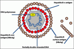
3 antigens: - HBsAg - HBeAg - HBcAg partially dsDNA DNA polymerase |
|
|
What is included in the panel for Hep B screening? What do each value represent when raised? |
1) HBSAg (shows that the patient is currently still infected by HBV, unless occult) 2) HBeAg (needs to interpreted with other results, but usually means that a high viral load is present) 3) IgM anti-Hbc (indicated an acute hep B infection or flare) 4) IgG anti-HBc (indicates chronic or long-standing hep B infection) 5) Anti-HBs (indicates immunity to hep B) 6) Anti-HBe (interpret with other results) 7) HBV DNA (will be in high amounts during replicative phase) |
|
|
How will a patient who is vaccinated with Hep B, but never infected by Hep B present for the panel? |
anti-HBc IgG or IgM will be negative, since there is no prior exposure HBsAg will be negative, since patient is not infected by Hep B currently anti-HBs will be positive, indicating protection |