![]()
![]()
![]()
Use LEFT and RIGHT arrow keys to navigate between flashcards;
Use UP and DOWN arrow keys to flip the card;
H to show hint;
A reads text to speech;
31 Cards in this Set
- Front
- Back
|
In which direction does the external and internal IC muscle run? |
External = hands in pockets. anteroinferiorly.
Internal = hands to mouth. Anterosuperiorly |
|
|
Which muscles elevate the sternum? |
Interchondral part of the internal intercostals. |
|
|
What muscle provides the primary elevation of the ribs? |
External IC. |
|
|
Where is the subclavian artery in relation to the anterior scalene muscle? |
Subclavian artery is deep to the anterior scalene muscle. |
|
|
What runs in each intercostal space? |
VAN |
|
|
Where is the brachial plexus? |
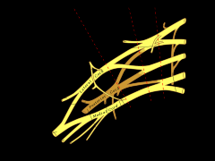
Behind the anterior scalene muscle. |
|
|
What nerves does the brachial plexus contain? |
C5-T1
Musculocutaneous Axillary Median Radial Ulnar |
|
|
What supplies the thorax? |
No plexus. Nerves from T2 to T12 run in the intercostal spaces between each rib, above the intercostal artery and nerve. |
|
|
What are the branches of the subclavian artery? |
1st part from medial border:
Vertebral artery
Internal thoracic artery
Thyrocervical trunk
2nd part:
Costocervical trunk
3rd part
Dorsal scapular artery
|
|
|
What does each thoracic dermatome represent? |
The course of the intercostal bundle including the nerve. |
|
|
Where do the SCV and IJV meet? |
At the sternoclavicular joint. |
|
|
What intercostal space articulates with the sternum? |
The 6th IC space |
|
|
Where does the Phrenic nerve radiate to? |
Shoulder - dermatome for C3,4 and 5 |
|
|
What muscles/structure are involved in quiet inspiration? |
Diaphragm. External ICs. ICh part of Internal ICS. |
|
|
What muscles are invovled in deep inspiration? |
Diaphragm, ICH part of Internal ICs, External ICs, Accesory muscles of breathing: Scalenes, SCM, Pect. Minor and Seratus posterior superior. |
|
|
What muscles are involved in quiet exiration? |
Elastic recoil (passive) |
|
|
What muscles are invovled in forced expiration? |
Abdo muscles, Iterosseous part of Int. ICs. Serratus posterior inferior. |
|
|
What is pleurisy? |
Inflammation to the pleura. |
|
|
What is a pleural effusion? |
Fluid within the pleural space |
|
|
What is pulmonary oedema? |
Fluid within the lung parenchyma itself - into the alveoli. |
|
|
Where is fluid from a pleural effusion likely to accumulate? |
In the pleural recess. |
|
|
At what point is the transverse fissue? |
4th rib |
|
|
Where does the oblique fissure run? |
Starts at the 6th rub and runs to L2/L4 posteriorly.
L2/4 is the point of the shoulder blades therefore is a useful marker between the upper and lower lobes |
|
|
At what level is the diaphragm on expiration? |
Just below the nipple = 4th intercostal space anteriorly. |
|
|
On which side of the body is the diaphragm higher/lower? |
It is higher as it is pushed up by the liver. |
|
|
What is the level of the diaphragm on the back? |
9th/10th IC space. |
|
|
Where would you do a thoracocentesis? ( to take a sample of fluid from the pleural space) |
Mid-axillary line 9th IC space.
Needle angles up to avoid the liver.
This area avoids the superior and inferior vascular bundles. |
|
|
Where would you insert a chest tube? |
Into the safe area of the chest = 4-5th IC space. Mid-axillary line.
Area under the armpit.
Avoid Pect. maj anteriorly and lat. dorsi posteriorly. High enough to avoid the liver and spleen. |
|
|
Describe a tension pneumothorax: |
Medical emergency. One way valve created. The whole lung is collapsed. Mediastinal shift. Lung deviation. |
|
|
Describe air trapping: |
Air trapping => loss of elastic recoil smaller airways are held upon by the alveoli around them. |
|
|
Describe emphysema: |
There is destruction of the lung parencyma. Breath in: larger airways are held open by cartilage. Small airways collapse upon breathing out.
The air within the lungs becomes hypoxic so drive to breath more air in but air is already there so volume area of gas exchange is very reduced.
Flattened diaphragm.
Causes R. sided heart failure due to vasoconstriction of hypoxic areas increased afterload on the R> side of the heart due to vasoC => R. ventricular heart failure. |