![]()
![]()
![]()
Use LEFT and RIGHT arrow keys to navigate between flashcards;
Use UP and DOWN arrow keys to flip the card;
H to show hint;
A reads text to speech;
27 Cards in this Set
- Front
- Back
- 3rd side (hint)
|
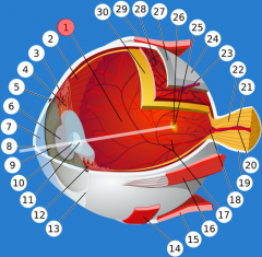
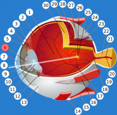
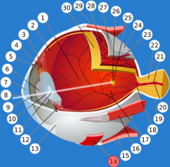
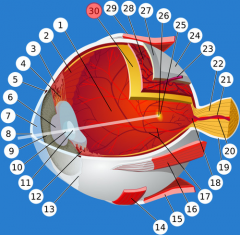
posterior chamber |
rear cavity filled with vitreous humor; maintains the shape of the eye |
|
|
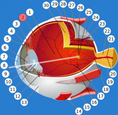
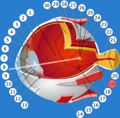
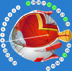
ora serrata |
serrated boundary between the ciliary muscle and the retina |
|
|
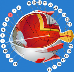
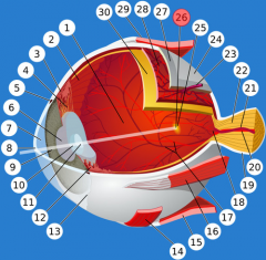
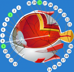
ciliary body |
ring of tissue encircling the lens |
|

|
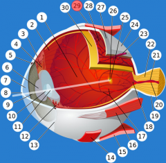
ciliary zonules |
ligaments connecting the ciliary body to the lens |
|
|
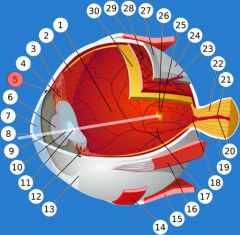
canal of Schlemm |
canal encircling the eye that returns aqueous humor to the blood |
|
|
pupil |
round central opening of the eye that controls the amount of light entering |
|
|
anterior chamber |
front cavity filled with aqueous humor; refracts light onto the retina |
|

|
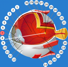
cornea |
transparent front part of the eyeball |
|

|
iris |
colored portion of the eye that controls pupil size |
|

|
lens |
flexible structure that changes shape to focus light on the retina |
|
|
ciliary processes |
posterior folds of the ciliary body that secrete aqueous humor |
|
|
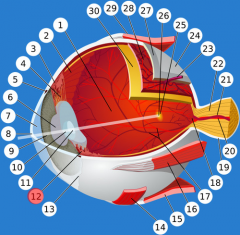
conjunctiva |
mucous membrane lining the eyelids and the anterior portion of the sclera |
|
|
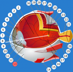
inferior oblique muscle |
bottom muscle that elevates and turns eye laterally; oculomotor |
|

|
inferior rectus muscle |
muscle that turns the eye down and medially; oculomotor |
|

|
medial rectus muscle |
muscle that turns the eye medially; oculomotor |
|

|
optic disc |
blind spot where the optic nerve leaves the eye |
|
|
dura mater |
outer layer of the meninges continuous with the sclera |
|

|
optic nerve (II) |
the nerve that carries neural impulses from the eye to the brain |
|

|
macula lutea |
posterior center of the retina with a high concentration of cones (clear, detailed vision) |
|
|
fovea centralis |
center of the macula lutea; highest concentration of cones |
|

|
sclera |
white part forming the outer covering of the eye (with the cornea) |
|

|
choroid |
highly vascular membrane between the retina and the sclera that provides oxygen and nourishment to the eye |
|
|
superior rectus muscle |
muscle that turns the eyeball upward and medially; oculomotor |
|
|
retina |
the light-sensitive inner surface of the back of the eye; contains rods and cones |
|

|
fibrous tunic |
1. sclera 2. cornea |
|
|
vascular tunic |
1. choroid 2. ciliary body 3. iris |
|
|
sensory tunic |
1. retina 2. optic nerve |