![]()
![]()
![]()
Use LEFT and RIGHT arrow keys to navigate between flashcards;
Use UP and DOWN arrow keys to flip the card;
H to show hint;
A reads text to speech;
127 Cards in this Set
- Front
- Back
|
Functional Classifications of Joints |
based on relative joint mobility |
|
|
Structural Classifications of Joints |
based on the way bones are held together |
|
|
Divisions of Functional Classifications of Joints |
synarthrosis, amphiarthrosis, diarthrosis |
|
|
Divisions of Structural Classifications of Joints |
fibrous, cartilaginous, synovial |
|
|
Synarthrosis |
-immovable joint -structures: suture, gomphosis, synchondrosis |
|
|
Amphiarthrosis |
-slightly movable -structures: syndemosis, smyphysis |
|
|
Diarthrosis |
-freely movable -structure: synovial |
|
|
Suture |
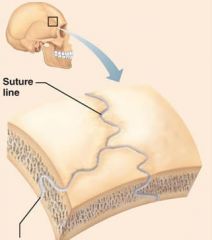
-dense regular connective tissue connects skull bones -function: synarthrosis |
|
|
Gomphosis |
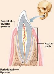
-periodontal membranes hold tooth to bony jaw (peg-in-socket joint) -function: synarthrosis |
|
|
Synchondrosis |
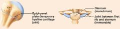
-hyaline cartilage plate between bones -function: synarthrosis |
|
|
Syndemosis |
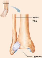
-dense regular connective tissue fibers between bones -function: amphiarthrosis |
|
|
Symphysis |
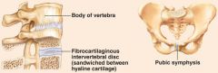
-fibrocartilage pad between bones -function: amphiarthrosis |
|
|
Synovial |
-bones joined by synovial fluid in a joint capsule/cavity
-function: diarthrosis |
|
|
Stabilizing Factors at Synovial Joints |
-shapes of articular surfaces (minor role) -ligament number and location (limited role) -muscle tone, which keeps tendons that cross the joint taut -important in reinforcing shoulder and knee joints and arches of the foot |
|
|
Fibrous |
-dense regular connective tissue holds together the ends of the bones and bone parts -no joint cavity -bones joined by dense fibrous connective tissue |
|
|
Cartilaginous Joints |
-pad of cartilage is wedged between the ends of bones -no joint cavity -bones joined by cartilage |
|
|
Plane Joint |
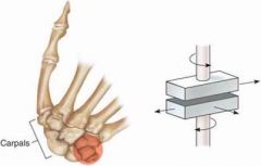
-nonaxial -relatively flat surfaces apposing each other; like a book on a table -allow only slipping or gliding moments |
|
|
Plane Joint Examples |

-intercarpal joint -intertarsal joint |
|
|
Hinge Joint |

-uniaxial -door hinge -permits angular movements in a single plane (like a mechanical plane) -flexion & extension |
|
|
Hinge Joint Examples |
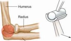
-humeroulnar joint (elbow) -interphalangeal joint |
|
|
Pivot Joint |

-uniaxial -door knob -permits only rotation -rotation of one bone around its own long axis |
|
|
Pivot Joint Examples |
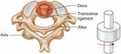
-proximal radioulnar joint (head of radius articulate with radial notch of ulna) -atlantoaxial joint (atlas and axis) |
|
|
Condyloid Joint |

-biaxial -mostly spheric convex curved concave and convex surface that is enlarged in on dimension, paired with a shallow concave cup -flexion & extension; abduction & adduction |
|
|
Condyloid Joint Examples |
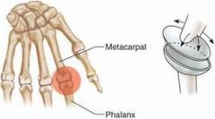
-radiocarpal joint (wrist) -metacarpophalangeal joint (knuckle) |
|
|
Saddle Joint |
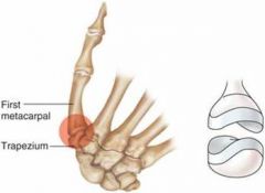
-biaxial -each member has a reciprocally curved concave and convex surface oriented at right angles to the other; similar to condyloid joint, but allows greater movement -flexion & extension; abduction & adduction |
|
|
Saddle Joint Examples |

carpometacarpal joint of the thumb |
|
|
Ball-and-Socket Joints |

-multiaxial -spheric convex surface paired with a concave cup -flexion & extension; abduction & adduction; rotation; circumduction |
|
|
Ball-and-Socket Joints Examples |
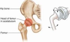
-glenohumeral joint (shoulder) -acetabulofemoral joint (hip) |
|
|
Flexion |
decreased in the angle between articulating bones, brings the bones closer together |
|
|
Extension |
increase in the angle between articulating bones |
|
|
Hyperextension |
continuation of extension beyond the normal extension |
|
|
Lateral Flexion |
-movement of the trunk sideways to the right or left at the wrist -movement of the trunk sideways to the right or left at the waist |
|
|
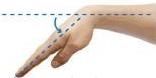
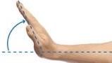
Abduction |
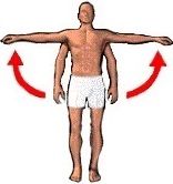
movement of a bone away from the midline |
|
|
Adduction |
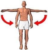
movement of a body part toward the body midline; movement that returns body parts to normal position |
|
|
Rotation |
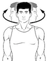
movement of a body part around its own axis |
|
|
Circumduction |
movement of a body part in a circle |
|
|
Supination |
rotation of the forearm so that the palm faces up |
|
|
Pronation |
rotation of the forearm so that the palm faces down |
|
|
Anterior Cruciate Ligament (ACL) |
prevents tibia from hyperextension (moving forward) on the femur |
|
|
Posterior Cruciate Ligament (PCL) |
prevents tibia from hyperflexion (moving backward) on the femur |
|
|
Medial Collateral Ligament (MCL) |
prevents excessive abduction and resists forces that would push the knee medially |
|
|
Lateral Collateral Ligament (LCL) |
prevents excessive adduction movement at the joint |
|
|
Meniscus |
half-moon shaped (fibro)cartilage lying between the knee joint |
|
|
Bursa |
small fluid-filled sac lined by synovial membrane |
|
|
Tendon Sheath |
layer of synovial membrane around a tendon |
|
|
Functions of Muscle Tissue |
-movement of bones or fluid (e.g., blood) -maintaining posture and body position -stabilizing joints -heat generation (especially skeletal muscle) |
|
|
Cardiac Muscle Tissue - Structure |
bifurcated (branching), striated cells fused at plasma membrane |
|
|
Cardiac Muscle Tissue - Function |
pumping blood in the circulatory system |
|
|
Cardiac Muscle Tissue - Location |
only in the heart |
|
|
Smooth Muscle Tissue - Structure |
long, spindle-shaped cells, each with a single nucleus |
|
|
Smooth Muscle Tissue - Function |
propulsion of substances along internal passageways |
|
|
Smooth Muscle Tissue - Location |
in walls of hollow organs throughout the body |
|
|
Skeletal Muscle Tissue - Structure |
long striated cells with multiple nuclei |
|
|
Skeletal Muscle Tissue - Function |
contraction for voluntary movements |
|
|
Skeletal Muscle Tissue - Location |
attached to bones and skin |
|
|
Myofilament |
-part of sarcomere -proteins -two types: actin (thin) and myosin (thick) |
|
|
Myofibril |
-long organelle inside a muscle -wrapped in sarcoplasmic retuiculum |
|
|
Myofiber |
-single muscle cell
-wrapped in endomysium |
|
|
Fascicle |
-bundle of myofibers bounded together by perimysium -portion of muscle |
|
|
Muscle |
-wrapped in epimysium -the whole organ |
|
|
Sarcoplasmic Reticulum |
-storage of calcium ions to be released when stimulated by an impulse -series of tubules -stores calcium -calcium moved from sarcoplasm into *insert term here* |
|
|
Endomysium |
connective tissue that wraps each individual myofiber |
|
|
Perimysium |
connective tissue that wraps bundles of muscle fibers - the "bundles" being known as fascicles |
|
|
Epimysium |
connective tissue that wraps the entire muscle |
|
|
Sarcolemma |
the cell membrane of a muscle fiber (myofiber) |
|
|
Sarcoplasm |
the cytoplasm of a muscle fiber, rich in oxygen-storing myoglobin protein |
|
|
Sarcomere |
contractile unit of a muscle fiber |
|
|
Transverse Tubules |
conduct the nerve impulse from the sarcolemma to the interior of the cell |
|
|
Sliding Filament Theory of Muscle Contraction |
a cycle of repetitive events that causes actin and myosin myofilaments to slide over each other, contracting the sarcomere and generating tension in the muscle |
|
|
Stored ATP |
reactant: ATP products: ADP; phosphate duration: 4-6 seconds |
|
|
Direct Phosphorylation |
reactant: creatine phosphate products: 1 ATP per creatine phosphate duration: 15-20 seconds |
|
|
Glycolysis |
anaerobic pathway reactant: glucose products: 2 ATP per glucose; lactic acid duration: ~60 seconds |
|
|
Cellular Respiration (aerobic) |
reactant: glucose; pyruvic acid; free fatty acids from adipose tissue; amino acids from protein catabolism products: 32 ATP per glucose; CO2; H2O duration: hours |
|
|
Lactic Acid Formation(Fermentation) |
anaerobic pathway reactant: glucose products: 2 ATP per glucose; lactic acid duration: 60 seconds or slightly more |
|
|
Why Would a Muscle Fatigue? |
occurs when ionic imbalances (K+, Ca2+, Pi) interfere with excitation-contraction coupling |
|
|
Contraction |
-generation of force -doesn't necessarily cause shortening of the fiber -shortening occurs when tension generated by cross bridges on the thin filaments exceeds forces opposing shortening |
|
|
Sliding Filament Model of Contraction |
-in the relaxed state, thin and thick filaments overlap only slightly -during contraction, myosin heads bind to actin, detach, and bind again, to propel the thin filaments toward the M line -as H zones shorten and disappear, sacromeres shorten, muscle cells shorten, and the whole muscle shortens |
|
|
Neuromuscular Junction |
-situated midway along the length of a muscle fiber -axon terminal and muscle fiber are separated by a space called the synaptic cleft -synaptic vesicles of axon terminal contain the neurotransmitter acetylcholine (ACh) -junction folds (motor end plates) of the sarcolemma contain ACh receptors |
|
|
Cross Bridge Cycle |
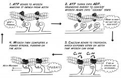
-cross bridge formation: myosin head attaches to actin -power stroke: myosin pulls actin toward the M line -cross bridge detachment: ATP attaches to myosin head and cross bridge detaches -cocking of the myosin head: energy from hydrolysis of ATP cocks the myosin head into the high-energy state |
|
|
Muscle Twitch |
response of a muscle to a single, brief threshold stimulus |
|
|
Phases of Muscle Twitch (in order) |
latent period period of contraction Refractory Period period of relaxation |
|
|
Latent Period |
-events of excitation-contraction coupling -time required for excitation |
|
|
Period of Contraction |
cross bridge formation; tension increases |
|
|
Refractory Period |
muscle has temporarily lost it's ability to be excited |
|
|
Period of Relaxation |
Ca2+ reentry into the sarcoplasmic reticulum; tension declines to zero |
|
|
Threshold Stimulus |
stimulus strength at which the first observable muscle contraction occurs |
|
|
Response to Change in Stimulus Strength |
-muscle contracts more vigorously as stimulus strength is increased above threshold -contraction force is precisely controlled by recruitment (multiple motor unit summation), which brings more and more muscle fibers into action |
|
|
Muscle Tone |
constant, slightly contracted state of all muscles; state of readiness |
|
|
Isotonic Contraction |
muscle changes in length and moves the load |
|
|
Concentric Contraction |
the muscle shortens and does work |
|
|
Eccentric Contraction |
the muscle lengthens as it does work (contracts) |
|
|
Isometric Contraction |
-tension increases to the muscle's capacity, but the muscle neither shortens nor lengthens -ex: holding a bag still and putting a notebook in |
|
|
Increasing Contractile Force Can Be Caused By Any of the Following: |
-large number of muscle fibers activated -large muscle fibers -high frequency of stimulation -muscle and sarcomere stretched to slightly over 100% of resting length |
|
|
Muscle Fiber Types |
-speed of contraction -metabolic pathways for ATP synthesis |
|
|
Speed of Contraction |
slow or fast according to: -speed at which myosin ATPases split ATP -pattern of electrical activity of the motor neurons |
|
|
Metabolic Pathways for ATP Synthesis |
-oxidative fibers: use aerobic pathway -glycolyic fibers: use anaerobic pathway |
|
|
Effects of Endurance Exercise |
-aerobic -lead to increased: muscle capillaries; number of mitochondria; myoglobin synthesis -results in greater endurance, strength, and resistance to fatigue -may convert fast glycolytic fibers into fast oxidative fibers |
|
|
Effects of Resistance Exercise |
-anaerobic -muscle hypertrophy due to increase in fiber size -increased mitochondria, myofilaments glycogen stores, and connective tissue |
|
|
Sprains |
ligaments are stretched or torn |
|
|
Cartilage Tears |
-due to compression and shear stress -cartilage rarely repairs itself, surgery often necessary |
|
|
Dislocations |
-occur when bones are forces out of alignment -accompanied by sprains, inflammation, and joint immobilization |
|
|
Subluxation |
partial dislocation of a joint |
|
|
Bursitis |
-an inflammation of a bursa, usually caused by a blow or friction -treated with rest and ice and, if severe, anti-inflammatory drugs |
|
|
Tendonitis |
-inflammation of tendon sheaths typically caused by overuse -symptoms and treatment similar to bursitis |
|
|
Arthritis |
-most widespread crippling disease in the US -symptoms: pain, stiffness, swelling of a joint -acute forms: caused by bacteria, treated with antibiotics -chronic forms: osteoarthritis, rheumatoid arthritis, and gouty arthritis |
|
|
Rheumatoid Arthritis (RA) |
-chronic inflammatory, autoimmune disease of unknown cause -usually arises between age 40-50, but may occur at any age; affects 3x as many women as men -signs and symptoms: include joint pain and swelling (usually bilateral), anemia, osteoporosis, muscle weakness, and cardiovascular problems |
|
|
Gouty Arthritis |
-deposition of uric acid crystals in joints and soft tissues, followed by inflammation -more common in men -typically affects the joint at the base of the big toe -if untreated, the bone ends fuse and immobilize the joint -treatment: drugs, plenty of water, avoidance of alcohol |
|
|
Lyme Disease |
-caused by bacteria, transmitted by the bites of ticks -symptoms: skin rash, flu-like symptoms, and foggy thinking -may lead to joint pain and arthritis -treatment: antibiotics |
|
|
Ligament |
-dense fibrous connective tissue attaching bones to together -reinforces joints |
|
|
Tendon |
-dense fibrous connective tissue attaching muscle to bone -reinforces the joint capsule as it spans a joint |
|
|
Articular Cartilage |
hyaline cartilage that covers the surface of all bones forming the joint |
|
|
Joint Cavity |
a space between the articulating bones and is filled with synovial fluid |
|
|
Articular Capsule |
two layers that enclose the joint cavity |
|
|
Fibrous Layer |
tough external layer that is composed of dense irregular connective tissue |
|
|
Synovial Membrane |
inner layer that is composed of loose connective tissue |
|
|
Special Characteristics of Muscle Tissue |
-excitability (responsiveness or irritability): ability to receive and respond to stimuli -contractility: ability to shorten when stimulated -extensibility: ability to be stretched -elasticity: ability to recoil to resting length |
|
|
Inversion |
a movement of the foot which causes the soles of the feet to face inwards |
|
|
Eversion |
a movement of the foot which causes the soles of the feet to face outwards |
|
|
Plantar Flexion |
movement of the foot in which the foot or toes flex downward toward the sole |
|
|
Dorsiflexion |
movement of the foot in which the foot or toes flex upward |
|
|
Z Disk |
formed between adjacent sarcomeres |
|
|
Aponeurosis |
a type of connective tissue that provides a point for a muscle to attach to a bone or cartilage |
|
|
End Plate |
large and complex terminal formation by which the axon of a motor neuron establishes synaptic contact with a striated muscle fiber |
|
|
Motor Unit |
made up of a motor neuron and the skeletal muscle fibers innervated by that motor neuron's axonal terminals |
|
|
Synaptic Cleft |
microscopic gap between neurons |
|
|
Acetylcholine |
substance that is released at the junction between neurons and skeletal muscle fibers, where it works as a neurotransmitter |
|
|
Acetylcholinesterase |
enzyme that breaks down the neurotransmitter acetylcholine at the synaptic cleft so the next nerve impulse can be transmitted across the synaptic gap |