Reading...
![]()
Play button
![]()
Play button
![]()
Use LEFT and RIGHT arrow keys to navigate between flashcards;
Use UP and DOWN arrow keys to flip the card;
H to show hint;
A reads text to speech;
62 Cards in this Set
- Front
- Back
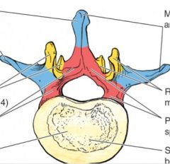
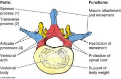
ID the following
spinous process transverse process areas of muscle attachment and movemnt areas that restrict movment areas that protect spinal cord area that supports body weight articular processes (4) vertebral arch vertebral body |
|
|
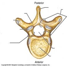
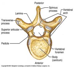
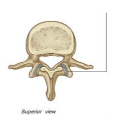
ID:
spinous process lamina vertebral arch vertebral foramen body pedicle superior articular process transverese process |
|
|
|
what is pars interatricularis
|
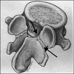
part of vertebra located between the inferior and superior articular processes of the facet joint. In the transverse plane, it lies between the lamina and pedicle.
|
|
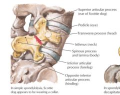
ID the :
superior articular process pedicle transverse process isthmus spinous process and lamina inferior articular process oppositie inferior articular process |
|
|
|
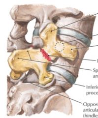
what is the difference between spondylolisis and spondylolisthesis
|
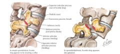
in spondylolisis the dog is wearing a collar
in spondylolisthesis the poor little scottie dog was decapitated |
|
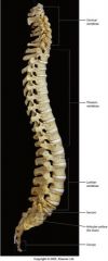
ID:
cervical vertebrae thoracic vertebrae lumbar vertebrae sacrum coccyx |
|
|
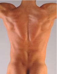
ID the following based on surface anatomy:
C2 C7 T3 T4 T7 L1/L2 L4 S2 |

C2: below skull
C7: base neck T3: spine scapula T4: sternal angle T7: inferior angle scapula L1/L2: end spinal cord L4: iliac crest, split aorta S2: PSIS |
|
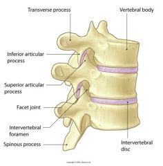
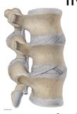
ID:
transverese process vertebral body interveterbral disc inferior articular process superior articular process facet joint intervertebral foramen spinous process |
|
|
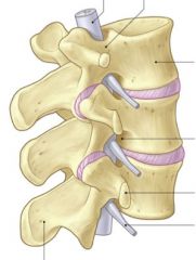
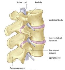
ID:
spinal cord pedicle vertebral body intervertebral foramen transverse process spinal nerve spinous process |
|
|
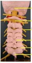
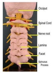
ID:
occiput spinal cord nerve root lamina facet spinous process |
|
|
|
what is the zygapophysial joint
|
a plane joint between superior and inferior articular process on vertebra
-angles of facet help dictate the amount of movement -horizontal facets favor rotation |
|

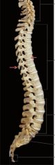
what is this pointing to?
what level vertebra is this? what type of movement does this allow? |
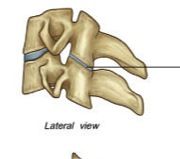
zygapophysial joing in Cervical vertebra (C3-C7)
30-45 degrees coronally off transverese plane, has lots of movement in all planes |
|
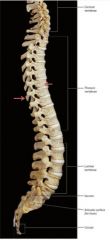
what is line pointing to?
what level vertebra is this? what type of motion does it allow? |
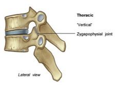
zygapophysial joint
thoracic vertebra upper is 45/60 degrees, mid - frontal plane, lower - frontal sagittal allows slight rotation, limited motion due to ribs |
|
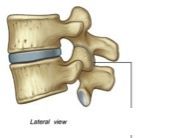
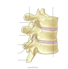
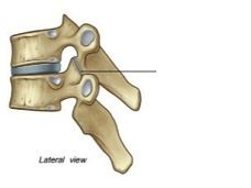
what is the line pointing to?
what level vertebra is this? what type of movement does it allow? |
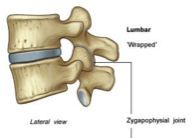
zygapophysial joint
lumbar vertebra joint faces medially, concave and allows more extension than flexion |
|
|
where do the most severe spondylosis injuries occur?
in what age group? |

between C4-C7
>55 yo |
|
|
which joints may degenerate and be a big factor in foraminal stenosis: a narrowing of the spinal foramen, the hole through which passes a spinal nerve as it exits the spine. It is usually a form of degenerative spine disease which occurs slowly over time with wear and tear of the spinal column. Arthritic changes of the spine, including herniated discs and bulging discs, soft tissue swelling and bony growth can all impinge on the formal foramen and compress the nerve within.
|

facet joints/zygapophyseal joints
this can cause radicular symptoms |
|
|
what is spondylosis?
where is it common? what age group? what are the sxs? |
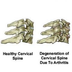
is a degenerative arthritis
is common in cervical and lumbar regions is most common > 55yo can compress nerves causing weakness, numbness, tingling and absence of reflex |
|

ID supraspinous ligament
where is it? what is it's purpose |

Dorsal to spinous process C7-sacrum
prevents flexion |
|

ID
nuchal ligament where is it |
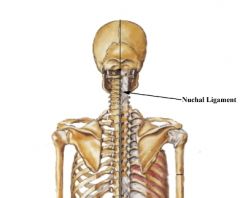
is a fibroelastic tissue in sagittal plane
runs from external occipital protuverance to spinous process of c-spine |
|

ID
ligamenta flava where is it what's it do |

connects the laminae
provides strong constant tension limits abrupt flexion and protects IV preserves the curves of the column |
|

ID
anterior longitudinal ligament it goes from where to where what does it do |
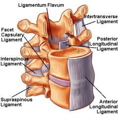
goes from anterior body of teh sacrum to the skull
prevents extension |
|
ID
posterior longitudinal ligament it goes from where to where what does it do |

is thin and weak and goes from posterior body of the sacrum to the skull, IV
it prevents flexion |
|

ID
interspinous ligament where is it what does it do |

is between spinous processes
prevents flexion, rotation |
|
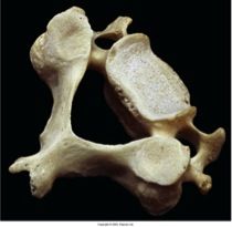
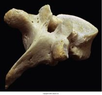
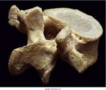
what vertebra is this?
ID: dens posterior articular facet for transverse ligament body vertebral foramen groves for atacment of alar ligaments superior articular facet foramen of transverese process transverese process inferior articular articular process lammina spinous process (bifid) |
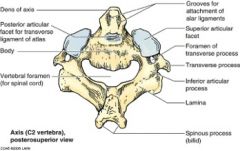
Axis
C2 vertebra |
|
|
what is the atlanto-occipital joint
what kind of joint is it what is it's function |

the joint between the atlas and occipital- occipital condyles articulate to lateral mass, superior articular facets of C1
function: flextion and extension, some lateral flexion, no rotation is a biaxial condyloid joint |
|
|
what is the atlanto-axial joint
what type of joint is it |
joint between axis and atlas- has 2 parts
articular facets of C1-C2 is a plane or gliding joint odontoid process of C2 and anterior arch of C1 is a medial pivot joint |
|

the transverese ligament of the atlas goes from where to where?
what is it's function? Find it on this picture! |

goes from lateral mass to lateral mass
holds the dens in place |
|
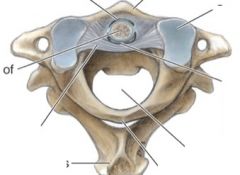
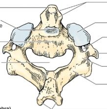
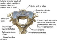
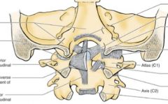
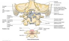
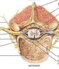
ID the following
anterior arch of atlas transverese ligament dens spinous process of axis vertebral canal posterior arc of atlas superior articular facet of atlas |
|
|
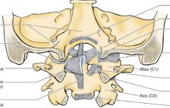
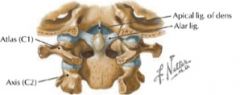
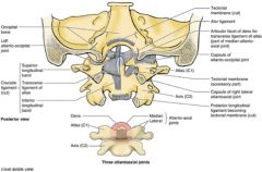
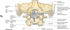
ID the following:
occipital bone atlanto-occipital joint transverese ligament inferior longitudinal band atlas axis posterior longitudinal ligament dens alar ligament |
|
|
what vertebrae is this?
how many of them are there? what is the size of the body? how many foramina? how big is the vertebral foramen? |

Cervical!
there are 7 with small bodies with 3 foramina and a large vertebral foramen. |
|

what vertebrae is this?
what is the spinous proccess like? what are the movements it makes? is it attached to ribs? does it have a transverse facet? is it stable? where does the spinous process point? |

Cervical
has a forked/bifid SP, is able to rotate, flex and extend. it does not attache to the ribs and has no transverese facet. It is the least stable of the vertebrae and it's spinous process points straight back. |
|

what vertebrae is this?
how many do we have? describe its: body size # foramina size of vertebral foramina SP primary movements attachments stability body shape |
Thoracic!
we have 12 describe its: body size - medium # foramina - 1 size of vertebral foramina - med SP - single, points down primary movements - flex/ext attachments- articulates with ribs at its transverese facets stability- the most stable vertebrae body shape - heart shaped |
|
what vertebrae is this?
how many do we have? describe its: body size/shape # foramina vertebral foramina SP primary movements clinical significance |

LUMBAR!
we have 5 describe its: body size/shape- large kidney bean # foramina - 1 small vertebral foramina SP- single that points straight back primary movements - flex/ext clinical significance - most prone to injury |
|
what is clinically significant in patients with down's syndrome in the atlas/axis joint?
ID the ligament this affects? |
In persons with Down syndrome, it is common to see congenital absence or laxity of the transverse atlas ligament
|
|
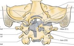
ID the following:
antlanto-occipital joint superior longitudinal band transverse ligament of the atlas inferior longitudinal band dens atlas axis alar ligament posterior longitudinal ligament becoming tectorial membrane |
|
|
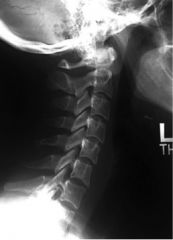
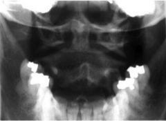
ID:
C1-C7 occiput dens facet joints intervertebral disc spaces inferior articular facets vertebral bodies T1 pedicles |

|
|
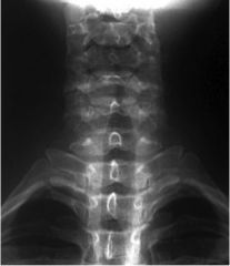
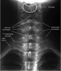
ID:
spinous processes T1 transverse process right 1st rib cervical transverse processes |
|
|
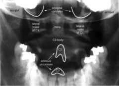
ID:
dens occipital condyles occiput lateral mass of C1 C2 body SP of C2/C3 |
|
|
|
at what point does the SP change direction in the thoracic vertebrae?
|

T4-T6 point downward to have tip line up half way between it's TP and the TP below it
|
|
|
at what level in thoracic vertebrae does the tip of SP line up with the TP of the vertebrae below it?
what else is significant at the begining of this? |
T7-T9
*T7 is also the apex of the kyphotic curve |
|
|
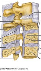
what accounts for 25% of height of vertebral column
where are these thickest |
vertebral disc
they get thicker as you go down the column and are thicker anteriorly in the cerfical and lumbar region (areas that have lordosis) |
|
|
what does the intervertebral disc do?
|
shock absorption, and allows ball bearing movment
|
|
|
what are the 2 parts of the intervertebral discs, what are they made of and what is their function?
|

annulus fibrosus is concentric rings of collagen in oblique fibers that sits around exterior of disc- it stable and withstands stretching
nuculus pulposus - center of disc made of watery type 2 collagen, acts as a semifluid fulcrum during movement |
|
|
when do you have least disc pressure?
most? |
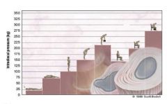
least= lying down
most= seated while beant forward while holding weight |
|
|
what are uncovertebral joints?
|
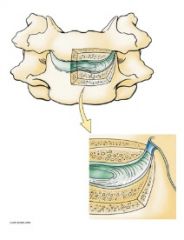
also called joints of luschka
b/w C3 and C7 are the lateral and posterolateral margins of IV discs w/ disc degeneration, uncinate proccess contact the body above it |
|
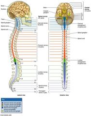
spinal nerves in each region are based on?
|

where they come out of vertebrae
8 cervical nerves (based on vertebrae they come out above- i.e. C1 is between skull and CV1, C4 is between CV3/4) 12 thoracic nerves (based on the vertebrae they exit BELOW ) 5 lumbar nerves (based on vertebrae they exit BELOW ) 5 sacral nerves (based on the vertebral body they exit BELOW) |
|
|
any damage to the spinal cord above C4/C5 results in
|

the brain can't communicate with the phrenic nerve or the diaphragm- aka no breathing
Phrenic nerve comes out of cervical plexus |
|
|
what level is the brachial plexus
|
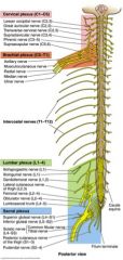
C5-T1
|
|
|
what level are intercostal nerves
|

|
|
|
what level is lumbar plexus
|

|
|
|
what level is sacral plexus
|

|
|
|
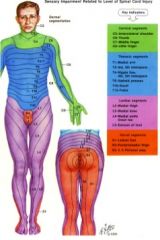
know basic dermatomes!
|
|
|
|
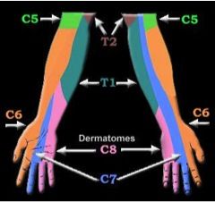
know hand and arm dermatomes
|
|
|
|
peripheral nerve innervation differs from dermatomes because of
|
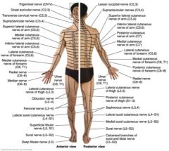
the plexi!
|
|
|
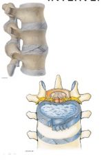
what are the jobs of the deep muscles of the back?
where are they? |
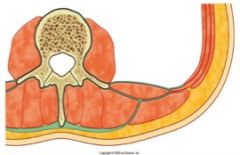
job= move, extend, rotate vertebral column
found posterior to lamina and transverse process and is surrounded by a fascia (shown in green) |
|
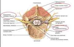
ID:
innervation of deep muscles of the back posterior ramus rami communicantes |
|
|
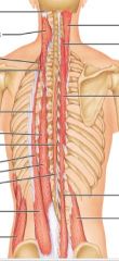
erector spinae
|

Iliocostalis, longissimus, spinalis
PA: Sacrum, iliac crest, SP DA: Ribs, TPs, SPs, mastoid process A: Extend and laterally bend vertebral column, extend head Assists w/ flexion of trunk??? (I love spaghetti) |
|

transversospinalis
|
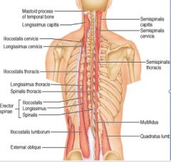
Semispinalis, multifidi, rotatores
PA: TPs DA: SPs above it A: C/L rotation, lateral flex, support low back |
|
|
sternoclaudomastoid
|
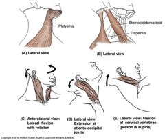
Spinal accessory (CN XI); sensory C2-C3
2 Unilateral action Lateral flex CL rotate 3 Bilateral actions Flex cervical/neck Extend head (AO jt) Combined Protrude head |
|
|
longus colli
|
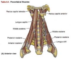
Bodies C1-C3 TP C3-C5 body C5-T3
Flex cervical vertebra N: VR C2-C6 |
|
|
longus capitis
|

Basilar part of occipital bone TP C3-C6
Flex head N: Ventral Rami C2-C3 |
|
|
what do the suboccipital triangle muscles do?
|
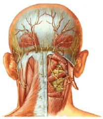
Extend head on C1
rotate head and C1 on C2; NS – Suboccipital n. (DPR C1) BS – occipital a. if they get tight they press nerve (specifically greater occipital) and arteries and can cause tension headaches |