![]()
![]()
![]()
Use LEFT and RIGHT arrow keys to navigate between flashcards;
Use UP and DOWN arrow keys to flip the card;
H to show hint;
A reads text to speech;
87 Cards in this Set
- Front
- Back
|
Areolar tissue |
Describe what tissue looks like. Clear saran wrap. Name cells ground substance fibers. Fibroblasts hydrolic acid Few scattered. What is the function of Areolar tissue? Holds things loosely |
|
|
Blood tissue |
Liquid connective tissue Hemocytoblasts plasma Few fibers Transports things through the body |
|
|
Fibrous tissue |
Rope like or stringy Fibroblasts hyaluronic acid many Holds things tightly |
|
|
Bone tissue |
Rock solid connective tissue Osteocytes hydroxyapatite Vary Supports and protects |
|
|
Adipose |
Fat Adipocytes hyaluronic acid Few fibers Protects insulates source of stored food |
|
|
Cartilage |
Rubbery Chrondrocytes chondroitin very many fibers Supports protects and gives shape to body parts |
|
|
Name parts of rat formed from epithelial tissue |
Skin Claws Hair Whiskers |
|
|
Name parts of rat formed from nervous tissue |
Brain Spinal cord |
|
|
Name type of connective tissue that forms specific parts like trachea tendóns |
Arelor Fibrous Adipose |
|
|
Name type of muscle tissue that forms specific parts like wall of ventral cavity lungs heart |
Cardiac Ventral cavity Vísceral |
|
|
Why is the trachea Formed from cartilage connective tissue rather than vísceral muscle tissue |
So it doesnt move or Block airways |
|
|
Why is the rat uterus Y shaped |
For múltiple pregancies |
|
|
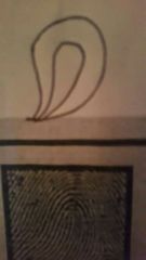
Define loop fingerprint |
|
|
|
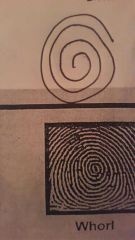
Define whorl |
|
|
|
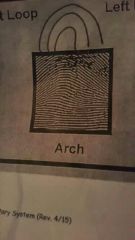
Define arch |
|
|
|
Sense receptors |
Krause. Ice water Ruffini. Hot water Pacinian. Pressure |
|
|
Cranial |
Covers back of skull |
|
|
Foramen magnum |
Large hole Spinal cord passes to connect to brain |
|
|
Parietal |
Cover top of skull |
|
|
Frontal |
Covers Forehead |
|
|
Temporal |
Sides of skull above ears |
|
|
Mastoid process |
Round marking behind ear |
|
|
Styloid process |
Long slender marking that connect to muscles of tongue |
|
|
Sphenoid |
Bat shaped |
|
|
Ethmoid |
Top of nasal cavity and medial wall of the eye socket |
|
|
Perpendicular plate of ethmoid |
Thin plate at center of ethmoid to which nasal septum attaches |
|
|
Malleus (ear bones ) |
Hammer shaped |
|
|
Incus (ear bones) |
Anvil shaped |
|
|
Stapes (ear bones ) |
Stirrup shaped |
|
|
Vomer |
Located at bottom center of nasal cavity |
|
|
Lacrimal |
Located in front of ethmoid on medial wall of eye socket |
|
|
Nasal concha |
Located in nasal cavity on either side of vomer |
|
|
Zygomatic |
Cheek bone |
|
|
Palatine |
Back part of the roof of the mouth |
|
|
Maxilla |
Upper jaw |
|
|
Mental foramen |
Holes through which nerves run to teeth |
|
|
Hyoid bone |
Horseshaped in neck Supports weight of tongue |
|
|
Laminae |
Thin flat plates |
|
|
Spina bifida |
Failure of láminae to unite |
|
|
Articular |
Connect to bones located above and below Allow vertebrae to sit one on top of another |
|
|
Thoracic vertebrae |
No transverse foramen Spinous process is long narrow and slanted downward |
|
|
Lumbar vertebrae |
No transverse foramen Spinous process is short thick and straight |
|
|
Structure of sacrum |
Body round disc Sacral canal opening at top Sacral foramen openings along sides Sacral hiatus space at bottom Sacral crests projections on back |
|
|
Sternum (breast bone) |
Manubrium Wide top |
|
|
Xiphoid process |
Cartilage at bottom Can snap off and go into liver causing hemorrhage if chest compressions are improperly performed |
|
|
True ribs |
1-7 connect directly to the sternum |
|
|
False ribs |
Pairs 8-12 do not connect directly to the sternum |
|
|
Floating ribs |
Pairs 11 & 12 are unattached at front |
|
|
Humerus |
Upper arm bone |
|
|
Head |
Round top that joints to scapula |
|
|
Anatomical neck |
Área below head |
|
|
Greater Tubercle |
Larger bump at head end for muscle attachment |
|
|
Lesser Tubercle |
Smaller bump at head end for muscle attachment |
|
|
Capitulum |
Round marking that joins to radius |
|
|
Trochlea |
Wider spool shaped marking that joins to ulna |
|
|
Epicondyle |
Markings on outside edges that join to muscles |
|
|
Olecranon fossa |
Indentation on posterior side |
|
|
Ulna |
Elbow bone |
|
|
Olecranon process |
Large curved projection at top that sits in olecranon fossa of humerus |
|
|
Styloid process |
Pointed slender projection at bottom for muscle attachment |
|
|
Funny bone |
Ulnar nerve runs across olecranon process |
|
|
Radius |
Flat headed bone on lateral side of foreman |
|
|
Carpals wrist bone |
Scaphoid Lunate Triquetrum Pisiform Trapezium Trapezoid Capitate Hamate |
|
|
Metacarpals |
Form body of hand |
|
|
Phalanges |
Finger bones |
|
|
Clavicle |
Collar bone |
|
|
Scapula |
Shoulder blade |
|
|
Acromion |
Highest marking that joins to clavicle |
|
|
Glenoid cavity |
Indentation that recieves head of humerus |
|
|
Coracoid process |
Lower marking for muscle attachment |
|
|
Spine |
Scapular spine Projection on back surface |
|
|
Supraspinous fossa |
Small surface above spine |
|
|
Infraspinous fossa |
Large surface below spine |
|
|
Fémur |
Thigh bone |
|
|
Condyles |
Round markings at bottom |
|
|
Patella |
Kneecap |
|
|
Tibia |
Shin bone Tougher on big toe side |
|
|
Medial malleolous |
Drop at bottom of bone on inner ankle side |
|
|
Fibula |
Fragile Smaller bone on little toe side |
|
|
Lateral malleolous |
Drop at bottom on outer ankle side |
|
|
Fibular heminmelia |
Congenital absence of fibula |
|
|
Tarsals (ankle bone) |
First second third cuneiforms Cuboid Navicular Talus Calcaneous. Heel bone |
|
|
Ilium (hip bone ) |
Wide top portion |
|
|
Ischium (hip bone ) |
Lower back portion |
|
|
Pubis |
Front portion |
|
|
Obturador foramen |
Large hole through which blood vessels passes |
|
|
Acetabulum |
Indentation that recieves head of fémur |