![]()
![]()
![]()
Use LEFT and RIGHT arrow keys to navigate between flashcards;
Use UP and DOWN arrow keys to flip the card;
H to show hint;
A reads text to speech;
43 Cards in this Set
- Front
- Back
|
Listand define the major structural components of a cell |
Plasma Membrane - forms a cells surface boundary Cytoplasm - material between the plasma membrane and the nucleus Cytoskeleton - a supportive framework of protein filaments and tubules Organelles - performs various metabolic tasks for the cell Inclusions - accumlated cell products; lipids and pigments Cytosol - clear gel between membrane and nucleus |
|
|
Name the fluids that occur inside and outside a cell |
IntracellularFluid (ICF) – Fluid within the cell ExtracellularFluid (ECF) – All body fluids not contained in the cells also called Tissue Fluid (located in between the cells) |
|
|
State the size range of human cells and explain why cell size is limited |
Micrometer (onemillionth) of a meter, a cell cannot attain UNLIMITED size. If a cell grew excessively large it would rupture like a water balloon. |
|
|
Identify the components of the plasma membrane that encloses each cell |
Membrane Lipids – Phospholipids, Cholesterol, Glycolipids MembraneProteins – Integral Protein’s,Glycoproteins, Peripheral Proteins, (functions include; Receptors, Enzymes,Channel Proteins, Carriers, Cell-Identity Markers, Cell-Adhesion molecules) |
|
|
State the functions of each molecular component of the plasma membrane |
Receptor – receive and bind chemical signals from other cells Enzymes – carry out chemical reactions at the membrane surface,degrading signal molecules after message is received and breaking downnutrients into forms the intestine can absorb Channel Proteins – (Ion Channel) tunnels through them that allow water and Hydrophilic solutes to enter or leave a cell. Carriers – actively bind a substance on one side of the membrane and release it on the other side (transport glucose, amino acids, sodium,potassium, calcium) |
|
|
State the functions of each molecular component of the plasma membrane (continued) |
Cell IdentityMarkers – Glycoproteins andglycolipids are a cell identification tags, genetically unique to an individual. Enables immune system to distinguish what belongs to one owns body and what does not, so it can attack such things as bacteria and parasites Cell-Adhesionmolecules – (CAM’s) are membrane proteins that link cells to each other and to extracellular material; they bind tissue together like coupling between railroad cars (sperm-egg binding) (immunecell-cancer cell) |
|
|
Describe the composition, appearance, and functions of the glycocalyx that coats each cell |
All cells are covered with a fuzzy carbohydrate coat called (Gylcocalyx)It consists of short chains of sugars belonging to the glycolipids, andglycoproteins. It also cushions the plasma membrane and protects it from physical and chemical injury. It also functions in cell identity, and thus the body’s ability to distinguish its own healthy cells from diseased cells (transfusions) |
|
|
Describe the structure and functions of microvilli, cilia, and flagella |
Microvilli – folds of cell membrane, may be associated with protein filaments Cilia – (primary cilium) are hair like processes about 7 – 10nm long, with axoneme core Flagella – single long structure with axoneme core , the whip like tail of a sperm (used to crawl) |
|
|
Name the different types of junctions that connect cells to each other, and describe their functions |
Cell Junctions –protein at the cells surface that link cells together and attach them to extracellular material. Tight Junction – completely encircles the epithelial cell near it supper end and joins it tightly to adjacent cells. Tight Junctions ensure that absorbed materials must pass through the epithelial cells and not between them.· Gap Junction – is formed by a ring of six proteins arranged some what like the segments of an orange. Nutrients pass from cell to cell-using gap junctions. |
|
|
Define selectively permeable membrane |
Selectivelypermeable membrane – membrane allows some substances to pass through but holds back others, especially those too large to pass |
|
|
Define active transport |
Active Transport – process that employs a protein and uses energy from ATP to move a solute through the membrane up its concentration gradient – that is, from one side where it is less concentrated to the side where it already is concentrated. |
|
|
Define Simple Diffusion |
is the net movement of particles from a place of high concentration to a place of low concentration, in other words, down a concentration gradient. |
|
|
Define Osmosis |
is the net movement of water through a selectively permeable membrane from the side where there is a relatively low concentration of solutes to the side where there is a higher solute concentration |
|
|
Facilitated Diffusion |
is the movement of a solute through a cell membrane, down its concentration gradient, with the aid of a carrier. It does not involve any ATP. |
|
|
Define Sodium-PotassiumK Pump |
binds 3 sodium ions from the ICF and ejects them from the cell, then binds two potassium ions from the ECF and releases these into the cell, repeats the process over and over using 1 ATP molecule. |
|
|
Define Vesicular Transport |
cells move larger particles or droplets of fluid through the membrane in bubble-like vesicles. |
|
|
Describe the cytoskeleton, its three components, and its functions |
Cytoskeleton – is a network of protein filaments and tubules, forms a very dense supportive web in the cytoplasm. Microfilaments – are about 6 nm thick, form a terminal web on the internal side of plasma membrane IntermediateFilaments – 8-10 nm thick, are thicker and stiffer that microfilaments, they contribute to the strength of the desmosomes and include the tough protein, keratin, fills the epidermis and gives strength to the skin· Microtubules – 25 nm thick, are hollow cylinders of protein. They hold organelles in place, from bundles that maintain cell shape and rigidity. |
|
|
Give some examples of cell inclusions and explain how inclusions differ from organelles |
Inclusions – two kinds, stored cellular products such as pigments,fats, globules, and glycogen and foreign bodies such as viruses and bacteria.Inclusions are never enclosed in a membrane and unlike organelles they are not essential to cell survival |
|
|
List the main organelles of a cell and explain their functions |
Nucleus – the largest organelle, and usually the only one visible with the light microscope, most cells only have one nucleus EndoplasmicReticulum – “little network within”they cytoplasm, interconnected channels called cisternae, enclosed by a membrane. Ribosomes – are small granules of protein, read coded genetic messages from the nucleus and assemble amino acids into proteins specified by the code (ribosomal ribonucleic acid – rRNA) Golgi Complex – is a small cluster of cisternae that synthesize carbohydrates and put the finishing touches on protein and glycoproteinsynthesis Lysosomes – is a package of enzymes enclosed in a membrane;digestive enzymes· Peroxisomes – not produced by the Golgi complex, abundant in liver and kidneys cells, primary function to breakdown fatty acids into two carbon molecules, also neutralize free radicals, detoxify alcohol and other drugs.· Mitochondria – organelles specialized for ATP synthesis Centrioles – is a short cylindrical assembly of microtubule sarranged in nine groups of three microtubules |
|
|
Define transcription and translation |
Transcription - step from DNAto mRNA – unzips the double helix (mirror image of the gene) Translation– The process of reading mRNA molecule and synthesizing the proteinen coded in its nucleotide sequence; step from mRNA to protein |
|
|
Describe the stages of a cell’s life cycle |
First Gap Phase(G1) – in an interval between cell division; the birth of two new cells from the parent cell and DNA replication Synthesis Phase – is the period in which a cell carries out DNAreplications; doubling its DNA content in preparation for the upcoming celldivision Second Gap Phase – brief interval, between DNA replication and cell division; finishes replicating its centrioles and synthesizes enzymes that control cell division Mitotic Phase – is the period in which the cell undergoes mitosis – it replicates its nucleus, divides its DNA into two identical sets (one pernucleolus) and pinches it in half to form two genetically identical daughter cells. DNA Polymerase – reads the base sequence on one strand and assembles nucleotides In the right order to make a complementary strand, most active in the Synthesis Phase |
|
|
Name the stages of mitosis and describe what occurs in each |
Prophase – chromosomes condense and nuclear envelopes break down Metaphase – chromosomes lie along midline of cell, some spindle fibers attach to centromeres and others to the plasma membrane Anaphase – centromeres divide into two; spindle fibers pull sister chromatids to opposite poles of cells Telophase – chromosomes gather at each pole of a cell; Cytokinesis – achieved by a motor protein pulling on the membrane skeleton; division of the cytoplasm |
|
|
Name the four primary classes of adult tissues |
Epithelia Connective Nervous Muscular |
|
|
Visualize the three-dimensional shape of a structure from a two-dimensional tissue section |
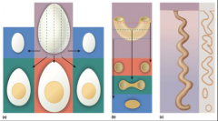
|
|
|
Interpret descriptive terms for various shapes of cells |
Squamous – thin,flat, scaly shape. Lines the esophagus and form the surface layer (epidermis)of the skin. Cuboidal –Squarish, and about equal in height and width. Good examples are found in the kidney tubules and liver. Columnar – distinctly taller than wide, as the inner lining cells of to mach and intestines. Polygonal - Having irregular,angular shapes with four, five, or more sides Stellate – having multiple pointed processes projecting from the body of a cell giving itsome what star like shape. Most nerve cells are Stellate. Spheroidal to ovoid– round to oval as in egg cells and white blood cells Discoidal – disc shaped as in red blood cells Fusiform – spindle shaped, elongated, with a thick middle and tapered ends, as in smooth muscle cells Fibrous – long,slender, and threadlike as in skeletal muscle |
|
|
Describe the properties that distinguish epithelium from other tissue classes |
Epithelium covers the body surface,lines body cavities, forms the external and internal linings of many organs,and constitutes most gland tissue |
|
|
Define basement membrane, basal surface, apical surface, and lateral surface, simple and stratified epithelium |
Basement Membrane - Between an epithelium and the underlying connective tissue is a layer called the basement membrane Basal Surface – the cell surface attached to the basement membrane Apical Surface –upper surface opposite of the basement membrane Lateral Surface –surface between the basal and apical surface Simple Epithelium –every cell touches the basement membrane StratifiedEpithelium – some cells rest on top of other cells |
|
|
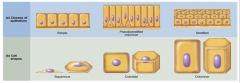
List and classify eight types of epithelium, distinguish them from each other, and state where each type can be found in the body |
Simple SquamousEpithelium – thin scaly cells (flat) – found in air sacs of lungs Simple Cuboidal Epithelium – squarish rounded cells – forms walls of kidney tubules-Simple Columnar Epithelium – tall narrow cells lines most of digestive track from stomach to anal Pseudostratified Columnar Epithelium – not reaching top – house goblet cells – found in large pathways in the respiratory system-Stratified Squamous Epithelium (Keratinized) – covered with dead cells, packed with keratin-Stratified Squamous Epithelium(Non-keratinized) – found in oral cavity, party of pharynx, esophagus, and vagina and anus -Stratified Cuboidal Epithelium – protective function; found on sweat glands, exocrine glands, male urethra Transitional Epithelium – urinary tract allows for stretching of the bladder as it fills |
|
|
Recognize Epithelial Types |
|
|
|
Describe the properties that most connective tissues have in common |
In most cases bind organs together, Is richly supplied with blood vessels; they are also the most abundant. |
|
|
Discuss the types of cells found in connective tissue |
Fiber type –Collagen Fibroblast are the most common cell type- Macrophages – engulf and destroy bacteria -Leukocytes – defense against bacteria, toxinsand other foreign agents -Adipocytes – fat cells Chondrocytes – cells of cartilage · Osteocytes – found in bone cells· Erythrocytes – red blood cells Leukocytes – combat infections |
|
|
Explain what the matrix of a connective tissue is and describe its components |
Extracellular material of a tissue. Composed of tissue fluid |
|
|
List 10 types of connective tissue |
Areolar:
Adipose: Reticular: Dense Regular: Dense Irregular: Elastic Connective Tissue: Hyaline: Elastic Cartilage: Fibrocartilage: Bone: Blood: |
|
|
Explain what distinguishes excitable tissues from other tissues |
Excitability is the characteristic of all living cells; nervous and muscular tissue. Electrical charge on the plasma membrane |
|
|
Name the cell types that compose nervous tissue |
Consist of neurons(nerve cells) Glial cells which protect and assist neurons |
|
|
Identify the major parts of a nerve cell |
· Neuron; has a cell body that houses the nucleus; several short filaments processes calleddendrites; then the axon/nerve fiber that sends signals out |
|
|
Name the three kinds of muscular tissue and describe the differences between them |
Skeletal – voluntary muscle Cardiac –involuntary Smooth – involuntary |
|
|
Distinguish between endocrine and exocrine glands |
Exocrine - release a secretion onto an epithelial surface and usually into the lumen (internal cavity) of another organ. (sweat, mammary,salivary glands, kidneys and liver) Endocrine - have no ducts, but instead, release their products into the bloodstream |
|
|
Compare modes of glandular secretion |
Merocrine – Release by exocytosis, Milk, Sugar Apocrine – coalesce from the cytosol into a single droplet, sweat glands (axillary) · Holocrine glands –break down then become secretion |
|
|
Describe the structure of mucous and serous membranes. |
Know that mucous membranes line cavities that open to the outside of the body and that serous membranes cover organs and line the thoracic and abdominal cavities |
|
|
Define hyperplasia, hypertrophy, and neoplasia |
Hyperplasia – tissue growth through cell multiplication Hypertrophy –Enlargement of preexisting cells Neoplasia – development of a tumor |
|
|
Define regeneration and fibrosis. Discuss scar tissue |
Regeneration:replacement of dead or damaged cells by the same type of cells as before Fibrosis:replacement of damage tissue with scar tissue Scar tissue – helps to hold an organ together |
|
|
Name and describe some modes and causes of tissue shrinkage and death |
Atrophy – shrinkage of a tissue through loss in a cell size or number. (Muscle shrinking) Necrosis – death of a tissue through trauma (infection or infarction) |