Reading...
![]()
Play button
![]()
Play button
![]()
Use LEFT and RIGHT arrow keys to navigate between flashcards;
Use UP and DOWN arrow keys to flip the card;
H to show hint;
A reads text to speech;
65 Cards in this Set
- Front
- Back
|
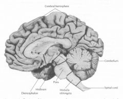
Anatomically, The nervous system is divided into what two systems, and what do they contain?
|
Central nervous system, consisting of the brain and spinal chord, located in the cranial cavity and spinal canal
Peripheral nervous system. consisting of cranial, spinal, and peripheral nerves that conduct impulses from efferent/motor nerves, and to afferent/sensory nerves, (centered in the CNS), as well as to and from ganglia nerve cell body collections, and specialized nerve endings. |
|
|
Functionally, the nervous sytem is divided into what two systems, and what are their functions?
|
Somatic Nervous System - somatic body parts of the CNS and PNS. Providing sensory and motor innervation to all parts of the body except viscera, smooth muscle, and glands.
Autonomic Nervous system - consisting of autonomic parts of the CNS and PNS. Provides efferent involuntary motor innervation to smooth muscle, the conducting system of the heart, and glands. Provides afferent sesnroy innervation from the viscera (pain and autonomic reflexes). |
|
|
What is the autonomic nervous system divided into?
|
Sympathetic nervous system
Parasympathetic nervous system and the Enteric Division, which serves the alimentary canal, and can function independently of the other two divisions of the ANS. |
|
|
what are supporting cells in the nervous tissue?
What are these CNS, and in the PNS? |
supporting cells are nonconducting cells that are in intimate apposition to the neurons. In the CNS they are known as neuroglia or just glia.
In the PNS they are called Schwan cells (insulators) and Satellite cells (PNS ganglia cells functioning in support) |
|
|
What are supporting cells called in nervous tissue of the Enteric division?
|
enteric Glial cells, despite being part of the PNS.
|
|
|
What are three functions of supporting cells in nervous tissue?
|
physical support, electrical insulation, and metabolic exchange
|
|
|
What are the specific effectors in internal organs effected by the autonomic nervous system, and what do they do?
|
Smooth muscle, whose contraction modifies diameter or shape of tubular or hollow viscera such as blood vessels, gut, gallbladder, and urinary bladder.
Cardiac conducting cells (purkinje fibers) located within the conductive system of the heart where the inherent frequency of depolarization of these fubers regulates the rate of cardiac muscle contraction and can be modified. glandular epithelium - in which synthesis, composition, and release of secretions can be modified. |
|
|
Explain the differences between the categories of neurons.
|
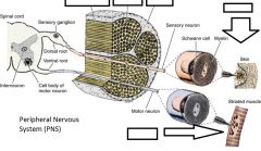
Sensory neurons - convey impules TO receptors to the CNS and are involved in somatic afferent (pain, temp, touch, proprioception which is nonconscious sensation of pain and placement) from the body, and visceral afferent nerve fibers which transmit similar information from mucous membranes, glands, and blood vessels.
Motor Neurons - convey impulses FROM the CNS or ganglia of effector cells. Processes of these neureons are included in somatic efferent (voluntary movement of skeletal muscle) and visceral efferent (transmit involuntary impulses to smooth muscle, cardia conducting cells 'purkinje fibers', and glands) nerve fibers. Interneurons - (aka intercalated neurons) form a communicating and integrating network between the sensory and motor neurons. 99% of neurons are connected to this network. |
|
|
What are three classifications for neurons based on processes from the cell body?
|
Multipolar - one axon and two or more dendrites. These are mainly motor neurons and interneurons. Basic impulse transmitter.
Bipolar neurons - have 1 axon and 1 dendrite. These are really only found in the retina of the eye and the ganglia of the vestibuloochlear nerve (cranial nerve VIII) of the ear and Unipolar - which has a single axon which divides close to the cell body. These are mainly found in the dorsal root ganglia and cranial nerve ganglia as sensory neurons connecting periphery directly to the CNS. |
|
|
What is the origin of the CNS neurons?
What is the origin of the PNS and ANS neurons? |
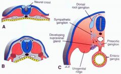
CNS neurons are derived from the neuroectodermal cells of the neural tube.
PNS and ANS come from the neural crest. |
|
|
a group of neurons with related function and connections are called a ____.
|
nucelus; these are the morphological and functional equivalents of ganglia in the PNS, but the nuclei are in the CNS.
|
|
|
gray matter contains
|
neurons
|
|
|
white matter contains
|
myelinated axons (which are formed by oligodendroglia)
|
|
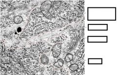
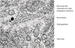
Name the structures
|
|
|
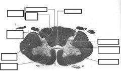
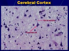
Identify the structures.
|
|
|
|
cell bodies of motor neurons that innervate striated muscle are located where?
|
the ventral (anterior) horn of the gray matter. This is an efferent neuron, carrying an impulse up to the spinal chord (CNS) from the ventral anterior root, which was from a spinal nerve of that segment of muscle, which was conveyed from the muscle.
Muscle >spinal nerve >root>horn>spinal chord/CNS |
|
|
Where are the cell bodies of sensory neurons located.
|
Sensory neuron cell bodies are located in the ganglia that lie on the dorsal root of the spinal nerve. these pseudounipolar cells are afferent and carry nerve impulses from the periphery (ie. pacinian corpuscle) up to the cell body in the dorsal root ganglion, and then finally transmitting this to the gray matter of the spinal chord.
|
|
|
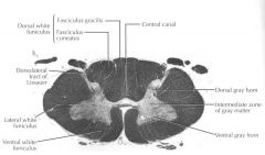
gray matter contains what?
|
neuronal cell bodies and processess.
astrocytes myoglia some oligodendroglia neurons are within nuclei - which requires Cresyl violet, nucelus violet, and myelin sheath blue to see. |
|
|
white matter contains what?
|
myelinated axons. (myelin being formed by oligodenroglia).
|
|
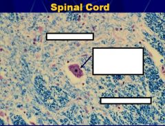
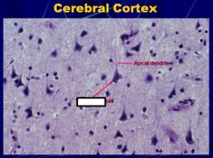
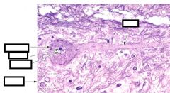
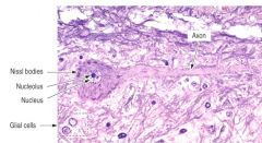
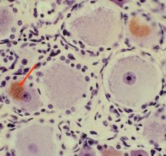
Identify the matter, and the cell with the arrow, AND describe the features of the cell.
|
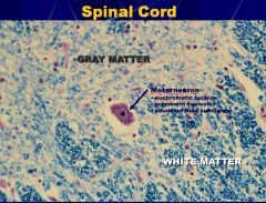
Motorneuron
euchromatic nucleus prominent nucleolus abundant Nissl substance |
|
|
What cell is found in the CNS but is derived from bone marrow?
|
Microglia is not derived from the neural crest like all of its neighbors, but actually develops from the bone marrow monocyte precursor cells along with other marcophages of the body.
|
|
|
Describe the types of glial cells.
|
Oligodendrocytes, small cells that are active in the formation and maintenance of myelin in the CNS.
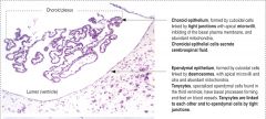
Astrocytes, morphologically eterogeneous cells that provide physical and metabolic support for the neurons of the CNS. They are fibrous in the white matter, and protoplasmic in the dark matter. Microglia, inconspicuous cells with small, dark, elongated nuclei that possess phagocytic properties. (formed in the bone marrow instead of neural crest) Ependymal cells, columnar cells that line the ventricles of the brains and the central canal of the spinal cord. Choroid Plexus cells, which are not in the text but mentioned in the lecture, and secrete CSF. |
|
|
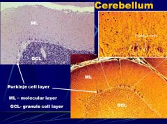
pukinje fibers are in the heart. cells, however, are neurons with very large dendritic sheets.
|
|
|
|
|
|
|
|
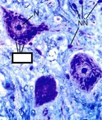
The neuron is a highly active cell as evidenced by a prominent nucleolus within a euchromatin nucleus (N). The glial cells nuclei (NN) are not as active and so their cytoplasm is not stained.
What is the box pointing to, what is it made up of, and what does it indicate? |

Neuronal cytoplasm is typically basophilic.
The basophilia can be seen to occur in clumps called Nissl bodies (NB). Nissl bodies are made up of RER and polysomes. These bodies reflect the high level of synthesis of membrane and secreted products. |
|
|
dendritic arborization = ____
|
dendritic arborization = dendritic tree
|
|
|
|
|
|
|
|
|
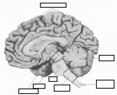
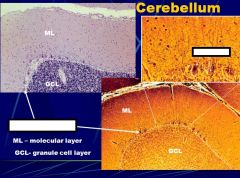
some neurons, such as the purkinje cells of the cerebellum, have protrusions that are called ___ ___ and are specialed regions of what?
|
dendritic spines; these are specialized forms of synaptic contact.
|
|
|
neurons typically have ___ axon(s).
|
one.
|
|
|
why do axons not stain with Nissl stains (cresyl violet)?
|
axons do not contain RER or ribosomes, which are the main components of Nissl bodies, (besides polysomes).
|
|
|
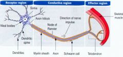
what are the terminal arborizations of axons called?
|
telodendria.
|
|
|
Both microtubules and neurofilaments are found in axons.
Describe each. |
Microtubules are important as tracks for fast anterograde and retrograde axonal transport.
Neurofilaments predominate, i.e. are more abundant than microtubules, and are believed important in maintaining axonal diameter |
|
|
What are the classifications for axonal transport?
|
Anterograde - to the axon
Retrograde - away from axon slow transport fast transport |
|
|
Describe the features of slow axonal transport.
|
anterograde only.
used for structural proteins tubulin, actin, microtubules, neurofilaments. (once they get there they're broken down by high Calcium levels 100x slower than fast transport. |
|
|
Describe the features of fast axonal transport.
|
its 100x fast than slow axonal transport.
it goes both directions, (anterograde and retrograde) Anterograde - takes membranous organelles and membrane-bound proteins away from cell body and toward axon terminals (like mitochonria, synaptic vessicles, and SER). Retrograde - takes worn organelles, endocytosed materials like growth factors toxins and viruses away from axon and sends them back to the cell body. |
|
|
What are some of the needs of fast axonal transport?
|
Microtubule dependent
Anterograde component uses kinesin as a motor protein Retrograde component uses dynein as a motor protein Substances that disrupt microtubules or that prevent their assembly will prevent or inhibit fast axonal transport (ie. ricin toxin used by nazis inihibits this process, injection by dipped and sharp tip of umbrella). |
|
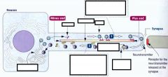
What is the process shown?
|
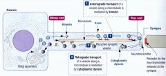
Fast Axonal Transport
|
|
|
Describe lipofuscin.
|
When retrogradely transported endosomes and multivesicular bodies reach the cell body, they fuse with lysosomes
Material is digested. Lipofuscin is material that cannot be digested. Accumulates with age and in some diseases |
|
|
What do rabies, tetanus, and ricin have in common?
|
Each of these have the ability to be transported retrogradedly to the cell body from an axon. Rabies virus binds to acetylcholine receptor of motor neurons innervating infected muscle cells, Retrogradely transported to cell body where it can replicate.
Toxins such as tetanus toxin, a protease, and ricin toxin are retrogradely transported |
|
|
Describe the major types of synapses.
|
Chemical synapses function through the release of neurotransmitters, are the most common, and can either be excitatory or inhibitory.
Electrical synapses involve the quick exchange of ions through gap junctions that allow neurons to act in unison. These are present in some regions of the CNS. |
|
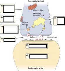
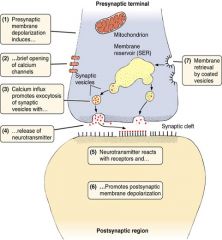
List the steps in this process.
|
|
|
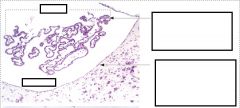
Describe this slide.
|
|
|
|
What are fibrous astrocytes involved in?
|
white matter
structural support for tracts of white matter GFAP - glial fibrillary acidic protein, are bundles of strong intermediate filaments, (present in protoplasmic astrocytes as well but less so) |
|
|
What are protoplasmic astorcytes involved in?
|
gray matter,
thick short bushy processess less GFAP secrete growth factors and act as support cells for neurons. MOST IMPORTANT - they're little feet extend to the basal lamina of the pia mater to form the glia limitans (relatively impermeable barrier surround the CNS - but not the blood-brain barrier) they do however induce capillaries to form the blood brain barrier |
|
|
What would you use antibodies to GFAP to find?
|
Astrocidic tumor
|
|
|
What is the most common type of primary brain tumor?
|
astrocystomas.
80-90% of all glial tumors in adults. anaplastic over time (less differentiation = malignancy) blood-brain barrier may be involved with preventing metastasis to other regions of body besides CNS. 8-10 month mean survival |
|
|
What do oligodendrocytes do?
|
myelinate axons similar to schwann cells, but do it in the CNS.
may myelinate 50 axons. may not myelinate some axons and instead act as satellite cells |
|
|
What disease has these characteristics?
Chronic demyelinating disease of the CNS Multiple, focal plaques of demylenization Optic nerve, which is myelinated by oligodendroglia, often affected. Prevalence 1 in 1000 in USA Pathogenesis undetermined May be autoimmune Antibodies to central myelin Characterized by remission and relapse CNS plaques (scars) formed by astrocytes |
Multiple Sclerosis (MS)
- Symptoms in declining order of frequency. Unilateral visual impairment. Diplopia (double vision). Paresthesias (pins and needles) Ataxia (unsteadiness) Vertigo (dizziness) Fatigue Paresis (muscle weakness) Dysarthria (speech problems) Mental disturbances |
|
|
when do microglia migrate into the CNS?
|
uring the development of CNS vasculature.
|
|
|
When do microglia have a name change?
|
Twice.
When the CNS is injured, the microglial nuclei enlarge and elongate. These cells are then called rod cells by pathologist Increased damage causes the microglia to become phagocytic. They are then called gitter cells by pathologists. |
|
|
In CNS injury, what usually occurs?
|
Astrocytes react to injury – gliosis (flocking of glial cells)
ends up forming a glial scar that inhibits axonal regeneration. ALSO, oligodendroglia express protein that prevents axonal growth |
|
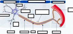
Fill in the blanks.
The big arrows are asking afferent or efferent. |
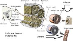
Efferent or motor nerves innervate muscle while afferent send a message from the periphs directly to the CNS.
|
|
|
Dorsal root ganglion contain what type of neuron and what do they send?
|
sensory neurons that signal pain and temperature, touch, pressure, vibration and proprioception.
|
|
|
|
|
|
What disease has the following characteristics?
Acute inflammatory polyradiculoneuropathy Affects PNS myelin only Usually bilateral, symmetrical, motor weakness Evidence of T-cells, plasma cells and macrophages around axons May be an autoimmune disease Severe cases can lead to death Most cases resolve in 2-4 weeks. |
Guillan-Barre´ Syndrome
|
|
|
What critical role do schwann cells play in peripheral nerve regneration?
|
become phagocytic while synthesizing essential growth promoting factors and cytokines, that hopefully a sniffing axon will find and penetrate original detached nerve fiber
|
|
|
what explain phantom limb pain?
|
tramatic Neuroma, where several months of regeneration and still nerve fibers were not properly re-established, resulting in tangled axonal processess (bushy looking)
|
|
|
A 62-year-old man has begun to notice some tingling and numbness in his right face, neck, and upper limb. He also believes he has become weaker in his right upper limb and has begun having difficulty doing fine motor tasks with his right hand.
After seeing a neurologist, who confirms the man’s deficits, he undergoes an MRI of his brain that reveals a 3cm mass in his left cerebral hemisphere. A biopsy is arranged and a malignant CNS tumor is diagnosed. What is the most common form of primary CNS tumor in adults? What type of intermediate filaments do the tumor cells express? What is this man’s prognosis? |
What is the most common form of primary CNS tumor in adults? astrocytoma
What type of intermediate filaments do the tumor cells express? Glial fibrillary acidic protein (GFAP) What is this man’s prognosis? 8-10 months survival |
|
|
what is chromatolysis?
|
the dissolution of nissl bodies that usually occurs during the axonal reaction, (occurring after axotomy (axon severed)).
|
|
|
What is the axonal reaction?
|
The axonal reaction occurs after axotomy (severed) in an attempt to prevent neuron death. It involved the swelling of the cell, a dissolution of Nissl bodies called chromatolysis, and the nucleus moving to an eccentric position, all within 24-48 hours.
|
|
|
Why would actin and tubulin genes be up-regulated in a neuron and synaptic transmitter genes be down regulated?
|
The axonal response, which produces morphological changes in a neuron in an attempt at regeneration.
|
|
|
In the axonal response. What happens at the proximal and distal ends of the severed neuron?
|
proximal portion degenerates back to the next node of Ranvier - retrograde degeneration
distal portion begins to degenerate because separated from synthetic machinery of the cell anterograde or Wallerian degeneration |
|
|
What is an axonal growth cone?
|
A motile structure that has a concentration of receptors for neurotrophic factors and laminin. This is in response to being recently severed, and searching for these substances in higher concentration may lead the cone directly to its severed member. (laminin found on basal lamina and schwann cells)
|