![]()
![]()
![]()
Use LEFT and RIGHT arrow keys to navigate between flashcards;
Use UP and DOWN arrow keys to flip the card;
H to show hint;
A reads text to speech;
43 Cards in this Set
- Front
- Back
- 3rd side (hint)
|
Define taxonomy. |
classification of organisms |
|
|
|
List the 5 orders commonly seen in veterinary practice.
|
1. Order Carnivora 2. Order Artiodactyls (even-toed hoofed animals) 3. Perissodactyla (odd-toed hoofed animals) 4. Order Rodentia 5. Order Lagomorpha
|
|
|
|
List the levels of organization.
|
cells, tissues, organs, and organ systems
|
|
|
|
List and describe the four main types of tissue.
|
1. Epithelial: sheets of cells that cover all of the internal and external surfaces of the body and line the cavities, organs, and form the glands
2. Connective: holds the structures of the body together; eg. Bone, cartilage, tendons, fat, etc 3. Muscle: specialized cells that shorten to produce movement when stimulated 4. Nervous: specialized cells that have ability to conduct electrical impulses |
|
|
|
Define histology.
|
Branch of anatomy that deals with microscropic structure and composition of tissues |
|
|
|
True or False: Most organs contain muscle tissue and helps bind the tissues within the organ together.
|
False
|
|
|
|
True or False: An organ will often contain all four primary tissue types.
|
True
|
|
|
|
What is the composition and function of cerebrospinal fluid?
|
Cerebrospinal fluid baths and protects the brain and spinal cord from the hard surface of the skull. CSF's chemical composition may be involved in regulation of certain autonomic functions such as respiration and vomiting. CSF is clear and slippery and circulates between layers of the meninges and through cavities inside of the brain and spinal cord. |
|
|
|
What is the composition and function of synovial fluid?
|
Synovial fluid is normally transparent and has the viscosity of medium-weigh motor oil. It lubricates the joint surface.
|
|
|
|
What is the composition and function of peritoneal and pleural fluid?
|
Both fluids are transudate and are thin, watery, fluid-containing little protiens that have been passed through a membrane.It creates a moist and slippery surface which reduces friction between adjacent organs and between organs and the cavity wall. Pleural fluid is a transudate in the thorax while peritoneal fluid is in the abdomen.
|
|
|
|
What is the structure and function of red blood cells?
|
Erythrocytes (RBC) carry oxygen from the lungs to the cells and tissues of the body. Usually mature mammalian RBC are round, biconcave (both sides are pushed in) disks that have no nucleus and stain red bc of the hemoglobin (binds with the O2). The biconcave disk shape serves 3 functions: 1. Deformable (but not elastic) and can take in water and swell without rupturing the membrane 2. Provides more membrane surface are for diffusion of O2 and CO2 to take place 3. Results in shorter diffusion distance in and out of the cell compared to a sphere
|
|
|
|
What is the structure and function of white blood cells?
|
Leukocytes (WBC)defend the body against foreign invaders and each 5 types of WBC have its own unique role in this defense. WBC usually do their job out in the tissues and use the peripheral blodd to travel from their site of production in the bone marrow to their site of activity (tissues). When accumulated in one place, they appear white or cream colored. They are any nucleated cell normally found in blood and are larger than RBC. |
|
|
|
What are the 5 types of WBC and their functions and structure?
|
1.neutrophil: cytoplasmic granules don't stain from blue alkaline or red acid stain; have polymorphonuclear shape (nuclei have many shapes and are segmented); is a phagocyte; most numerous WBC in dog, cat, and horse
2.eosinophils: cytoplasmic granules stain red and polymorphonucleur; attracted to and inhibit local allergic and anaphylactic reactions and some phagocytosis 3.basophils: c.g. stain blue; polymorphonuclear; contain histamine and heparim; initiate immune and allergic reaction--heparin acts as localized anticoagulant to keep blood flowing to injured or damaged area 4.monocytes: largest WBC in circulation; pleomorphic nucleus (can be round or of many diff. shapes) but does not split up into distinct segments; abundant cytoplasm that stains gray-blue and may contain vacuoles of various sizes; major phagocytic cell; monocytes in bloodstream 5.lynphocytes: normally predominant WBC in circulation of pigs and ruminants; only WBC with phagocytic cap.; most live in lymphoid tissues and constantly recirculate btwn tissue and blood;no granules in cytoplasm; nucleus is round or oval and not segment--a. B lymphocyte are antibody production and humoral immunity( when recognize antigen, transform into plasma cells that release antibodies) b. T lymphocytes- produce lymphokine thru helper T.L. which is activates k.t.l and responsible for cell-mediated immunity (where killer T.L. destroy cells during c.m.i. |
|
|
|
What is the structure and function of platelets?
|
Thrombocytes (platelets)are pieces of cytoplasm that have been isolated and release from giant, multinucleated cells (megakaryocytes)in bone marrow; are round and have numerous small, purple granules scattered thruout cytoplasm; granules contain clotting factors neccesarry for blood to clot; usually smaller than RBC; essential part of hemostasis and 3 specific fx: 1. maintainance of vascular integrity 2. platelet plug formation 3. stabilization of hemostatic plug by contributing to process of fibrin formation
|
|
|
|
What are the functions of blood and lymph?
|
Blood is a connective tissue and its 3 main fx are transport, regulation, and defense. 1.Carries O2, nutrients, and other essential compounds to every living cell (hemoglobin carries O2 and nutrients and etc dissolved in plasma); waste products of cellular metabolism (primarily CO2) away from cells to waste disposal organs (lungs, kidneys); hormones from endocrine glands to target organs; WBC from bone marrow to tissues; platelets to site of damage to a vessel wall
2. Aids in regulation of body temp, tissue fluid content, regulation of blood pH (7.4) 3.WBC provide denfese against foreign invaders thru phagocytosis and, in addition to platelets, 13 clotting factors in blood are necessary for blood to clot Lymph: 1. removes excess tissue fluid 2.waste material transport 3.large protein, esp. enzymes, transport to blood from their cell of origin (proteins are too large to enter venous circulation directly) |
|
|
|
Describe the functions and structures of the lymphatic system including its role in the immune system.
|
fx: 1. removal of excess tissue fluid 2.waste material transport 3. Filtration of lymph 4.protein transport
structures: 1.lymph nodes: small, kidney-shaped structures located at various points along the lymph vessels--lymph flows into the node in afferent vessels and leaves in efferent vessels--lymph will pass thru at least one lymph nosde that will put lymphocytes into the lymph and as lymph passes thru the node, tissue macrophages acts as filters to remove microorganism or other foreign matter 2.spleen: has both lymphatic and hematological fx; tongue shaped and located on left side of abdomen; soft tissue interior divided into red (consists of blood vessels, tissue macrophages, and blood storage spaces/sinuses) and white (consists of localized areas of lymphoid tissue)pulp; spleen acts as a reservoir for blood when the animal is at rest and does not need larger amounts of oxygen; fx:1.blood storage in red pulp 2.removal of foreign material from circulation by tissue macrophages in red pulp 3.removal of dead, dying, and abnormal RBC by tissue macrophages in red pulp 4.lymphocyte cloning in white pulp during an immune response thymus:located in caudal neck and cranial thoracic region lying on either side of trachea;where T lymphocytes processed tonsils:nodules of lymphoid tissue in an epithelial surface that are not covered with a capsule; found all over the body; peripheral lymphoid tissue in which mature lymphocytes live Gut-associated lymph tissue: lymphoid tissue found in the lining of the intestines; where B lymphocytes are processed |
|
|
|
Describe the process of glycolysis.
|
Glycolysis is a metabolic pathway that is found in the cytoplasm of cells in all living organisms and does not require oxygen. The process converts one molecule of glucose into two molecules of pyruvate, and makes energy in the form of two net molecules of ATP. Four molecules of ATP per glucose are actually produced; however, two are consumed for the preparatory phase. 2 NADH are produced when the triose sugars are oxidized. The overall reaction can be expressed this way:
Glucose + 2 ATP + 2 NAD+ + 2 Pi + 4 ADP → 2 pyruvate + 2 ADP + 2 NADH + 4 ATP + 2 H2O |
|
|
|
Describe the process of cellular respiration.
|
Def: the cellular process of taking in O2 and producing ATP in the mitochondria; Aerobic respiration requires oxygen in order to generate energy (ATP). It is the preferred method of pyruvate breakdown from glycolysis and requires that pyruvate enter the mitochondrion to be fully oxidized by the Krebs cycle. The product of this process is energy in the form of ATP (Adenosine Triphosphate), by substrate-level phosphorylation, NADH and FADH2. The reducing potential of NADH and FADH2 is converted to more ATP through an electron transport chain with oxygen as the "terminal electron acceptor". Most of the ATP produced by cellular respiration is by oxidative phosphorylation, ATP molecules are made to the chemiosmotic potential driving ATP synthase. Respiration is the process by which cells obtain energy when oxygen is present in the cell.
About 30 ATP molecules can be made per glucose during cellular respiration (2 from glycolysis 2 from the Krebs cycle, and about 26 from the electron transport system). However, such conditions are not realized due to losses as the cost of moving pyruvate into mitochondria. 1. Produces acetyl-CoA from pyruvate inside the mitochondrial matrix. This oxidation reaction also releases carbon dioxide as a product. In the process one molecule of NADH is formed per pyruvate oxidized 2. Krebs cycle: When oxygen is present, acetyl-CoA is produced from pyruvate; gets oxidized to CO2 while at the same time reducing NAD to NADH. NADH can be used by the electron transport chain to create further ATP as part of oxidative phosphorylation. To fully oxidize the equivalent of one glucose molecule two acetyl-CoA must be metabolized by the Krebs cycle. Two waste products, H2O and CO2 are created during this cycle 3. electron transport system: electron transport chain that establishes a proton gradient (chemiosmotic potential) across the inner membrane by oxidizing the NADH produced from the Krebs cycle. ATP is synthesised by the ATP synthase enzyme when the chemiosmotic gradient is used to drive the phosphorylation of ADP. 1. pyruvic acid is first altered in the transition reaction by removal of a carbon and two oxygens (which form carbon dioxide). When the carbon dioxide is removed, energy is given off, and NAD+ is converted into the higher energy form NADH. Coenzyme A attaches to the remaining 2-C (acetyl) unit, forming acetyl Co-A. 2. Kreb's Cycle: acetyl-CoA is acted on and the potential energy in its chemical bonds is transferred to other molecules equaling 2 additional 2 ATP and 2 additional carrier molecules; electron transport chain: carrier mol. generated by glycolyis and Kreb cycles are acted upon to produce 28 ATP molecules; hydrogen ions are pumped out of the mitochondria and H+ electron is passed along a series of rections with cytochromes embedded in mitochondrial membrane; e- lose their energy as they progress thru series of rx. final electron acceptor is O2. engergy is transferred into ATP with assistance from enzyme ATP synthase. Through glycolysis, Krebs cycel, and e- transport chain produce total of 32 ATP, 2 ATP used at several steps so net yeild from one mol. of glucose is 30 ATP mol. |
|
|
|
Identify the parts of the skeletal system and discuss functions and interrelationships.
|
2 categories: appendicular skeleton (consists of bones of the limbs, shoulders, and pelvic) and axial skeleton (bones of the skull, spine, ribs, and sternum). The axial s. forms the central supporting structure for body's organs and provide protection for delicate structures such as the brain, spinal cord, heart, and lungs.
|
None
|
|
|
List the 3 major classes of joints and explain the structure and function of each.
|
1.synarthrosis: a joint that allows no movement of the bones
2.amphiarthrosis:allows only a limited amount of movement 3.diarthrosis:allows a wide range of mvmt |
None
|
|
|
Compare and contrast the three types of muscle tissue, their structures, and functions.
|
1.skeletal: (fx)move bones, generate heat; multiple nuclei; striated; long, thin fiber cell shape; voluntary control
2.cardiac: (fx)pump blood; single nuclei; striated; branched cell shape with intercalated disks; involuntary control--specialized to generate and conduct electrical impulses and undergo consistent rhythmic contractions 3.smooth: (fx)produce mvmt in internal organs and structures; single nuclei; nonstriated; spindle cell shape; involuntary control--nerves and hormones control its contractions |
None
|
|
|
Describe the mechanism of muscle contraction.
|
Skeletal muscle signaled to contract by nervous system. Skeletal muscle myofibrils have thin actin filaments (composed of 2 strands of actin and 2 strands of a thin protein called tropomyosin twisted together) arranged side by side in a cylinder and partly interdigitating with thick myosin filaments; each myosin filament has may heads or cross-bridges (consists of pieces of protein that stick out from the core of the filament and reach toward teh actin mol.); actin strands have areas called active sites , believed to be ADP molecules; tropomyosin mol. thought to block the active sites on actin mol. when muscle is at rest; loosely attached to tropomyosin mol. are mol. of troponin (help bind the tropomyosin and actin mol together to initiate contraction.
1. Nerve cells cary electrical signals from brain->down spinal cord->out along the nerves of body and end at skeletal muscle fibers at a site called neuromuscular junction. Nerve cell releases neurotransmitter which initiates an electrical curent in the sacrolemma. The excitatory signal is carried inside the muscle cell by the T-tubule system which contacts all the myofibrils in the cell. Electrical changes in the T-tubules cause the sarcoplasmic reticulum to release Ca+. In presence of higher than normal Ca+, there is a change in the tropomyosin mol. which uncovers binding sites on the actin mol. are uncovered which allows the head of the myosin mol. to interact with the active sites of the actin (after ATP binds to the myosin head and the myosin head splits the ATP into ADP and phospate--breaking the bond releases energy for bending process) 2.When the myosin mol. binds to the active site, the myosin mol. bends just below the head so that head pulls the actin filament in the direction of the bend. 3.Once the bending is completed and actin filament has slid over the myosin filament, the myosin mol. detaches from actin mol. 4.myosin mol. straightens again and is brought into contact with the next active site on the actin molecule 5. Process of attaching, bending, and detaching is repeated rapidly so thin filament is pulled over thick filament shortening the overall length of myofibrils and resulting in muscle contraction |
None
|
|
|
Discuss the functional groupings of muscles.
|
1.supraspinatus muscle: extends shoulder
2.infraspinatus: flexes the shoulder joint 3.pectoral: adducts the leg (pulls it medially) 4.trapezius: abducts the leg (pulls it laterally) and elevates it 5.biceps brachii:flexes elbow, extends shoulder 6.brachialis: flexes elbow 7.triceps brachii:extends elbow 8.gluteal muscle-superficial, middle, and deep: to extend and abduct hip 9.biceps femoris: extends hip, stifle, and tarsus 10.semitendinosus:extends hip, flexes stifle, extends tarsus 11.semimembranosus:extends hip 12.quadriceps femoris: includes rectus femoris, vastus lateralis,vastus intermedius, and vastus medialis, which fuse distally to form one tendon of the quadriceps in which the patella is embedded; powerful extensor of the stifle, flexs hip 13.cranial tibial (cranial tibial,peroneus longus,digital extensors): flex tarsus, extend digits 14.gastrocnemius: forms main compotent of common calcanean tendon (Achilles tendon); primarily extends tarsus, flexes stifle |
None
|
|
|
Define the terms relating to bone structure and movement.
|
Bone exist in two forms:1.cancellous bone-somewhat irregularly arranged group of bony material plates (trabeculae) found in the bone marrow cavity; provides a framework upon which the bone marrow material can perform its function; aslo includes spinelike pieces of boney material called spicules; spicules and trabeculae are arranged along lines of stress and force; provide strength and arrangement of trabeculae and spicules are loose enough to act as a shock-absorbing tissue
2.compact: more highly structured series of bone layers found in the outer portion of the bone; composed of a series of tubelike structures arranged so that the tube shafts are parallel to the bone shaft; in center of each tube is a space called the haversian canal where blood vessels, lymphatic vessels, and nerves travel thru compact bone -each bone layer is called a lamella -interspersed btween the lamellae at random intervals are small spaces called lacunae |
None
|
|
|
Describe the structure and functions of each of the components of the digestive system.
|
1.Oral Cavity: consists of lips, teeth (I,C,P,M), gums, tongue, oropharynx, and associated salivary glands(fx)prehend the food and initiate mechanical (tearing, shredding,crushing, and grinding of food) and chemical (enzymes secreted by the body breakdown diff. components (sugars, proteins, fats, etc.) digestion
2.Pharynx:muscular walled area that leads to digestive system and resp. system; serves as a conduit to transport food and liquid from oral cavity to esophagus 2.Esophagus: muscular tube that transports food from pharynx to stomach; bulk is a muscle tissue that contracts in a coordinated fashion to push food in teh aborad direction 3.Stomach: bean-shaped hollow muscular organ; break down large molecules to smaller ones so they can be absorbed in small intestines (protiens -> amino acids); absorb some ions, water, and lipid soluable compounds; simply a food storage cavity 4.Small Intestine:includes the duodenum, jejunum, and ileum; has folds (plicae circulara) and projections (villae and microvillae) to increase surface area to absorb nutrients; where vast majority of digestive occurs in jejunum including degrading peptides into amino acides, lipids into fatty acids and glycerol, and carbs into simple sugars 5.Large Intestine: absorb the remaining water from indigestible food matter, stores these unusable food matter (wastes) and then eliminates the wastes from the body 6.Rectum and Anus:final straight portion of the large intestine in some mammals, and the gut in others, terminating in the anus; rectum acts as temporary storage facility for feces; as rectal walls expand due to material, stretch receptors from nervous systems stimulate desire to defecate 7.Liver:major role in metabolism and has a number of functions in the body, including glycogen storage, plasma protein synthesis, and detoxification 8.Pancreas:both exocrine (secreting pancreatic juice containing digestive enzymes) and endocrine (producing several important hormones, including insulin, glucagon, and somatostatin). |
None
|
|
|
Compare and contrast the structures of the stomach and large intestines of carnivores versus herbivores.
|
fx for both stomachs are the same: breaks down large molecules (such as from food) to smaller ones so that they can eventually be absorbed from the small intestine;can produce and secrete about 2 to 3 liters of gastric acid; absorbing some ions, water, and some lipid soluble compounds such as alcohol, aspirin, and caffeine; simply a food storage cavity
carnivore/simple stomach: roughly j-shaped; essentially a large, dilated tube with 6 regions: esophageal region (most cranial and contain cardia), cardia (contains mucous glands that secrete mucus but not digestive enzymes), fundus (forms bulk of stomach, contains most of the gastric glands that produce digestive gastric juices which contains large amounts of HCl and pepsin (digestive enzyme) that begins breakdown of protien; lining of stomach secretes a thick layer of mucus that protects it from harsh acid and pepsin), body (distensible area situated in middle portion of stomach), antrum, and pyloric antrum (muscle sphincter, the pylorus, which helps regulate the passage of food from stomach into small intestines) compound/ruminant stomach: swallow their food and regurgitate it back thru esophagus into mouth to chew on it again (called rumination); diff. bec. designed to digest plant material; have 1 stomach with four chambers--1.reticulum; smallest; mucosa has a honeycomb pattern of folds that fx to increase surface area and provides greater area for absorption of nutrients 2.rumen:largest part of forestomach; takes up most of left side; most imp. part of ruminant stomach bc where fermentation occurs (bacteria enzymes act on plant material to break down the polysaccharide cellulose--the primary part of plant fiber-and volatile fatty acid, proteins, and vitamins are produced that can be used) 3.omasum: ball-shaped cavity with numerous muscular folds; VFAs not absorbed in rumen are absorbed in omasum; bicarbonate ions and some moisture are removed from ingesta 4.abomasum; known as "true stomach" and very similar to simple stomach in organization and fx; first 3 chambers called forestomach |
None
|
|
|
Describe the structure and digestive functions of the salivary glands, pancreas, and liver.
|
salivary glands: located thru out oral cavity (tongue, inside of lips and cheeks, below tongue); are very small, consisting of cluster of cells that produce saliva and a small tube called a duct that carries the saliva from secreting cells to oral cavity's surface; fx:1.moistens food to make it easier to chew and form a bolus 2.keeps lining of oral cavity moist 3.wash particles over the taste buds for sensation of taste 4.kills some, but not many, bacteria
pancreas:composed mostly of clumps of glandular secretory tissue and tubules carrying pancreatic digestive secretions containing digestive enzymes to small intestine liver:plays a major role in metabolism and has a fx in glycogen storage, plasma protein synthesis, and detoxification; produces bile, an alkaline compound which aids in digestion, via the emulsification of lipids;a soft, pinkish-brown "boomerang shaped" organ |
None
|
|
|
Describe the structural and functional characteristics of the organs of the upper and lower respiratory tracts.
|
upper:consists of the nostrils (where most air enters; constructed mostly of cartilage and thick stratified sqamous epithelium)-> nasal passages (air passing thru upper resp. system is moistened by secretions of goblet cells (so alveoli don't dry out), filtered by cilia and mucus on surface and warmed by heat radiating from the nasal mucosa)->pharynx (work with pharynx to keep food from entering resp. system), larynx (prevents food and liquid from entering lower resp. tract; composed of cartilage plates of varying sizes covered w/a layer of pseudostratified columanar epithelium), and trachea (series of rings of cartilage called tracheal rings that are connected by soft connective tissue to form at tube--c shaped
lower:comprised of lungs that contain bronchi, bronchioles, resp. bronchioles, alveoli, blood vessels, nerves, lymphatics, and connective tissue that holds them all together; most parts designed to simply transport air from outside the body to lungs; alveoli are primary functional units of resp. system and are responsible for talking O2 from air and transferring to bloodstream and help body eliminate wastes from blood by transferring wastes from bloodstream to air that is exhaled; as you travel down bronchial tree, diameter decreases; thin layer of fluid is present in alveolus and contains a substance, surfactant, that contains a lipid that helps decrease surface tension of the fluid lining alveoli and resp. bronchioles and allows lungs to expand with less effort; surface of lungs is covered with thin layer of elastic connective tissue and a single layer of flattened epithelial cells, mesothelial cells, called the pleura; btwn the lungs and thoracic cavity is pleural space and contains only a very small amount of watery fluid that lubricates the lungs so they can slide over the thoracic wall->diaphragm |
None
|
|
|
Describe how the respiratory system supplies oxygen to cells, including both the physical and chemical processes involved in respiration.
|
physical:when animal breathes in, the diaphragm contracts and moves caudally (increases length of thoracic cavity); external muscles and intercostal muscles help by pulling ribs cranially and laterally which moves sternum farther from spine (increases depth of thoracic cavity); both increases volume of thorax and increase generates negative pressure which causes air to enter lungs via upper resp. system until pressure of alveoli equals that of outside air--this is inspiration and an active process; expiration is mostly a passive process--muscles pulling on ribs and diaphragm relax and elastic nature of intercostal muscles and diaphragm cause them to return to resting position which decreases volume of thoracic cavity; elastic tissue cause lungs to snap back into resting position and create positive pressure and the air moves out of the lungs and into the atmosphere
chemical:purpose of gas exchange is to take O2 into body and eliminate CO2 from body; gas exchange is the diffusion of gas from area of higher conc. to area of lower conc.; O2 moves from the air, dissolves in the alveolar wall, travels across the wall, and crosses the wall of the capillary where it dissolves into the blood and is taken into RBC for distribution to cells; C02 travels in opposite direction; each gas acts independantly of each other and can occur at the same time in the same alveolus; gas exchange is a passive process of diffusion; several factors govern gas exchange including physical characteristics of gases, rate and depth of respiration, and structure of alveoli |
None
|
|
|
Explain how the process of respiration is controlled.
|
resp. center is area of nerve cells located in the brain's medulla oblongata and pons; various parts control inspiration, expiration, and rate and depth of breathing; these nerve cells must coordinate muscles that expand the chest, open the larynx, and end the inspiration to allow passive expiration to occur
most of the resp. center's regulation is chemical in origin; level of CO2 in blood is crucial to control of resp.; it is a waste gas produced as a byproduct of cellular resp. and metabolism ans as the level of CO2 increases , breathing becomes more frequent and deep to eliminate more CO2; resp. center has sensors that detect level of CO2 in blood vessels passing thru it and is call chemosensitive area hypothalamus: signals an area of resp. center called panting center which increases rate of resp., which increases replacement of alveolar air with new, cooler air to cool dog off |
None
|
|

Identify the heart's gross anatomy, including the pericardium and valves.
|
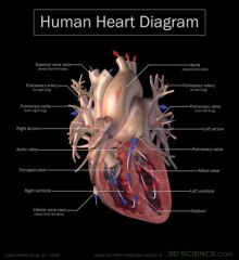
De-O2 blood enters the right atrium -thru tricuspid valve-->right ventricle-thru pulmonary semi-lunar valve into pulmonary arteries (one for each lung)-->lungs-thru pulmonary veins with O2 rich blood--> left atrium -thru bicuspid/mitrial valve-->left ventricle-thru aortic semi lunar valve into aorta-->body-deO2 blood and C02 rich blood pool in venous system which consists of two major veins: the superior vena cava (roughly speaking from areas above the heart) and the inferior vena cava (roughly speaking from areas below the heart-->empty into the right atrium
|
None
|
|
|
Describe the structural and functional differences among the vessels of the circulatory system.
|
artery:muscular blood vessels that carry blood away from the heart; with the exception of the pulmonary and umbilical arteries, carry oxygenated blood; outermost layer is known as the tunica externa
veins:carries blood toward the heart;majority of veins in the body carry low-oxygen blood from the tissues back to the heart; the exceptions being the pulmonary and umbilical veins which both carry oxygenated blood;Veins are essentially hollow tubes that collapse when not filled with blood |
None
|
|
|
Describe the fetal circulatory system.
|
Blood from the placenta is carried to the fetus by the umbilical vein. About half of this enters the fetal ductus venosus and is carried to the inferior vena cava, while the other half enters the liver proper from the inferior border of the liver. The branch of the umbilical vein that supplies the right lobe of the liver first joins with the portal vein. The blood then moves to the right atrium of the heart. In the fetus, there is an opening between the right and left atrium (the foramen ovale), and most of the blood flows from the right into the left atrium, thus bypassing pulmonary circulation. The majority of blood flow is into the left ventricle from where it is pumped through the aorta into the body. Some of the blood moves from the aorta through the internal iliac arteries to the umbilical arteries, and re-enters the placenta, where carbon dioxide and other waste products from the fetus are taken up and enter the woman's circulation.[25]
Some of the blood from the right atrium does not enter the left atrium, but enters the right ventricle and is pumped into the pulmonary artery. In the fetus, there is a special connection between the pulmonary artery and the aorta, called the ductus arteriosus, which directs most of this blood away from the lungs (which aren't being used for respiration at this point as the fetus is suspended in amniotic fluid). |
None
|
|
|
Describe the process of mitosis.
|
process of nuclear division in a living cell by which chromosomes are exactly replicated and the two copies distributed to identical daughter nuclei
1.interphase: chromosomes are dispersed in the nucleus and appear as a network of long, thin threads or filaments, called the chromatin; chromosomes replicate themselves to form pairs of identical sister chromosomes; DNA is synthesized Actual mitosis: 2.prophase: chromatid contract and coil but remain attached to one another at centromere; nucleolus disappear; spindle begins to form; centrioles separate and move apart and radiating bundles of fibers, appear around them;some sets of fiber run from one centriole to the other--these are the spindle fibers 3.metaphase:chromosomes congregate at midway btwn two ends to which the spindle tapers--where the whole cell will divide when nuclear division is completed; the ends of the spindle are the poles to which the chromatids will migrate. The chromatids are attached to the spindle fibers at the centromeres 4.anaphase: the two chromatids of each chromosome separate and move to opposite poles, as if pulled along the spindle fibers by the centromeres 5.telephase: new nuclear envelopes form around the two groups of daughter chromosomes, the new nucleoli begin to appear, and the spindle fibers disappear. The chromosomes uncoil to assume their dispersed distribution within the interphased nucleus. Cytokinesis, which may begin before or after mitosis is completed, finally separates the daughter nuclei into two new individual daughter cells. |
None
|
|
|
Describe the process of meiosis.
|
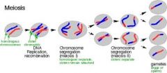
process of nuclear division in a living cell by which the number of chromosomes is reduced to half the original number;occurs only in the process of gametogenesis--when the gametes, or sex cells (ovum and sperm), are being formed.
just before meiosis each chromosome replicates to form two identical copies in the form of strands called chromatids joined together at a point called the centromere. In the first stage of meiosis, called the reduction division, the members of each pair of homologous chromosomes lie side by side and crossing over occurs. Each member of the pair then moves away from the other toward opposite ends of the dividing cell, and two nuclei, each with the haploid number of double-stringed chromosomes, are formed. Thus at the beginning of the second meiotic sequence, called the equational division, each cell nucleus contains one chromosome from each homologous pair and each chromosome is of two strands that are identical (except where crossing over has occurred). Then the chromosomes separate into their single strands which move toward opposite ends of the dividing nucleus. The result of meiotic division is four cells, each haploid, with one chromosome of each pair. |
None
|
|
|
Describe transcription.
|
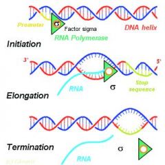
the process through which a DNA sequence is enzymatically copied by an RNA polymerase to produce a complementary RNA. i.e.-- it is the transfer of genetic information from DNA into RNA; the stretch of DNA that is transcribed into an RNA molecule is called a transcription unit; as in DNA replication, transcription proceeds in the 5' → 3' direction (i.e. the old polymer is read in the 3' → 5' direction and the new, complementary fragments are generated in the 5' → 3' direction). Transcription is divided into 3 stages: initiation: begins with the binding of RNA polymerase to the promoter in DNA
elongation:mRNA transcription can involve multiple RNA polymerases, so many mRNA molecules can be produced from a single copy of the gene. This step also involves a proofreading mechanism that can replace an incorrectly added RNA molecule termination:involves cleavage of the nascent transcript, followed by template-independent addition of As at its new 3' end, in a process called Poly-adenylation |
None
|
|
|
Describe translation.
|
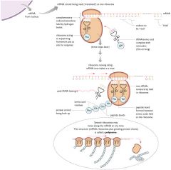
occurs in the cytoplasm where the ribosomes are located; Ribosomes are made of a small and large subunit which surrounds the mRNA. In translation, messenger RNA (mRNA) is decoded to produce a specific polypeptide according to the rules specified by the genetic code; process that converts an mRNA sequence into a chain of amino acids that form a protein; translation proceeds in four phases: activation: the correct amino acid (AA) is joined to the correct transfer RNA (tRNA)
initiation:involves the small subunit of the ribosome binding to 5' end of mRNA with the help of initiation factors (IF), other proteins that assist the process elongation:occurs when the next aminoacyl-tRNA (charged tRNA) in line binds to the ribosome along with GTP and an elongation factor termination:happens when the A site of the ribosome faces a stop (nonsense) codon (UAA, UAG, or UGA). When this happens, no tRNA can recognize it, but releasing factor can recognize nonsense codons and causes the release of the polypeptide chain. |
None
|
|

List organelles present in cells and describe their function.
|
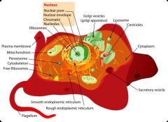
cytoskeleton: maintains cell shape,protect the cell, enables some cell motion (using structures such as flagella and cilia), and plays important roles in both intra-cellular transport (the movement of vesicles and organelles) and cellular division
nucleus: enclosed by a double membrane (commonly referred to as a nuclear envelope), with pores; contains most of the cell's genetic material; main fx: control gene expression and mediate the replication of DNA during the cell cycle Endoplasmic reticulum: facilitation of protein folding and the transport of synthesized proteins in sacs called cisternae; rough ER-ribosomes only bind to the ER once it begins to synthesize a protein destined for sorting; smooth ER-synthesis of lipids, metabolism of carbohydrates and calcium concentration, drug detoxification, and attachment of receptors on cell membrane proteins; known for its storage of calcium ions in muscle cells Golgi bodies: integral in modifying, sorting, and packaging macromolecules for cell secretion (exocytosis) or for use within the cell. It primarily modifies proteins delivered from the rough endoplasmic reticulum, but is also involved in the transport of lipids around the cell, and the creation of lysosomes; it packages and labels "items" and then sends them to different parts of the cell. Vesicles may be specialized for various purposes. mitochondria: double membranes ; where aerobic respiration takes place and production of ATP; contain their own DNA and are only formed by the fission of other mitochondria |
None
|
|
|
What is the distinguishing factor between different amino acids?
|
R-group
|
None
|
|
|
What is the nasal septum?
|
separates the left and right airways in the nose
|
None
|
|
|
What is the longest cervical vertebra and another name for it?
|
second cervical vertebra (C2); also called the axis
|
None
|
|
|
What are functions of bone?
|
1.points of attachment for muscles
2.levers for muscle action 3.strengthen your body against injury 4.protect your internal organs 5.metabolic functions |
|
|
|
What are the 3 types of joints based on joint structure and their functions?
|
1.fibrous: bones are connected to each other by fibrous tissue; connect bones of the skull to each other at wavy junctures know as sutures; most joints have little motion and, in other words, are synarthoses
2.cartilaginous: united by cartilage; may be synarthroses or amphiarthroses depending on amount of mvmt allowed; eg.growth plate in a long bone and pelvic symphysis (btwn the two halves of pelvic lies a plate of fibrocartilage that acts as a shock absorber and allows for a small amount of mvmt during childbirth. 3.synovial: most common type; the articulating bones are separated by a fluid-filled space called a joint cavity; found btwn most of the long bones in the body and have the greatest flexibility; their diarthroses; has articular cartilage (spongy consistency that helps absorb shock and its glassy smooth surface helps reduce friction on joint surface) |
|