Reading...
![]()
Play button
![]()
Play button
![]()
Use LEFT and RIGHT arrow keys to navigate between flashcards;
Use UP and DOWN arrow keys to flip the card;
H to show hint;
A reads text to speech;
206 Cards in this Set
- Front
- Back
- 3rd side (hint)
|
Humeroulnar joint: Radius articulates with _____ of the humerus, radial head fits into the _____; Ulna articulates with _____ of the humerus, olecranon fits into the ____, coronoid process fits into the _____.
|
Capitulum; radial fossa; trochlea; olecranon fossa: coronoid fossa.
|
|
|
|
Radius pivots about the _____ on the ulna during _____ and _____.
|
Radial notch; supination; pronation.
|
|
|
|
The radius/ulna articulates with carpal bones.
|
Radius.
|
|
|
|
Which styloid process is smaller, radius/ulna? Hence, which is more possible, adduction or abduction of the hand?
|
Ulnar styloid process is smaller. Adduction is more possible
|
|
|
|
_____ membrane runs between radius and ulna. It runs _____ and _____. What is the purpose of this membrane?
|
Interosseous membrane; obliquely and inferiorly. To transmit forces received from the hands by radius to ulna.
|
|
|
|
Transmission of force in the case of a fall (on outstretched hands).
|
Metacarpals - carpals - radius - interosseous membrane - ulna - humerus - scapula - clavicle -thorax.
|
|
|
|
Which bone in the hand has a hook?
|
Hamate.
|
|
|
|
Pisiform is a _____ bone in the tendon of _____.
|
Sesamoid; flexor carpi ulnaris.
|
|
|
|
Bones of hand:
|
Phalanges; metacarpals; carpal bones.
|
|
|
|
List the carpal bones:
|
(Some Lunatics Try Positions That They Cannot Handle)
1st row (radius to ulna): scaphoid; lunate; triquetrium; pisiform. 2nd row (radius to ulna): trapezium; trapezoid; capitate; hamate. |
|
|
|
Most commonly fractured carpal bone? What may happen as a result?
|
Scaphoid - avascular necrosis of the proximal fragment may result.
|
|
|
|
Fall on outstretched hand may cause dislocation of the _____. Which nerve most likely to be affected?
|
Lunate; median nerve.
|
|
|
|
Glenohumeral joint: ______ cavity of the scapula, which is deepened by ______.
|
Glenoid cavity; glenoid labrum.
|
|
|
|
Articular surfaces of glenohumeral joint covered by _____ cartilage.
|
Hyaline.
|
|
|
|
Glenohumeral joint type?
|
Synovial ball and socket joint.
|
|
|
|
What produces viscous fluid to lubricate synovial joint?
|
Synovial membrane.
|
|
|
|
Glenohumeral joint: Synovial membrane attaches to which structure?
|
Margins of articular surfaces.
|
|
|
|
Glenohumeral joint: Fibrous capsule attached to the margin of the _____ on the scapula, and the _____ of the humerus.
|
Glenoid fossa; anatomical neck.
|
|
|
|
Glenohumeral joint is strengthened by:
|
Rotator cuff muscles.
|
|
|
|
Ligaments in glenohumeral joint?
|
Glenohumeral (medial, inferior, superior); coracohumeral; coracoacromial; transverse humeral.
|
4 ligaments.
|
|
|
Attachments of glenohumeral ligament:
|
Glenoid labrum at supraglenoid tubercle - anatomical neck of humerus.
|
|
|
|
Attachments of coracohumeral ligament:
|
Coracoid process - anterior greater tubercle of humerus.
|
|
|
|
Attachments of coracoacromial ligament:
|
Coracoid process - acromion.
|
|
|
|
Attachments of transverse humeral ligament:
|
Greater tubercle - lesser tubercle.
|
|
|
|
Which ligament in the glenohumeral joint holds the biceps tendon?
|
Transverse humeral ligament.
|
Attachment is between greater and lesser tubercle.
|
|
|
Which ligament in the glenohumeral joint prevents superior displacement?
|
Coracoacromial ligament.
|
Attachment is between coracoid process and acromion.
|
|
|
Blood supply of glenohumeral joint:
|
Anterior and posterior circumflex arteries, branches of suprascapular artery.
|
|
|
|
Nerve supply of glenohumeral joint:
|
Suprascapular, axillary.
|
|
|
|
Glenohumeral joint is weakest _____, because _____.
|
Inferiorly, rotator cuff is deficient.
|
|
|
|
Glenohumeral joint is prone to dislocation when _____.
|
Abducted.
|
|
|
|
Frozen shoulder (adhesive capsulitis) means?
|
Adhesive fibrosis and scarring of the joint capsule, rotator cuff, and deltoid, resulting in difficulty in abducting arm.
|
|
|
|
The _____ tendon is particularly vulnerable to calcification due to _____?
|
Supraspinatus; poor blood supply.
|
|
|
|
Humeroulnar (elbow joint): between _____ of humerus and _____ of ulnar, _____ of humerus and _____ of radius.
|
Trochlea, trochlear notch; capitulum, head.
|
|
|
|
Which structure of the ulna prevents hyperextension?
|
Olecranon.
|
|
|
|
Proximal radioulnar joint: between _____ of radius and _____ of ulna.
|
Head; radial notch.
|
|
|
|
Humeroulnar joint type:
|
Synovial hinge joint.
|
|
|
|
Proximal radioulnar joint type:
|
Synovial pivot joint.
|
|
|
|
Ligaments of humeroulnar joint:
|
Radial collateral ligament; ulnar collateral ligament; anular ligament of radius.
|
3 ligaments.
|
|
|
Attachments of radial collateral ligament:
|
Lateral epicondyle - annular ligament.
|
|
|
|
Attachments of ulnar collateral ligament:
|
Medial epicondyle - coronoid process, olecranon.
|
|
|
|
Attachments of anular ligament of radius:
|
Encircles head of radius - coronoid process.
|
|
|
|
Blood supply of humeroulnar joint:
|
Anastomosis around elbow joint derived from brachial, radial and ulnar arteries.
|
Anastomosis around elbow joint derived from _____.
|
|
|
Fracture of radial head is common with a fall on the outstretched hand. Radiologically, this fracture may not be visible, but what other signs may suggest such a fracture?
|
Area of lucency - fluid filling the synovial cavity may elevate the fatty pads, showing a "fat pad sign".
|
|
|
|
Overuse strain on the common flexor and extensor origins at the epicondyles may result in:
|
Epicondylitis.
|
|
|
|
Articulation of the radiocarpal (wrist) joint proximally and distally:
|
Proximally: radius and articular disc of distal radioulnar joint.
Distally: proximal carpal bones EXCEPT PISIFORM. |
|
|
|
Radiocarpal joint type:
|
Synovial ellipsoid/condyloid joint.
|
|
|
|
Difference between synovial membrane and fibrous capsule of wrist joint:
|
Synovial membrane does not communicate with distal radioulnar joint or intercarpal joints; fibrous capsule surrounds wrist joint and attached to distal ends of radius, ulna and proximal carpal joints EXCEPT pisiform.
|
|
|
|
Ligaments of radiocarpal joint:
|
Palmar ligament; dorsal ligament; ulnar collateral ligament; radial collateral ligament.
|
4 ligaments.
|
|
|
Sternoclavicular joint type:
|
Saddle.
|
|
|
|
Acromioclavicular joint type:
|
Plane.
|
|
|
|
Distal radioulnar joint type:
|
Pivot.
|
|
|
|
Intercarpal joint type:
|
Plane.
|
|
|
|
Carpometacarpal joint type:
|
2-5: plane; 1: Saddle.
|
|
|
|
Why is carpometacarpal joint 1 different from the rest of the carpometacarpal joints?
|
Separate articular cavity, saddle joint instead of plane joint.
|
|
|
|
Metacarpophalangeal joint type:
|
Condyloid.
|
|
|
|
Interphalangeal joint type:
|
Hinge.
|
|
|
|
Axioappendicular (extrinsic shoulder) muscles:
|
Posteriorly: Trapezius, levator scapulae, rhomboid major, rhomboid minor, latissimus dorsi.
Anteriorly: Pectoralis major, pectoralis minor, subclavius, serratus anterior |
|
|
|
Nerve, attachments and actions of trapezius:
|
Nerve: Spinal accessory nerve (CN11).
Attachments: Medial 1/3 of superior nuchal line, external occipital protuberance, ligamentum nuchae, C7-T12 spinous processes - Lateral 1/3 of clavicle, scapula spine and acromion. Action (on scapula): Superior rotation, retraction, elevaton, depression. |
|
|
|
Nerve, attachments and actions of levator scapulae:
|
Nerve: cervical nerve, dorsal scapular nerve.
Attachments: C1-C4 Transverse process - Medial border of scapula superior to spine. Action (on scapula): Superior rotation, elevation. |
|
|
|
Nerve, attachments and actions of rhomboid major:
|
Nerve: Dorsal scapular nerve.
Attachments: C7-T1 spinous process - Medial border of scapula at root of spine. Action (on scapula): Retraction. |
|
|
|
Nerve, attachments and actions of rhomboid minor:
|
Nerve: Dorsal scapular nerve.
Attachments: T2-T5 spinous process - Medial border of scapular inferior to spine. Action (on scapula): Inferior rotation. |
|
|
|
Nerve, attachments and actions of latissimus dorsi:
|
Nerve: thoracodorsal nerve.
Attachments: T7-T12 spinous process, lower 3-4 ribs, iliac crest - intertubercular sulcus of humerus (on the anterior surface!). Action (on arm): Extension, Adduction, Medial rotation. |
|
|
|
Nerve, attachments and actions of pectoralis major:
|
Nerve: lateral and medial pectoral nerve.
Attachments: Anterior medial 1/2 of clavicle (clavicular head)/Sternum and costal cartilage 1-6 (sternocostal head) - Lateral lip of intertubercular sulcus on humerus. Action (on arm): Flexion (if extended), extension (if flexed), adduction, medial rotation. |
|
|
|
Nerve, attachments and actions of pectoralis minor:
|
Nerve: medial pectoral nerve.
Attachments: Ribs 3-5 - Corocoid process. Action (on scapula): Depression, retraction. |
|
|
|
Nerve, attachments and actions of subclavius:
|
Nerve: nerve to subclavius.
Attachments: 1st costal cartilage and rib - Inferior medial 1/3 of clavicle. Action (on clavicle): Depression. |
|
|
|
Nerve, attachments and actions of serratus anterior:
|
Nerve: long thoracic nerve.
Attachments: External surface of lateral part of ribs 1-8 - anterior medial border of scapula. Action (on scapula): Protraction, superior rotation. |
|
|
|
Winged scapula is due to:
|
Paralysis of serratus anterior due to injury to long thoracic nerve (vulnerable to injury as it is a supplies serratus anterior on the superior surface).
|
|
|
|
Scapulohumeral (intrinsic shoulder) muscles:
|
Deltoid; Rotator cuff muscles ( Subscapularis, Supraspinatus, Infraspinatus, Teres minor); Teres major.
|
|
|
|
Nerve, attachments and actions of deltoid:
|
Nerve: Axillary nerve.
Attachments: Lateral 1/3 of clavicle, spine and acromion of scapula - deltoid tuberosity on humerus. Action (on arm): Abduction, Flexion/Extension, Medial/Lateral Rotation (due to both anterior and posterior fibres). |
|
|
|
Muscles used in abduction of arm:
|
1st 15 deg by supraspinatus, rest by deltoid.
|
|
|
|
With a dysfunctional supraspinatus, what will be observed?
|
Tilting of body to passively abduct arm to 15 degree, before deltoid takes over.
|
|
|
|
Nerve, attachments and actions of subscapularis:
|
Nerve: Upper and lower subscapular nerve.
Attachments: Subscapular fossa - Lesser tubercle of humerus. Action (on arm): Medial rotation. |
|
|
|
Nerve, attachments and actions of supraspinatus:
|
Nerve: Suprascapular nerve.
Attachments: Supraspinous fossa - Superior facet of greater tubercle. Action (on arm): 1st 15 degree of abduction. |
|
|
|
Nerve, attachments and actions of infraspinatus:
|
Nerve: Suprascapular nerve.
Attachments: Infraspinous fossa - middle facet of greater tubercle (posteriorly). Action (on arm): Lateral rotation. |
|
|
|
Nerve, attachments and actions of teres minor:
|
Nerve: axillary nerve.
Attachments: Middle part of lateral border of scapula - inferior facet of greater tubercle. Action (on arm): Lateral rotation. |
|
|
|
Nerve, attachments and actions of teres major:
|
Nerve: Lower subscapular.
Attachments: Posterior surface of inferior angle of scapula - Medial lip of intertubercular sulcus of humerus (anterior surface). Action (on arm): Medial rotation, Adduction. |
|
|
|
At about ____ degree of abduction, the greater tuberosity of the humerus comes into contact with the lateral edge of the acromion. Any more elevation of there arm is accomplished by scapular rotation.
|
120.
|
|
|
|
Anterior arm muscles and nerve supply:
|
Biceps brachii; Brachialis; Coracobrachialis.
Musculocutaneous nerve. |
|
|
|
Nerve, attachments and actions of biceps brachii:
|
Nerve: musculocutaneous nerve.
Attachments: Supraglenoid tubercle (long head), coracoid process (short head) - Radial tuberosity. Action (on shoulder and humeroulnar joint): Flexion (only long head on shoulder). Action (on radioulnar joint): Supination. |
|
|
|
Nerve, attachments and actions of brachialis:
|
Nerve: musculocutaneous nerve.
Attachments: Anterior surface of humerus - Ulnar tuberosity. Action (on humeroulnar joint): Flexion. |
|
|
|
Nerve, attachments and actions of coracobrachialis:
|
Nerve: musculocutaneous nerve.
Attachments: Coracoid process - midshaft of humerus. Action (on shoulder joint): Flexion, Adduction. |
|
|
|
Posterior arm muscles and nerve supply:
|
Triceps brachii. Radial nerve.
|
|
|
|
Nerve, attachments and actions of triceps brachii:
|
Nerve: radial nerve.
Attachments: Infraglenoid tubercle (long head), superior to radial groove (lateral head), inferior to radial groove (medial head) - Olecranon. Action (on shoulder and humeroulnar joint): Extension |
|
|
|
Biceps tendon reflex is done by:
|
Tapping the tendon at cubital fossa.
|
|
|
|
Significance of biceps tendon reflex:
|
Integrity of the C5/6 spinal segments and the musculocutaneous nerve.
|
|
|
|
Triceps tendon reflex is done by:
|
Tapping the tendon at olecranon.
|
|
|
|
Significance of triceps tendon reflex:
|
Integrity of the C6-8 spinal segments and the radial nerve.
|
|
|
|
Clinical significance of long head of biceps brachii:
|
Enclosed in a synovial sheath that moves back and forth in the intertubercular sulcus - wear and tear can course shoulder pain and inflammation.
|
|
|
|
Forearm muscles:
|
Anterior: Superficial/intermediate layer (Flexor carpi ulnaris; Palmaris longus; Flexor carpi radialis; Pronator teres; Flexor digitorum superficialis); Deep layer (Flexor digitorum profundus; Flexor pollicis longus; Pronator quadratus)
Posterior: Superficial layer (Brachioradialis; Extensor carpi radii longus; Extensor carpi radii brevis; Extensor digitorum; Extensor digiti minimi; Extensor carpi ulnaris; Anconeus); Deep layer (Supinator; Abductor pollicis longus; Extensor pollicis longus; Extensor pollicis brevis; Extensor indicis). |
Anterior: Superficial/intermediate layer (5), Deep layer (3).
Posterior: Superficial (7), Deep layer (5). |
|
|
General innervation of anterior (flexor) compartment of forearm muscles:
|
All median nerve except FCU and medial half of FDP.
|
|
|
|
Nerve, attachments and actions of flexor carpi ulnaris:
|
Nerve: ulnar nerve.
Attachments: Olecranon/medial epicondyle - via pisohamate ligament (extension of FCU) to hamate hook and metacarpal 5. Action (on wrist): Flexion, Adduction. |
|
|
|
Nerve, attachments and actions of palmaris longus:
|
Nerve: median nerve.
Attachments: Medial epicondyle - palmar aponeurosis. Action (on wrist): Flexion. |
|
|
|
Nerve, attachments and actions of flexor carpi radialis:
|
Nerve: median nerve.
Attachments: Medial epicondyle - metacarpal 2. Action (on wrist): Flexion, Abduction. |
|
|
|
Nerve, attachments and actions of pronator teres:
|
Nerve: median nerve.
Attachments: Medial epicondyle - Lateral midshaft of radius. Action (on radioulnar joint): Pronation. |
|
|
|
Nerve, attachments and actions of flexor digitorum superficialis:
|
Nerve: median nerve.
Attachments: Medial epicondyle, coronoid process - Middle phalanges 2-5. Action (on fingers): Flexion, middle phalanges 2-5. |
|
|
|
Nerve, attachments and actions of flexor digitorum profundus:
|
Nerve: median nerve (lateral 1/2); ulnar nerve (medial 1/2).
Attachments: Ulna (proximal anterior surface and interosseous membrane) - Distal phalanges 2-5. Action (on fingers): Flexion, distal phalanges 2-5. |
|
|
|
Nerve, attachments and actions of flexor pollicis longus:
|
Nerve: median nerve.
Attachments: Radius (anterior surface of interosseous membrane) - Distal phalanx 1. Action (on distal phalanx 1): Flexion. |
|
|
|
Nerve, attachments and actions of pronator quadratus:
|
Nerve: median nerve.
Attachments: Distal anterior surface of radius and ulna. Action (on distal radioulnar joint): Pronation. |
|
|
|
General innervation of posterior (extensor) compartment of forearm muscles:
|
ALL Radial nerve.
|
|
|
|
Nerve, attachments and actions of brachioradialis:
|
Nerve: radial nerve.
Attachments: Lateral supra-epicondylar ridge - Lateral distal part of radius. Action (on radioulnar joint): Flexion in mid-prone position. |
|
|
|
Nerve, attachments and actions of extensor carpi radii longus:
|
Nerve: radial nerve.
Attachments: Lateral supra-epicondylar ridge - Metacarpal 2. Action (on wrist): Extension, Abduction. |
|
|
|
Nerve, attachments and actions of extensor carpi radii brevis:
|
Nerve: radial nerve.
Attachments: Lateral epicondyle - Metacarpal 3. Action (on wrist): Extension, Abduction. |
|
|
|
Nerve, attachment and actions of extensor digitorum:
|
Nerve: radial nerve.
Attachments: Lateral epicondyle - Extensor hood 2-5. Action (on fingers): Extension, 2-5. |
|
|
|
Nerve, attachment and actions of extensor digiti minimi:
|
Nerve: radial nerve.
Attachments: Lateral epicondyle - Extensor hood 5. Action (on fingers): Extension, 5 only. |
|
|
|
Nerve, attachment and actions of extensor carpi ulnaris:
|
Nerve: radial nerve.
Attachments: Lateral epicondyle - Metacarpal 5. Action (on wrist): Extension, Adduction. |
|
|
|
Nerve, attachment and actions of anconeus:
|
Nerve: radial nerve.
Attachments: Lateral epicondyle - Olecranon. Action (on radioulnar joint): Abducts ulna during pronation. |
|
|
|
Nerve, attachment and actions of supinator:
|
Nerve: radial nerve.
Attachments: Lateral epicondyle, supinator crest on ulna - Lateral surface of proximal 1/3 of radius. Action (on radioulnar joint): Supination. |
|
|
|
Nerve, attachment and actions of abductor pollicis longus:
|
Nerve: radial nerve.
Attachments: Ulna - Metacarpal 1. Action (on metacarpal 1): Abduction. |
|
|
|
Nerve, attachment and actions of extensor pollicis longus:
|
Nerve: radial nerve.
Attachments: Ulna - Distal phalanx 1. Action (on distal phalanx 1): Extension. |
|
|
|
Nerve, attachment and actions of extensor pollicis brevis:
|
Nerve: radial nerve.
Attachments: RADIUS - Proximal phalanx 1. Action (on proximal phalanx 1): Extension. |
|
|
|
Nerve, attachment and actions of extensor indicis:
|
Nerve: radial nerve.
Attachments: Ulna - Extension hood 2. Action (on fingers): Extension, 2 only. |
|
|
|
Which muscle is the most powerful supinator? When is it especially effective?
|
Biceps brachii; when the arm is flexed.
|
|
|
|
Combinations of which muscles result in the movement of the wrist?
|
Flexor/extensor carpi radialis/ulnaris.
|
|
|
|
Muscles of thenar eminence:
|
Abductor pollicis brevis; Flexor pollicis brevis; Opponens pollicis.
|
|
|
|
General innervation of thenar eminence:
|
Median nerve.
|
|
|
|
Origin of thenar eminence muscles:
|
Flexor retinaculum; scaphoid/trapezium tubercles.
|
|
|
|
Muscles of hypothenar eminence:
|
Abductor digiti minimi; Flexor digiti minimi brevis; Opponens digiti minimi.
|
|
|
|
General innervation of hypothenar eminence:
|
Ulnar nerve.
|
|
|
|
Origin of hypothenar eminence muscles:
|
Flexor retinaculum; hook of hamate OR pisiform.
|
|
|
|
Other intrinsic hand muscles:
|
Lumbricals; Dorsal interossei; Palmar interossei; Adductor pollicis.
|
|
|
|
General innervation of other intrinsic hand muscles:
|
Mostly ulnar nerve except lateral 1/2 of lumbricals.
|
|
|
|
Innervation of lumbricals:
|
Lateral 1/2: median nerve; Medial 1/2: ulnar nerve.
|
|
|
|
Attachments of lumbricals:
|
Tendon of flexor digitorum profundus - Extensor hood of 2-5.
|
|
|
|
Actions of dorsal and palmar interrosei.
|
Dorsal: abducts; Palmar: adducts.
|
DAB, PAD.
|
|
|
Describe the brachial plexus:
|
Somatic plexus formed from the anterior rami of C5-T1.
|
|
|
|
Branches of the brachial plexus:
|
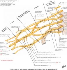
Roots - Trunks - Divisions - Cords - Terminal Branches (Musculocutaneous, Median, Radial, Ulnar).
|
|
|
|
Cause and presentation of waiter's tip position/Erb's palsy:
|
Injury to superior parts of the brachial plexus from an increase in angle between the neck and the shoulder.
Presents as medially rotated arm, extension of elbow and flexion of wrist. |
|
|
|
Law which states that nerves that supply the muscles acting on a joint also supplies the joint:
|
Hilton's law.
|
|
|
|
Origin of axillary nerve:
|
Posterior cord of brachial plexus.
|
|
|
|
Course of axillary nerve:
|
Enters quadrangular space with posterior circumflex humeral artery, winds round surgical neck of humerus deep to deltoid, gives rise to superior lateral cutaneous nerve of arm.
|
|
|
|
Motor supply of axillary nerve:
|
Deltoid; Teres minor.
|
|
|
|
Clinical importance of axillary nerve:
|
Important not to injure when giving intramuscular injections into the deltoid; injured by fracture at the surgical humeral neck.
|
|
|
|
Origin of musculocutaneous nerve:
|
Lateral cord of brachial plexus.
|
|
|
|
Course of musculocutaneous nerve:
|
Pierces coracobrachialis, descends between biceps brachii and brachialis, pierces deep fascia above elbow to continue as lateral cutaneous nerve of forearm.
|
|
|
|
Motor supply of musculocutaneous nerve:
|
Anterior arm flexors (BBC).
|
|
|
|
Sensory supply of hand:
|
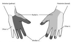
Median, radial and ulnar nerve.
|
|
|
|
Origin of radial nerve:
|
Posterior cord of brachial plexus.
|
|
|
|
Course of radial nerve:
|
Exits triangular interval (between long head of triceps, humeral shaft and inferior margin of teres major), run in radial groove (with profunda brachii artery), pierces inter-muscular septum, continues in anterior compartment of arm, passes anteriorly to lateral epicondyle, divide into deep and superficial branches.
|
|
|
|
Terminal branches of radial nerve:
|
Deep branch (motor): winds around radius, pierce supinator, continues as posterior interosseous nerve (running with posterior interosseous artery).
Superficial branch (cutaneous): winds lateral around radius, pierces supinator, continues to hand, passing anatomical snuffbox. |
|
|
|
Motor supply of radial nerve:
|
All posterior arm and forearm muscles.
|
|
|
|
Possible injury sites of radial nerve:
|
Axilla, humeral shaft, neck of radius.
|
|
|
|
Fracture at midshaft (radial groove) will paralyse:
|
Wrist down. Triceps (movement at the elbow) not affected as innervation is received higher up.
|
|
|
|
Wrist drop:
|
Loss of ability to extend wrist and fingers, unable to flex strongly for power grip.
|
|
|
|
Origin of ulnar nerve:
|
Medial cord of the brachial plexus.
|
|
|
|
Course of ulnar nerve:
|
Runs medial to axillary artery, in middle of arm, penetrates intermuscular septum and enters posterior compartment, passes elbow posterior to medial epicondyle, enters anterior compartment of forearm by passing between FCU heads, descends between FCU, medial to ulnar artery, at wrist pass between tendon of FCU and FDP, enters hand superior to flexor retinaculum in Guyon's canal (between pisiform and hook of hamate).
|
|
|
|
Motor supply of ulnar nerve:
|
Forearm: FCU and medial 1/2 of FDP; Intrinsic hand muscles: all except thenar eminence and lateral 1/2 of lumbricals.
|
|
|
|
Possible injury site of ulnar nerve:
|
Posterior to medial epicondyle, wrist.
|
|
|
|
Describe the ulnar paradox:
|
Higher the lesion, the less obvious the clawing deformity. In wrist lesions, FDP is not paralysed, causing flexion of the terminal phalanges. In higher lesions, FDP is paralysed, decreasing flexion of terminal phalanges, making the claw less obvious.
|
|
|
|
Describe the ulnar claw:
|
Atrophy of interossei and lumbricals on phalanges 4-5 but unopposed action of extensors and FDP.
Hyperextension of 4-5 digit at metacarpophalangeal joints and hyper flexion at interphalangeal joints. |
|
|
|
Guyon/Ulnar canal syndrome:
|
Compression of ulnar nerve at Guyon's canal, leading to weakness of hand muscles (except thenar muscles and lateral 1/2 of lumbricals).
Loss of sensation of medial 1.5 fingers. |
|
|
|
Froment's sign is due to the paralysis of:
|
Adductor pollicis.
|
|
|
|
Origin of median nerve:
|
Median root (from medial cord) and lateral root (from lateral cord) of brachial plexus.
|
|
|
|
Course of median nerve:
|
Runs lateral to brachial artery before crossing over to medial side (no motor supply to arm), run through cubital fossa below bicipital aponeurosis, medial to brachial artery, enter forearm between heads of pronator teres, continues between FDS and FDP, enters wrist behind palmaris longus tendon, passes through carpal tunnel deep to flexor retinaculum.
|
|
|
|
Motor supply of median nerve:
|
All anterior forearm muscles except FCU and medial 1/2 of FDP; thenar muscles, lumbricals 1-2.
|
|
|
|
Possible sites of injury of median nerve:
|
Elbow (supracondylar); wrist (carpal tunnel).
|
|
|
|
Presentation of median nerve injury at supracondylar fracture:
|
Benediction sign: loss of flexion at interphalangeal joints 2-3, loss of flexion of wrist, loss of pronation of forearm.
|
|
|
|
Presentation of median nerve injury at carpal tunnel:
|
Ape-like hand: loss of function of thenar muscles and 1/2 lumbricals.
Loss of sensation of lateral 3.5 digits. |
|
|
|
How to distinguish a lesion of nerve root T1 from lesion of ulnar nerve:
|
Lesion of T1 motor root lesion results in claw hand without sensory loss.
|
|
|
|
Myotomes of upper limb:
|
Shoulder C5, Flexors of forearm C5-6, Extensors of forearm C6-8 (triceps: C7-8), Forearm muscles C7-8, Intrinsic muscles of hand T1.
|
|
|
|
Axillary artery is the continuation of _____ artery at the _____.
|
Subclavian; lateral border of 1st rib.
|
|
|
|
2nd part of axillary artery is deep to which muscle?
|
Pectoralis minor.
|
|
|
|
3rd part of axillary artery terminates at which muscle?
|
Teres major (inferior border).
|
|
|
|
Branches of 1st part of axillary artery:
|
Superior thoracic artery.
|
|
|
|
Branches of 2nd part of axillary artery:
|
Thoraco-acromial artery, lateral thoracic artery.
|
|
|
|
Branches of 3rd part of axillary artery:
|
Subscapular artery, posterior circumflex humeral artery, anterior circumflex humeral artery.
|
|
|
|
Scapular anastomoses between subclavian and axillary artery:
|
Subclavian to 3rd part of axillary artery. To bypass 2nd part of axillary artery to ensure adequate blood flow regardless of position of the arm.
|
|
|
|
Anastomosis is deficient (in the arm) between:
|
Subscapular and profunda brachii artery.
|
|
|
|
Origin of the brachial artery:
|
Continuation of axillary artery beyond the inferior border of teres major.
|
|
|
|
Course of the brachial artery:
|
Medial side of proximal arm, courses with the median nerve and the brachial vein, enters cubita fossa, divides into ulnar and radial arteries.
|
|
|
|
Significant branches of the brachial artery:
|
Profunda brachii artery, accompanies radial nerve in radial groove.
|
|
|
|
Significance of brachial artery:
|
Brachial pulse can be felt on the median arm between biceps and triceps - compression of the brachial artery can be used to measure blood pressure.
|
|
|
|
Course of radial artery:
|
Arises in the cubital fossa, runs deep to (anterior compartment) brachioradialis, lateral to flexor carpi radialis, winds around radius laterally and crosses floor of anatomic snuffbox.
|
|
|
|
Course of ulnar artery:
|
Arises in the cubital fossa, runs in posterior forearm, resting on deep muscles lateral to the ulnar nerve, becomes superficial in the distal forearm, runs between FCU and FDS superior to flexor retinaculum, passes through Guyon's canal between pisiform and hook of hamate.
|
|
|
|
Termination of radial and ulnar arteries form:
|
Superficial (ulna) and deep (radial) palmar arches which then give branches to fingers.
|
|
|
|
Anterior and posterior interrosseus arteries are branches of the radial/ulnar artery?
|
Ulnar.
|
|
|
|
The radial pulse can be felt:
|
Lateral to FCR.
|
|
|
|
The ulnar pulse can be felt:
|
Lateral to FCU.
|
|
|
|
Cephalic and basilic vein originate from the:
|
Dorsal venous network of hand.
|
|
|
|
Course of the cephalic vein:
|
Winds around the lateral border of the forearm, ascends in the deltopectoral groove (between the deltoid and the pectoralis major), pierces deep fascia to drain in the axillary vein in the deltopectoral triangle.
|
|
|
|
Course of the basilic vein:
|
Winds around the median border of the forearm, ascend the medial side of biceps, pierces deep fascia to join venae comitantes of the brachial artery, which forms the axillary vein at the inferior border of teres major.
|
|
|
|
Communicating vein between cephalic and basilic vein is called:
|
Median cubital vein.
|
|
|
|
Median cubital vein is a branch of the:
|
Cephalic vein.
|
|
|
|
Clinical significance of median cubital vein:
|
For drawing blood, IV injections, and blood transfusions.
|
|
|
|
The median cubital vein lies on which structure and why is this important?
|
Bicipital aponeurosis. Care has to be taken not to puncture the bicipital aponeurosis else the brachial artery and the median nerve may be injured.
|
|
|
|
Cephalic and basilic veins are _____ veins.
|
Superficial.
|
|
|
|
Deep veins that accompany major arteries are are paired _____.
|
Venae comitantes.
|
|
|
|
Breasts extend from:
|
Horizontally: Lateral border of sternum to midaxillary line.
Vertically: 2nd to 6th rib. |
|
|
|
What structures are underlying the breasts?
|
Retromammary space, then pectoral fascia (deep).
|
|
|
|
What is the name of the part of the breast that extends upwards and laterally into the axilla?
|
Axillary tail.
|
|
|
|
Passage of milk:
|
Lobe - lobules of mammary glands - lactiferous ducts.
|
|
|
|
What is the name and function of dilated lactiferous ducts?
|
Lactiferous sinus, stores milk before secretion.
|
|
|
|
Ligaments that separate the lobules and attach the mammary glands to the dermis are called:
|
Cooper's ligaments.
|
|
|
|
Mammary glands are:
|
Modified sweat glands.
|
|
|
|
Breasts enlarge during pregnancy and menstruation due to:
|
Branching of lactiferous ducts.
|
|
|
|
What pathology can occur to Cooper's ligaments?
|
Peu de orange: puffy skin between dimpled pores due to cancerous invasion and fibrosis, causing shortening and pulling of Cooper's ligaments.
|
|
|
|
Flow summary/lymphogenic metastasis of lymph from breast (axillary lymph nodes):
|
Anterior (pectoral), posterior (subscapular), lateral (humeral) lymph nodes - Central nodes - Apical nodes - Subclavian lymphatic trunk - Right lymphatic duct/Thoracic duct - Junction of jugular and subclavian vein.
|
|
|
|
Lymph from medial part of breast is also drained by:
|
Parasternal lymph nodes.
|
|
|
|
Cancer from the breast can also metastasise to the:
|
Vertebrae or brain.
|
|
|
|
Structures located in the axilla:
|
Axillary artery, vein and brachial plexus nerves.
|
|
|
|
Structures located in the cubita fossa:
|
Tendon of biceps brachii, Brachial artery, Median nerve (deep to bicipital aponeurosis).
Superior to bicipital aponeurosis: median cubital vein. |
TAN.
|
|
|
Structures located in the carpal tunnel:
|
Tendons of a few flexors (FDS, FDP; FPL), Median nerve.
|
|
|
|
Structures located in Guyon's canal:
|
Ulnar artery and nerve. Superficial to flexor retinaculum between pisiform and hook of hamate.
|
|
|
|
Muscles that constitute the anatomical snuffbox:
|
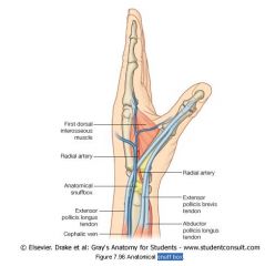
Extensor pollicis longus, Extensor pollicis brevis, Abductor pollicis longus.
|
|
|
|
Structures that pass the anatomical snuffbox:
|
Cephalic vein, radial artery and nerve.
|
|
|
|
What is the pulse felt in the anatomical snuffbox?
|
Radial pulse.
|
|
|
|
Long thoracic nerve from which spinal segment?
|
C5-C7.
|
|