Reading...
![]()
Play button
![]()
Play button
![]()
Use LEFT and RIGHT arrow keys to navigate between flashcards;
Use UP and DOWN arrow keys to flip the card;
H to show hint;
A reads text to speech;
33 Cards in this Set
- Front
- Back
|
A 14-year-old boy has a straddle injury to the perineum. Physical examination reveals ecchymosis limited to the penis and scrotum. The fascia that contains the extravasated blood is?
|
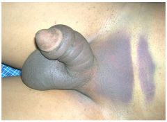
Colle's fascia. The corporal bodies are surrounded by tunica albuginea. Buck's fascia surrounds the corpora cavernosa and corpus spongiosum. If the injury to the urethra is contained within Buck's fascia the hematoma will remain confined to the shaft of the penis. Rupture through Buck's fascia leads to extension of the hematoma into the scrotum. The classic presentation of a urethral injury contained within Colles' fascia is that of a butterfly hematoma of the perineum. Superiorly the extravasation can extend to the clavicles where Scarpa's fascia, the abdominal extension of Colles' fascia, attaches.
|
|
|
50. When we’re in cysto and mention that we’re at the level of the vessels while looking at the ureters, what vessels are we talking about?
|
We’re talking about the bifurcation of the common iliac vessels into the internal and external iliacs, which the ureter passes OVER.
|
|
|
Which of the pelvic bones is more commonly injured during trauma, and why does the Urologist care?
|
The pubis is more commonly injured during trauma as it is the thinnest of the pelvic bones. It can potentially cause injury to the vagina, urethra, and/or bladder. A fracture of all four pubic rami is known as a “straddle fracture”.
|
|
|
What is Colles fascia? With what is it continuous, and why is it important to the Urologist?
|
A superficial fascial layer of the perineum; superiorly continuous with Scarpa’s fascia of the anterior abdomen, anteriorly with the dartos muscle of the penis and scrotum, and laterally with the fascia lata of the thigh. It is fused to the ischiopubic rami laterally and to posterior edge of Urogential diaphragm/perineal membrane. Important because in case of injury/infection (e.g. Fournier’s), fascial attachments limit spread and result in a butterfly-shaped hematoma that can also track into the anterior abdomen to the clavicles and around the flank to the back
|
|
|
How do men and women differ with respect to the bladder neck?
|
MEN: radially oriented inner longitudinal fibers pass through the internal meatus to become continuous with the inner longitudinal layer of smooth muscle in the urethra. The middle layer of muscle also forms a circular preprostatic sphincter, responsible for continence at the level of the bladder neck (even if the striated sphincter is damaged)
WOMEN: the middle layer is not as robust and many question its existence entirely. Also, the female bladder neck (unlike men) possesses little adrenergic activity. 50% of continent women will have urine enter the proximal urethra during a cough. |
|
|
What is thought to cause vesicoureteral reflux?
|
Insufficient submucosal ureteral length and poor detrusor backing as the ureter tunnels through the bladder to the ureteral orifice.
|
|
|
What are the dimensions of a normal prostate?
|
A normal prostate weighs approx. 18 g. and measures 3 cm. in length, 4 cm. in width, and 2 cm. in depth.
|
|
|
Where are the nerves found on the prostate? At the apex, at what clock positions are the nerves located?
|
Posterolaterally. Located at the 5- and 7-o’clock positions.
|
|
|
What percentage of the prostate is composed of glandular elements and fibromuscular stroma?
|
70% glandular, 30% fibromuscular stroma
|
|
|
The prostatic urethra is closest to what surface of the prostate?
|
Anterior
A good way to think about this is how Dr. Teigland describes doing a TURP, as if you are working inside of a globe, remembering that you don't have to take as much tissue off on the 12'oclock dissection because you are closer to the capsule on that side. |
|
|
What are the zones of the prostate and what is the prevalence of prostate cancer at each of these zones?
|
Transition zone: surrounds urethra proximal to Ejacul Ducts; where BPH arises and 20% of prostate cancers originate
Central zone: surrounds Ejacul Ducts and projects under the bladder base; where 1 to 5% of prostate cancers originate Peripheral zone: constitutes the bulk of the apical, posterior, and lateral aspects of the prostate; where 70% of prostate cancers originate. Also the most common area affected by chronic prostatitis |
|
|
What is the main arterial supply of the prostate and how does it enter the prostate? Why is this important to know?
|
Inferior vesical artery which branches into the urethral and capsular arteries. The urethral arteries penetrate the prostatovesical junction posterolaterally and approach the bladder neck in 1-5 o’clock and 7-11 o’clock positions. During resection, the most significant bleeding is encountered at the bladder neck at the 4- and 8-o’clock positions.
|
|
|
. What is the lymphatic drainage of the prostate?
|
Obturator and internal iliac nodes
|
|
|
How does sympathetic and parasympathetic innervation travel to the prostate?
|
Through the cavernous nerves, following branches of the capsular arteries.
|
|
|
What surrounds the membranous urethra? What is its nerve supply?
|
The external (striated) urethral sphincter. Pudendal nerve as well as a branch of the sacral plexus that runs on the pelvic surface of the levator ani.
|
|
|
What is the anatomic pathway of the vas?
|
Runs posterior to vessels of the cord and through the inguinal canal. Emerges in the pelvis lateral to the inferior epigastric vessels. At internal ring, diverges from the testicular vessels and passes medial to all structures to reach base of prostate posteriorly.
|
|
|
1.)Does the seminal vesicle store sperm?
2.)What is the blood supply of the vas deferens and seminal vesicles? 3.)What is the lymphatic drainage? |
1.)No, but it contributes the largest portion of fluid to the ejaculate.
2.)Vesiculodeferential artery (branch of superior vesical artery). 3.)External and internal iliac nodes. |
|
|
Do the corpora cavernosa communicate and what encloses them?
|
Yes, they are separated by a septum that becomes pectiniform distally, so their vascular spaces freely communicate. They are enclosed by tunica albuginea (made of collagenous fibers primarily)
|
|
|
Where is the anterior urethra most dilated and most narrow?
|
It is most dilated in its bulbar and glanular segments (fossa navicularis) and most narrow at the external meatus
|
|
|
What are the branches of the common penile artery (from the internal pudendal artery) and where do they course?
|
-Bulbourethral artery enters the spongiosum from its posterolateral border. It supplies urethra, spongiosum and glans
--Cavernosal artery pierces the corporal body and supplies the cavernous sinuses. --Dorsal artery of the penis passes between the crus penis and pubis to supply the dorsal surface of the corporal bodies. **NOTE: The penile arteries are highly variable in their course. |
|
|
What contains the blood around the penis during a penile fracture?
|
Buck’s fascia.
|
|
|
What must be anesthetized during a penile block?
|
The dorsal nerve of the penis and the small branches from the perineal nerve supplying the ventrum of the penis near the urethra and as far as the glans. This is why we do a ring block and put anesthetic at the 10 and 2 o’clock positions superior to the penis.
|
|
|
List the layers of the testes and the spermatic cord and heir derivation from the abdominal wall.
|
-Skin
-Dartos à Scarpa’s fascia -External spermatic fascia à External oblique fascia -Cremasteric muscle/fascia à Internal oblique muscle -Internal spermatic fascia à Transversalis fascia -Parietal & visceral tunica vaginalis à Peritoneum -Tunica Albuginea |
|
|
What supplies the anterior wall of the scrotum?
|
External pudendal vessels & ilioinguinal and genitofemoral nerves
|
|
|
T/F: The anterior vessels and nerves cross the midline raphe of the scrotum.
|
False. That is why transverse or midline raphe scrotal incisions are most appropriate.
|
|
|
Where does the epididymis attach to the testicle?
|
Posterolateral at the upper pole, you will also have a lateral sulcus which tells you which is lateral and which is medial.
|
|
|
What are the branches of the testicular artery?
|
Internal artery, inferior artery and capital artery to the head of the epididymis. The level of these branching varies in up to 80% of patients
|
|
|
In performing a testicular biopsy, where is the most appropriate location?
|
Medial or lateral surface of the upper pole- risk of vascular injury is lowest in this location because the testicular arteries insert on the anterior, medial and lateral portions of the lower pole and anterior segment of the upper pole.
|
|
|
What are the two routes by which visceral innervation to the testis and epididymis travel?
|
--Renal and aortic plexus through the gonadal vessels
--Pelvic plexus in association with the vas deferens (some afferent/efferent nerves can cross midline) |
|
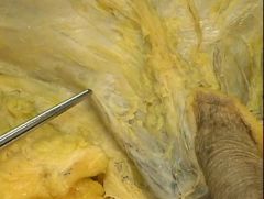
What is this a picture of?
|
The examiner is holding up the external spermatic fascia that is a continuation of the external abdominal oblique musculature. You can see it as it covers the cord there.
|
|
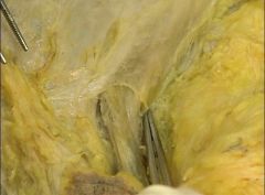
What is this a picture of?
|
This shows the external inguinal ring as the fibers cross and the cord enters from underneath.
|
|
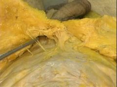
What is shown?
|
This is really a great view of the inguinal canal from above and how it exits at the superficial ring and you see how the exterrnal obliqu fascia sort of melds and continues as the external spermatic fascia that overlies the cord.
|
|
|
What does the inguinal canal carry?
|
Males: the spermatic cord, females: the round ligament. It carries the ilioinguinal nerve in both sexes.
|