Reading...
![]()
Play button
![]()
Play button
![]()
Use LEFT and RIGHT arrow keys to navigate between flashcards;
Use UP and DOWN arrow keys to flip the card;
H to show hint;
A reads text to speech;
76 Cards in this Set
- Front
- Back
|
The transverse/thoracic plane divides the mediastinum into the superior and inferior, what vertebrae level does this occur?
|
The T4/T5 level which is also equal with the sternal angle
|
|
|
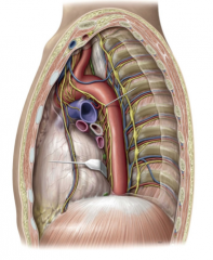
What are the components of the anterior mediastinum?
|
Thymus (in children)
Internal thoracic vessels Connective tissue, fat, lymph nodes |
|
|
What occurs to the thymus after childhood?
|

Fatty atrophy
|
|
|
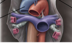
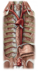
What are the contents of the superior mediastinum from anterior to posterior?
|
Thymus (in children)
SVC and brachiocephalic veins Aortic arch and branches Nerves (vagus and phrenic) Trachea Esophagus |
|
|
The vagus nerve travels lateral to which vessel?
|

The internal carotid artery
|
|
|
Which nerves travel posterior and anterior to the hilum of the lung?
|
The phrenic travels anterior and the vagus travels posterior.
|
|
|
Where is the azygous arch located?
|

At the T4 level just superior to the transverse thoracic plane.
|
|
|
What lymph drainage is located on the left side of the superior mediastinum?
|
The Thoracic duct
|
|
|
What vessels travel along the phrenic nerve?
|

The pericardiophrenic artery and vein (branches of the internal thoracic artery) These travel within the fibrous pericardium superficial to the parietal pericardium.
|
|
|
What three vessels branch from the aortic artery?
|
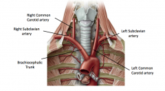
Brachiocephalic Trunk
Left Common Carotid Artery Left Subclavian Artery |
|
|
What are the two main branches of the brachiocephalic trunk?
|

The Right Common Carotid Artery and the Right Subclavian artery
|
|
|
What are the contents of the posterior mediastinum?
|
Thoracic Aorta
Nerves (Vagus and Phrenic Esophagus Azygous veins Thoracic Duct |
|
|
Describe the aortic arch.
|
Begins and ends within the transverse thoracic plane. Arches posteriorly and to the left over the pulmonary trunk to which it is attached through the ligamentum arteriosum (embryonic remnant ductus arteriosus). Starts from the ascending aorta and ends with the descending aorta.
|
|
|
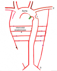
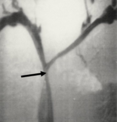
What is coarctation of the aorta?
|

A congenital condition where the aorta is constricted limiting the blood supply to the lower body. Commonly occurs distal to the ligamentum arteriosum.
|
|
|
What is rib notching?
|
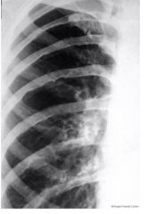
During coarctation of the aorta collateral blood flow increases to provide blood to the lower limbs including the anastamoses between the posterior and anterior intercostal arteries. The enlargement of these arteries can cause wear and tear to the ribs.
|
|
|
The smooth muscle in the ligamentum arteriosum can congenitally be present in the aortic arch-what is this condition called?
|
Coarctation of the aorta
|
|
|
What is an aortic aneurysm?
|
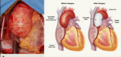
A life-threatening condition caused by a pathological dilation of the aorta. Eventually the walls of the vessel are weakened risking rupture
|
|
|
What is an aortic dissection (dissecting aneurysm)?
|
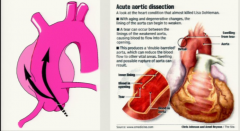
Condition in which the inner layer of the vessel wall develops a tear which allows blood to enter the middle layer of the vessel wall, casing the layers to separate.
|
|
|
What is the relation of the left primary bronchus and the aortic arch?
|
The aorta branches over the left primary bronchus
|
|
|
What are the branches of the superior vena cava? What are the branches of those branches?
|
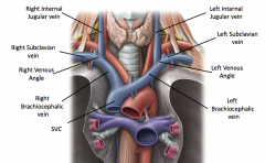
The superior vena cava is a combination of the left and right brachiocephalic veins. The brachiocephalic veins branch into the internal jugular and subclavian on both the right and left side of the body.
|
|
|
Where does the azygous vein drain?
|

Into the superior vena cava
|
|
|
What is SVC syndrome?
|
Restriction of the superior vena cava which restricts blood return to the heart which can cause dyspnea, swollen face, and coughing. Mostly caused from lung cancer.
|
|
|
What spinal roots are contained in the phrenic nerve?
|
C 3,4,5 keeps the diaphragm alive.
|
|
|
How does the phrenic nerve enter the thorax?
|
On the anterior surface of the anterior scalene muscle
|
|
|
What thoracic innervation does the phrenic nerve supply?
|

The motor and sensory innervation to the diaphragm and somatic sensory innervation to the mediastinal pleura and pericardium.
|
|
|
What nerve travels with the pericardiophrenic artery and nerve?
|

Phrenic nerve
|
|
|
Where does the vagus nerve travel through the neck?
|

Superficial to the carotid artery, but deep to the internal jugular vein.
|
|
|
Which nerve provides parasympathetic autonomic innervation to the thoracic and abdominal organs?
|

Vagus nerve-Cranial nerve X
|
|
|
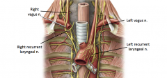
Where do the right and left vagus nerve branch near the aortic arch?
|

The left branches to form a loop under the aortic arch passing posterior to the ligamentum arteriosum now known as the left recurrent laryngeal nerve whereas the right loops under the right subclavian artery now known as the right recurrent laryngeal nerve. These both travel back to the larynx
|
|
|
What do the fibers of the left and right laryngeal nerves originate?
|

Both branches of the vagus nerve
|
|
|
What does the left vagus nerve become after the esophageal plexus?
|

The anterior vagal trunk
|
|
|
What does the right vagus nerve become after the esophageal plexus?
|

The posterior vagal trunk
|
|
|
Where do the anterior and posterior vagal trunks exit the thorax?
|

The esophageal hiatus of the diaphragm
|
|
|
What does sympathetic innervation do to the heart? What does parasympathetic innervation do to the heart?
|
Sympathetic: Increase HR and force of contraction
Parasympathetic: Decrease HR and force of contraction |
|
|
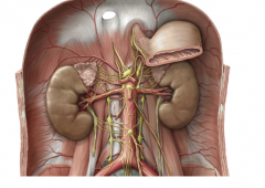
What is the sympathetic trunk?
|

A chain of sympathetic ganglia that lie parallel to the vertebral column
|
|
|
Which splanchnic nerves innervate the heart, lungs, and trachea?
|

T1-T4
|
|
|
How are the splanchnic nerves below T4 classified?
|

-Greater Splanchnic nerve T5-T9
-Lesser Splanchnic nerve T10-T11 -Least Splanchnic nerve T12 |
|
|
Where are the cell bodies of the sympathetic nervous system located?
|
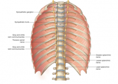
Preganglionic neurons are in the lateral horns of the spinal cord (T1-L2) and postganglionic neurons are located in sympathetic ganglia
|
|
|
Where are sympathetic ganglia?
|
Paravertebral within the sympathetic trunks and prevertebral will be in the abdominal cavity on branches of the abdominal aorta
|
|
|
Describe the location of the esophagus in the thoracic cavity
|
It descends into the thorax between the trachea and the vertebral column located posterior to the left atrium. It exits the thorax through the esophageal hiatus at T10.
|
|
|
What is the innervation and blood supply of the esophagus?
|

Innervation by the esophageal plexus supplied by the vagus nerve and it is supplied by branches of the thoracic aorta
|
|
|
What constitutes the upper third of the esophagus?
|

Skeletal muscle which gradually is replaced by smooth muscle
|
|
|
What are the three constrictions of the esophagus?
|
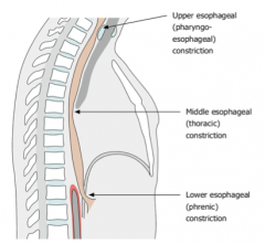
Upper constriction (cervical/pharyngoesophageal) at the larynx
Middle constriction (thoracic) at aortic arch and left main bronchus Lower (diaphragmatic/phrenic) at esophageal hiatus |
|
|
What is a hiatal hernia?
|
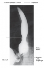
A protrusion through the stomach through the esophageal hiatus in the diaphragm. This can cause acid reflux and nausea and can be treated with smaller meals, control of acid secretion or surgery.
|
|
|
What is a TEE?
|
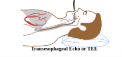
A transesophageal Echocardiogram where a transducer is lowered through the esophagus to ultrasound the heart. This is possible through the location of the esophagus posterior to the left atrium
|
|
|
What nerves exit with the esophagus through the esophageal hiatus?
|
The anterior and posterior vagal trunks-continuations of the vagus nerve
|
|
|
Where do the azygous veins originate?
|

They stem from ascending lumbar veins which unlike azygous veins drain into the IVC.
|
|
|
Where is the azygous vein located?
|

On the right side of the vertebral bodies against the posterior thoracic wall
|
|
|
What branches of the azygous vein are located on the left?
|
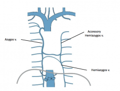
The Hemiazygous and the Accessory Hemiazygous veins which eventually drain into the azygous vein
|
|
|
Where does the azygous vein drain?
|
Into the SVC after it arches over the root of the lung at the T4 level
|
|
|
What drains the left intercostal veins?
|

The 4th -8th are drained by the accessory hemizygous vein and the 9th-11th are drained by the hemizygous vein
|
|
|
What is the thoracic duct?
|
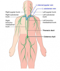
A lymphatic duct arising from the cisterna chyli in the abdomen where it ascends through the aortic hiatus and empties into the left venous angle between the LJV and the subclavian vein
|
|
|
What upper lymphatic trunks drain into the thoracic duct before it empties into the venous system?
|

The left brochomediastinal, subclavian, and jugular lymphatic ducts
|
|
|
What empties into the right venous angle?
|

The right lymphatic duct which receives lymph from the right bronchomediastinal, subclavian, and jugular trunks.
|
|
|
Where is the thoracic duct located in relation to the azygous vein?
|
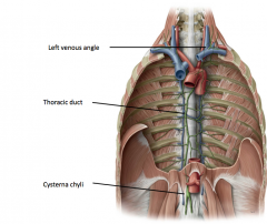
It is located between the azygous vein and the aorta.
|
|
|
What are the branches of the Descending aorta?
|
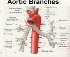
Intercostal arteries (3-11), subcostal artery (T12), bronchial arteries, and esophageal arteries
|
|
|
What type of autonomic innervation is supplied by the vagus nerve?
|
PARASYMPATHETIC!! Along with CN 3, 7, and 9 and Sacral levels 2, 3, and 4
|
|
|
What is the difference between white and grey rami communicans?
|
The white rami carry preganglionic fibers to the ganglia from the lateral horns of the spinal cord. The grey rami carry postganglionic fibers from the ganglia to the VPR. Only T1-L2 spinal nerves have both white and grey rami communicans (others only have grey)
|
|
|
What are the three options for sympathetic preganglionic axons?
|
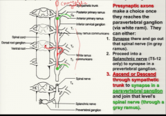
They can...
1) synapse in the paravertebral sympathetic ganglion and exit the same level 2) Ascend or descend through the sympathetic trunk and synapse at a paravertebral ganglion at another level exiting through a grey ramus at a higher or lower level 3) Proceed through the paravertebral ganglion to synapse at a prevertebral ganglion where it is called a splanchnic nerve (T5-T12) |
|
|
Do all spinal nerves have white and gray ramus communicans?
|
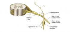
No all spinal nerves have gray communicans to connect them to the sympathetic chain, but only levels T1-L2 have white rami communicans
|
|
|
What nerves form which prevertebral ganglia?
|
Greater Splanchnic nerve-Celiac Ganglion
Lesser Splanchnic nerve-Superior Mesentric ganglion Least Splanchnic nerve-Aorticorenal ganglion |
|
|
What is the division between the thoracic and abdominal cavities?
|
The respiratory diaphragm
|
|
|
Where is the tendon located on the superior side of the respiratory diaphragm?
|

In the center surrounded by a muscular perimeter
|
|
|
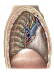
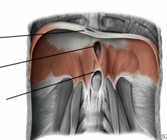
Where are the openings located in the respiratory diaphragm?
|

Caval opening-T8
Esophageal hiatus-T10 Aortic hiatus-T12 |
|

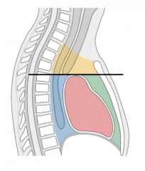
Name the structures highlighted in the image?
|
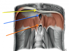
Caval opening (yellow)
Esophageal hiatus (blue) Aortic hiatus (orange) |
|
|
Which structures are found only in the superior mediastinum?
|
Thymus (in children)
SVC and brachiocephalic veins Trachea |
|
|
What structures are found only in the posterior mediastinum?
|
Azygous veins
|
|
|
Why is the left brachiocephalic vein longer?
|
It has to cross over the midline of the body to enter the SVC
|
|
|
What is a result of an aortic dissection?
|
A false lumen
|
|
|
What is the technical definition of an aneurysm?
|
When the diameter of a vessel is 1.5x its normal size
|
|
|
Where are aortic aneurysms most common?
|
The thoracic aorta
|
|
|
Where does the respiratory diaphragm attach inferiorly?
|
To the perimeter of the thoracic outlet
|
|
|
What exits through the caval opening?
|
The inferior vena cava
|
|
|
What drains into the azygous vein besides the posterior intercostal veins and the left vessels?
|
The ascending lumbar vein
|
|
|
Which sympathetic ganglia have white rami comunicans?
|
T1-L2
|
|
|
What levels do the splanchnic nerves stem from?
|
T5-T12
|