Reading...
![]()
Play button
![]()
Play button
![]()
Use LEFT and RIGHT arrow keys to navigate between flashcards;
Use UP and DOWN arrow keys to flip the card;
H to show hint;
A reads text to speech;
90 Cards in this Set
- Front
- Back
|
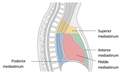
What are the boundaries of the mediastinum?
|
Anterior: Sternum
Posterior: Vertebral bodies Superior: Superior thoracic aperture Inferior: Diaphragm |
|
|
What divides the mediastinum into superior and inferior portions?
|

The transverse Thoracic plane (T4/T5 level) extension through the sternal angle
|
|
|
Which rib level is equal to the transverse thoracic plane
|

Rib 2
|
|
|
What are the divisions of the inferior mediastinum?
|

Posterior, Middle (heart), and anterior
|
|
|
What are the contents of the middle mediastinum?
|

The pericardium, heart, roots of the great vessels, and nerves and smaller vessels.
|
|
|
What are the great vessels?
|

The superior vena cava
Ascending Aorta Pulmonary Trunk |
|
|
What are the two parts of the pericardium?
|

The fibrous pericardium and the serous pericardium.
The serous pericardium is further subdivided into the parietal and visceral (epicardium) layers. |
|
|
What is the fibrous pericardium composed of?
|
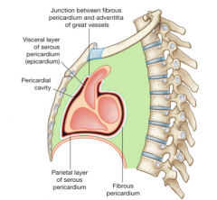
Dense fibrous connective tissue surrounding the entire heart and blends with the CT around the great vessels and attaches to the diaphragm inferiorly.
|
|
|
What is another name for the visceral layer of the pericardium?
|
Epicardium
|
|
|
What is the transverse pericardial sinus?
|

A reflection of the parietal pericardium which separates the outflow and inflow vessels.
|
|
|
What are the inflow and outflow vessels of the heart?
|

The inflow are the SVC, IVC, and pulmonary veins whereas the outflow channels are the aorta and the pulmonary trunk.
|
|
|
What two sinuses are formed by the reflections of parietal pericardium?
|

The transverse pericardial sinus and the oblique pericardial sinus. These are the areas where the visceral and parietal pericardia are continuous around the great vessels
|
|
|
What nerve innervates the pericardium?
|

The phrenic nerve (C3,4,5)
|
|
|
What supplies blood to the pericardium?
|
The pericardiacophrenic arteries and veins which are branches from the internal thoracic artery and vein (mammary artery and vein)
|
|
|
What is pericarditis?
|
Inflammation of the pericardium presenting as sharp pain posterior to the sternum and a friction rub upon auscultation. This can lead to pericardial effusion or cardiac tamponade
|
|
|
What is cardiac tamponade?
|
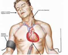
An acute accumulation of fluid or air in the pericardial cavity which compresses the heart, reduces venous return and cardiac output. This is a life-threatening condition.
|
|
|
What is the treatment for cardiac tamponade?
|

Pericardiocentesis-since the fibrous pericardium has little to no give any fluid build up is dangerous. This can be done posterior to the xiphoid process aiming at the left shoulder.
|
|
|
What are the layers of the heart wall?
|

Epicardium (visceral pericardium)
Myocardium Endocardium |
|
|
True or False: The myocardium is thicker in the atria?
|
False, the ventricles will have thicker muscle because they are responsible for pumping blood.
|
|
|
What composes the endocardium?
|
Endothelial cells
|
|
|
What are the chambers of the heart?
|

The Right Atrium->
The Right Ventricle-pulmonary veins-lungs-pulmonary arteries- The Left Atrium-> The Left Ventricle |
|
|
What are the two circuits of the circulatory system?
|
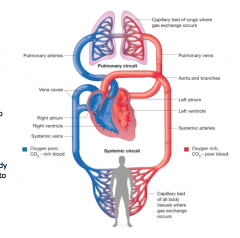
The pulmonary circuit which pumps blood to and from the lung to increase oxygen collection of Hb, and the systemic circuit which pumps blood to the rest of the body.
|
|
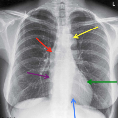
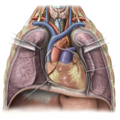
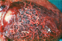
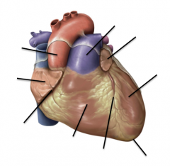
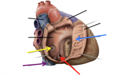
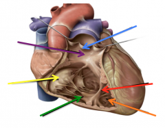
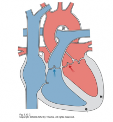
Name the regions highlighted in the image.
|

Aortic Arch "knob" (yellow)
SVC (red) Right Atrium (purple) Right Ventricle (blue) Levt Ventricle (green) |
|
|
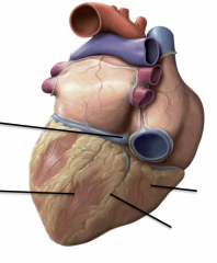
What are the borders of the heart on the surface anatomy?
|

Right Superior: 3rd Costal Cartilage at sternal margin
Right Inferior: 5th/6th costal cartilage at the sternal margin Left Superior: 2nd ICS at sternal margin Left inferior (apex) 5th ICS at mid clavicular line |
|
|
What are the surface anatomy location of the chambers?
|
The right atrium: 3-5th costal cartilages and sternal boarder
Right Ventricle: Deep to sternum and left 4-6th costal cartilages Left Ventricle: Left 3-5th ribs toward mid clavicular line |
|
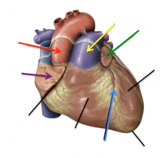
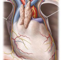
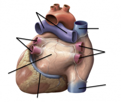
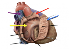
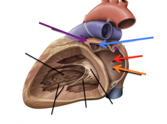
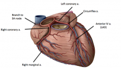
Name the highlighted regions in the image
|

Ascending Aorta (red)
Right Auricle (purple) Anterior Interventricular Sulcus (blue) Left Auricle (green) Pulmonary Trunk (yellow) |
|
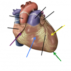
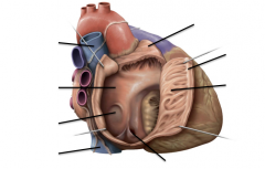
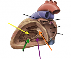
Name the highlighted regions in the image
|

Coronary Sulcus (purple)
Right Ventricle (blue) Apex (green) Left Ventricle (yellow) |
|
|
What is represented by the anterior interventricular sulcus?
|
The interventricular septum deep to this depression
|
|
|
What chambers are concurrent with the auricles?
|

The atria
|
|
|
Where is the coronary sulcus present?
|

It wraps around the heart posteriorly and is inferior to the right atrium, it does not cross the great vessels.
|
|
|
Why is the ascending aorta located to the right of the pulmonary trunk when seen from an anterior view?
|

Because it passes behind the pulmonary trunk and switches sides even though it is coming from the left ventricle
|
|
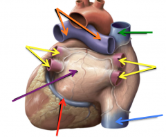
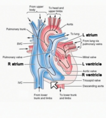
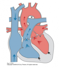
Name the highlighted regions in the image
|

Pulmonary arteries (orange)
Pulmonary veins (yellow) Left atrium (purple) Coronary Sinus (red) SVC (green) and IVC (blue) |
|
|
What does the coronary sulcus house?
|
The coronary sinus-the vein that sits in that depression
|
|
|
Why does the pulmonary artery bifurcate?
|

To supply both lungs
|
|

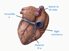
Name the highlighted regions in the image
|
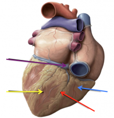
Left Ventricle (yellow)
Right Ventricle (blue) Posterior Interventricular Sulcus (red) Crux of the heart (purple) |
|
|
What is indicated by the crux of the heart?
|
The intersection of the four chambers of the heart where the coronary sinus empties into the right atrium and intersects with the posterior interventricular sulcus.
|
|

Name the highlighted regions in the image
|
Opening of the coronary sinus (red)
IVC (purple) Pectinate muscles (blue) Fossa Ovalis (yellow) |
|
Name the highlighted regions in the image
|

SVC (purple)
Interatrial septum (yellow) Right auricle (red) Crista terminalis (blue) |
|
|
Where are pectinate muscles located?
|
On the anterior wall of the right atrium
|
|
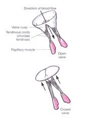
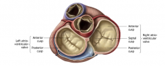
Name the highlighted regions in the image
|

Tricuspid Valve (yellow)
Papillary Muscles (green) Trabeculae Carneae (orange) Septomarginal Trabeculae (moderator band) (red) Pulmonary Valve (blue) Conus Arteriosus (purple) |
|
|
Where is the moderator band located?
|

In the right ventricle attached to the papillary muscles which helps coordinate the electric system of the heart with the valve contraction
|
|

Name the highlighted regions in the image
|
Left Auricle (purple)
Pectinate muscles (blue) Valve of the Foramen Ovale (red) Interatrial septum (orange) |
|
Name the highlighted regions in the image
|

Trabeculae Carnae (yellow)
Papillary Muscles (green) Chordae Tendinae (purple) Bicuspid/Mitral valve (orange) |
|
|
What are the active valves of the heart?
|
The atrioventricular valves including the tricuspid valve of the right ventricle and the bicuspid valve of the left ventricle. These valves need a papillary contraction to move
|
|
|
What are leaflets of the AV valves?
|

Anterior and Posterior (the right AV valve also has a septal leaflet)
|
|
|
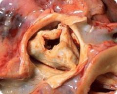
Where are the semilunar valves located?
|

One between the right ventricle to the pulmonary artery and another between the left ventricle and the aorta.
|
|
|
True or False: Semilunar Valves are active
|
False they are passive valves and have no muscle to hold them closed. They function based on pressure differences.
|
|
|
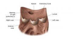
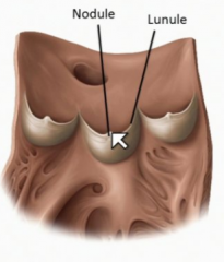
What is the name of the thickened edge and the point of the leaflets of the semilunar valve?
|

The lunule and the nodule respectively
|
|
|
Which valve has an anterior valve: pulmonary or aortic?
|
The pulmonary valve, the aortic valve has a right left and posterior cusp.
|
|
|
Where are beginnings of the coronary arteries?
|
Above the right and left cusps of the aortic valve
|
|
|
Where is the cardiac skeleton located?
|

Separates the four valves of the heart and gives a place of attachment for the valves and cardiac muscle fibers.
|
|
|
What provides insulation for the conduction system of the heart?
|
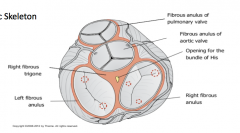
The cardiac skeleton
|
|
|
What are the three cusps of the pulmonary valve?
|
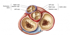
Right, Anterior and left cusps
|
|
|
Which cusps do the coronary arteries come from?
|
Superior to the left and right cusps
|
|
|
Where do you position the stethoscope to listen to the valve functionality?
|

Downstream from each valve
|
|
|
Where do you listen to the aortic valve?
|

Right 2nd intercostal space at the sternal margin
|
|
|
Where do you listen to the pulmonary valve?
|
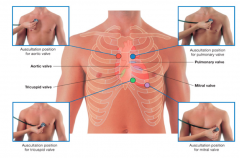
Left 2nd intercostal space at the sternal margin
|
|
|
Where do you listen to the tricuspid valve?
|

Left 5th intercostal space at the sternal margin
|
|
|
Where do you listen to the mitral valve?
|

Left 5th intercostal space at the midclavicular line
|
|
|
What is valve stenosis?
|
Condition in which the opening of the valve is narrowed due to calcifications or scarring on the valve leaflets
|
|
|
What is valve insufficiency?
|
Condition in which a valve does not close completely, allowing blood to regurgitate backwards through the valve.
|
|
|
What causes heart murmurs?
|
Turbulent blood flow caused by either blood being forced through a narrow passage or traveling backwards through an insufficient valve
|
|
|
What is Mitral Valve Prolapse?
|
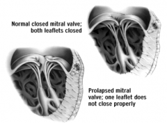
A condition in which one or both leaflets of the mitral valve prolapse (evert) into the left atrium, allowing blood to regurgitate back into the left atrium during systole. This causes a midsystolic clicking sound
|
|
|
What is the initiator of cardiac contraction?
|
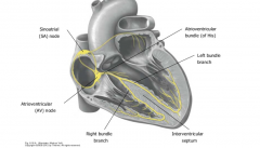
The SA node located where the SVC enters the right atrium. This is the pacemaker of the heart and sets the HR at 60-70 bpm
|
|
|
Where is the AV node located?
|

Superior to the interventricular septum
|
|
|
How do the ventricles receive their depolarization?
|

Through the AV bundle or the bundle of His-it does not travel directly from the atria.
|
|
|
How is the health of the electrophysiology of the heart measured?
|
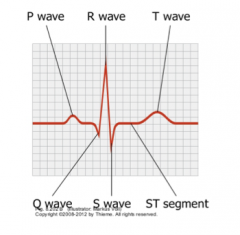
Through an electrocardiogram (EKG/ECG)
|
|
|
What are the characteristics of early diastole?
|
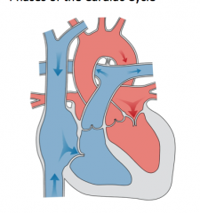
The atria and the ventricles are relaxed
All valves closed atria filling |
|
|
What are the characteristics of late diastole?
|
AV valves open
Ventricles Fill SA node initiates atrial contraction |
|
|
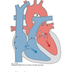
What are the characteristics of early systole?
|
AV valves close
AV node initiates ventricular contraction |
|
|
What are the characteristics of late systole?
|
Semilunar valves open
Ventricles Contract |
|
|
What is the left coronary artery?
|
A short artery arising from the aorta traveling between the left atria and the pulmonary trunk. This divides into the LAD and the circumflex artery
|
|
|
What is the Right coronary artery?
|

It travels around the right side of the heart in the coronary sulcus and gives a branch to the SA node, a branch to the right margin (feeds right ventricle), and posterior IV branch
|
|
|
What is another name for the Left Anterior Descending artery?
|

The anterior interventricular branch of the Left coronary artery
|
|
|
What supplies blood to the SA node?
|

It is commonly a branch from the right coronary artery but could also be supplied by the circumflex artery (branch of the left coronary artery)
|
|
|
What supplies the posterior interventricular artery?
|
60-70% of people are right arterial dominant (PIV arises from the right coronary artery)
15% arises from the circumflex artery (left arterial dominance) 15% is shared |
|
|
If someone is left arterial dominant what does this mean?
|
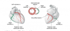
The posterior interventricular artery arises from the circumflex artery (a branch of the left coronary artery) and supplies the AV node of the heart. The entire interventricular septum is supplied by the left coronary artery in these people.
|
|
|
Which veins drain directly into the right atrium?
|
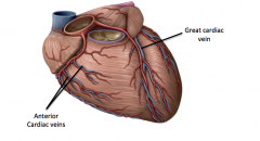
The coronary sinus and the anterior cardiac veins
|
|
|
What vein accompanies the LAD?
|

The great cardiac vein which drains the left and right ventricles and interventricular septum on the anterior side.
|
|
|
What vein accompanies the Posterior interventricular artery?
|
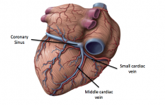
The middle cardiac vein
|
|
|
What vein accompanies the right coronary artery?
|

The small cardiac vein
|
|
|
What is the effect of the sympathetic nervous system on the heart?
|
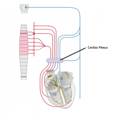
Increases the heart rate and the force of contraction
Causes dilation of the coronary arteries |
|
|
What is the effect of the parasympathetic nervous system on the heart?
|

Arising from the Vagus nerve this decreases the heart rate and the force of contraction. Constricts the coronary arteries
|
|
|
What detects ischemia of heart muscle?
|

Visceral Afferent nerves
|
|
|
What are the common sites of coronary artery blockage?
|
LAD 40-50%
RCA 30-40% Circumflex artery 15-20% |
|
|
What is the difference between acute and subacute damage to the cardiac tissue?
|
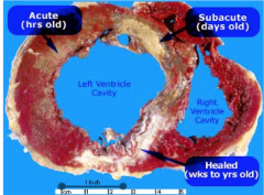
Acute is only hours old, but is still apparent through discoloration whereas subacute is days old and appears completely necrotic.
|
|
|
What is atherosclerosis?
|
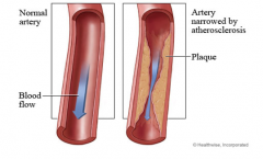
Narrowing of the arteries due to fatty deposits in the vessel wall.
|
|
|
What is a coronary angiogram?
|
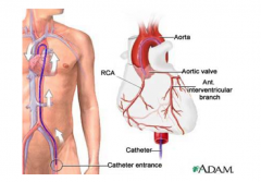
A catheter threaded through the femoral artery inserting radiopaque dye into the aorta and visualized as it travels through the coronary arteries.
|
|
|
What are two main options of a CABG?
|
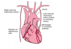
A coronary artery bypass graft can be done using a saphenous vein harvested from the leg or attached directly from the mammary artery
|
|
|
What is referred pain?
|
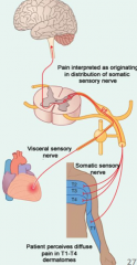
Noxious stimuli from an internal organ are perceived as superficial pain because the afferent fibers carrying the visceral afferent nerves also carry the dermatome nerves. "Cardiac pain" therefore arises from the T1-T4 spinal levels
|