Reading...
![]()
Play button
![]()
Play button
![]()
Use LEFT and RIGHT arrow keys to navigate between flashcards;
Use UP and DOWN arrow keys to flip the card;
H to show hint;
A reads text to speech;
97 Cards in this Set
- Front
- Back
|
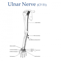
The Ulnar nerve contains nerve fibers from which VPR?
|
C7, C8, and T1
|
|
|
What are the attachment points for the coracobrachialis muscle?
|
Proximal: Coracoid process of the scapula
Distal: Mid humerus |
|
|
What nerve pierces the coracobrachialis?
|

Musculocutaneous
|
|
|
What motions does the coracobrachialis muscle contribute to?
|

The flexion and adduction of the glenohumeral joint
|
|
|
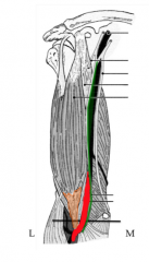
What are the attachments of the biceps brachii?
|
Proximal: Long Head-Supraglenoid tubercle of the scapula
Short head-Coracoid process Distal: Radial Tuberosity |
|
|
What motions does the biceps brachii contribute to?
|

Flexion of the shoulder and elbow
Supination of the elbow joint |
|
|
What nerve runs between the groove formed by the medial epicondyle and the olecranon?
|
The ulnar nerve-hitting this causes the "funny bone" sensation in the medial 1.5 digits.
|
|
|
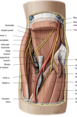
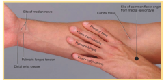
What structures will you find in the cubital fossa?
|
TAN!
Tendon: Biceps Brachii Artery: Brachial Artery Nerve: Median Nerve (does not innervate the arm) |
|
|
The musculocutaneous nerve runs deep to_________and superficial to _________.
|

Biceps Brachii
Brachialis |
|
|
What motions does the brachialis muscle contribute to?
|
Flexion at the elbow joint
|
|
|
What are the attachments of the Brachialis muscle?
|

Proximal: Mid-Humerous
Distally: Ulnar Tuberosity |
|
|
Once the musculocutaneous nerve passes distally to the cubital fossa what changes?
|
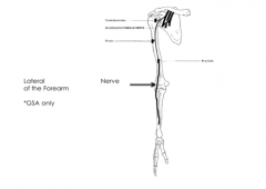
It only carries sensory fibers and then becomes the lateral cutaneous nerve of the forearm.
|
|
|
What is the Popeye Deformity and what causes this?
|
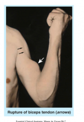
Rupture of the tendon of the long head of the biceps from the supraglenoid tubercle. This rolls up on itself to form a ball and must be fixed surgically
|
|
|
What is the primary motion of the triceps muscle?
|

Extends at the Elbow
|
|
|
What is the Anconeous?
|
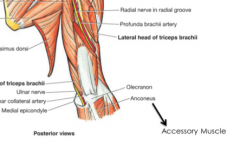
An accessory muscle of extension
|
|
|
What nerve and vessel will you find between the lateral and long head of the triceps?
|

The radial nerve and the deep brachial artery.
|
|
|
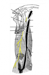
Describe the musculocutaneous nerve
|

Innervates the coracobrachialis, biceps brachii, and brachialis containing fibers from C5-7
|
|
|
What muscles are innervated by the axillary nerve?
|
The Deltoid and the Teres Minor
|
|
|
What muscles are innervated by the radial nerve?
|

Triceps, brachioradialis, Extensor carpi radialis longus-gives off a deep and superficial (cutaneous) branch. Then the deep branch innervates the extensor carpi radialis brevis and pierces the supinator muscle before transforming into the Posterior Interosseous nerve
|
|
|
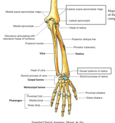
What is the coranoid fossa?
|
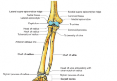
A shallow dip on the anterior distal humerus that houses the coranoid dip of the ulna when the elbow is flexed.
|
|
|
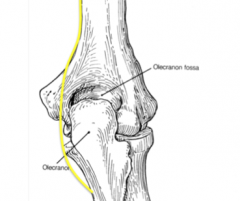
What is the olecranon?
|

A proximal tuberosity of the ulna that prevents hyperextension of the elbow joint when it fits into the olecranon fossa of the humerus
|
|
|
In a healthy elbow a line drawn between the medial epicondyle, ___________, and ____________ should be straight.
|
The Hueter line between the olecranon and lateral epicondyle should be straight
|
|
|
What is Volkmann's Ischemic Contracture?
|
A fracture above the condyles (supracondylar) can compromise the nerves and vessels. If untreated the tight fascia of the forearm will cut off the nerves and vessels around this injury with swelling. The result will be a flexed elbow, wrist, and joint extension of the MCP (knuckle) joint.
|
|
|
How many synovial joints are contained within the elbow joint?
|

3
-Humeroulnar -Humeroradial -Proximal Radioulnar joint |
|
|
What is the proximal radioulnar joint?
|

An articulation between the head of the radius and the radial notch of the ulna. This allows for rotation of the radial head within the cuff formed by the annular ligament and the radial notch of the ulna.
|
|
|
What is the ulnar collateral ligament?
|
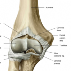
Provides stability to the radioulnar joint. A thick triangular band on the medial side of the elbow joint. This is a common baseball tear (UCL tear) that is repaired by the "Tommy John" surgery.
|
|
|
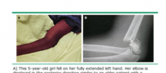
What is subluxation?
|

Proximal incomplete temporary dislocation at the head of the radius-Nursemaid's elbow
|
|
|
What action causes a nursemaid's elbow?
|
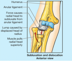
When a child is suddenly lifted by the upper limb when the forearm is pronated-the immature radial head slips out of the socket moving distally and partially out of the annular ligament.
|
|
|
What are the borders of the cubital fossa?
|

Superiorly: Imaginary line connecting medial and lateral epicondyles
Medially: Pronator Teres Laterally: Brachioradialis |
|
|
What is the bicipital aponeurosis?
|

A dense connective tissue sheath that comes from the tendon of the biceps brachii and swings medially blending in with the deep fascia of the arm
|
|
|
What are the attachments of the brachioradialis?
|

Lateral/Distal humerus to the radius (proximal to the styloid process)
When the forearm is mid-potonated it is a powerful flexor (stabbing muscle) |
|
|
What are the heads of the pronator teres?
|

The Humeral and the Ulnar head
|
|
|
What is the action of the pronator teres?
|
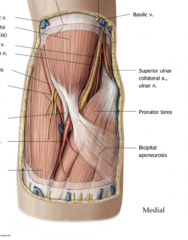
Flexor of the forearm
|
|
|
What nerve passes through the two heads of the pronator teres?
|

Medial nerve
|
|
|
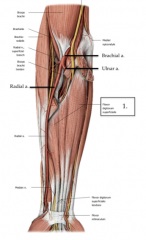
What are the two branches of the brachial artery?
|
The radial artery (travels laterally) and the ulnar artery (travels medially)
|
|
|
What are the insertions of the pronator teres?
|

Medial epicondyle humerus/ulna to the shaft of the radius
|
|
|
What is the movement of the pronator quadratus?
|
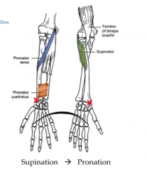
This bracelet muscle is responsible for pronation of the forearm
|
|
|
What are the movements the wrist is capable of?
|
Flexion, Extension, Abduction (Radial deviation), and Adduction (Ulnar deviation)
|
|
|
Besides connecting the ulna and radius-what are some other purposes of the interosseous membrane?
|
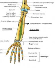
An attachment point for deep forearm muscles acting on the hand and a way to transfer energy and prevent a break of either bone.
|
|
|
What is the distal radio-ulnar joint?
|
The joint between the ulnar and radius before they articulate with the carpal bones. An articular cartilage disc prevents the ulnar from articulating with the carpal bones of the wrist
|
|
|
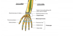
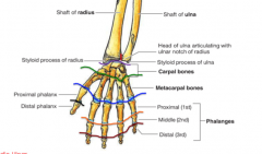
Which two carpal bones articulate with the radius?
|

The Schaphoid and Lunate
|
|
|
What are the eight bones of the wrist from Proximal to distal, lateral to medial?
|

Schapoid-Lunate-Triquetrum-Pisiform
Trapezium-Trapezoid-Capitate-Hamate |
|
|
What is the name of the metacarpals articulating with the carpal bones?
|

Carpometacarpal joints (CMC)
|
|
|
What are the MCP joints?
|

The metacarpophalangeal joints between the metacarpals and the proximal phalanges
|
|
|
How many different kinds of interphalangeal joints are there?
|

In the thumb there is only one-in all other fingers there are proximal and distal IP joints.
|
|
|
The superficial forearm muscles have the same proximal attachment-where?
|
The medial epicondyle known as the common flexor tendon (inflammed known as Golfers elbow)
|
|
|
What is the distal attachment of the pronator teres?
|

The radius and pronates the forearm
|
|
|
What is the flexor carpi radialis?
|
A superficial muscle of the forearm which inserts on the CFT and attaches at the second and third metacarpals. This laterally deviates the forearm and flexes the wrist.
|
|
|
What is the Palmaris Longus?
|

A superficial forearm muscle medial to the flexor carpi radialis. This attaches at the medial epicondyle to the palmar aponeurosis (fan of connective tissue near the base of the palm). This is involved in the flexion of the wrist
|
|
|
What is the Flexor Retinaculum?
|

A tight dense connective tissue band around the anterior of the wrist which prevents tendons from popping out if you flex the wrist.
|
|
|
If you need to harvest a flexion muscle from the forearm-which would you use?
|

The Palmaris Longus-it is missing in 10% of the population, so you can function without it and it can be used in the face.
|
|
|
What is the flexor carpi ulnaris?
|

A superficial forearm muscle that inserts into the medial epicondyle to the pisiform/hook of the hamate/5th medicarpal. This flexes the wrist and medially deviate the wrist (ulnar deviation)
|
|
|
What are the four superficial flexors of the forearm?
|

From Medial to Lateral: Flexor carpi ulnaris, palmaris longus, flexor carpi radialis, and pronator teres
|
|
|
What is the flexor digitorum superficialis?
|
This intermediate anterior forearm muscle. It originates from the medial epicondyle and the oblique line of the radius to the base of the middle phalanges of digits 2-5 (forms four tendons). It flexes the wrist and the digits proximal interphalangeal joints (PIP)
|
|
|
What innervates the flexor digitorum superficialis?
|

The Median Nerve
|
|
|
What are the attachments of the flexor digitorum profundis?
|

The anterior surface of the ulna to the base of the distal phalanx.
|
|
|
What are the actions of the FDP?
|
Flexion of the wrist
Flex at the MCP joints Flex at the PIP joints Only flexor of the Distal Interphalangeal joints (DIP) |
|
|
What nerve innervates FDP?
|

The median nerve innervates digits 2-3 (pointer and middle)
The ulnar nerve innervates digits 4 and 5 (ring and pinky) |
|
|
The median nerves runs between which two muscles of the forearm?
|

Deep to the FDS and superficial to the FDP
|
|
|
What is the Flexor Policis Longus?
|
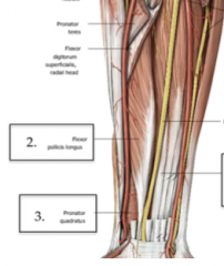
A flexor of the thumb which inserts into the distal phalanx of the thumb.
|
|
|
What are the three deep anterior muscles of the forearm?
|

Flexor Digitorum Profundis
Flexor Policis Longus Pronator Quadratus |
|
|
The median nerve gives off a branch just distal to the cubital fossa-why?
|
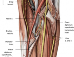
This nerve known as the anterior interosseous nerve innervates the radial half of FDP, and the flexor pollicis longus and pronator quadratus
|
|
|
Why does the FDS split nearing the base of the middle phalanges?
|
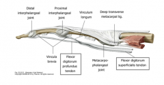
This allows the FDP tendon to continue to the base of the distal phalanges
|
|
|
How do you isolate the PIP joint?
|
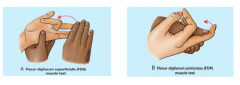
Hold the middle phalange and try to bend the finger. This tests the FDP muscle.
|
|
|
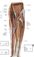
Tell the story of the median nerve
|
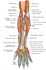
A terminal branch from the medial and lateral cord of the brachial plexus. It does not innervate anything in the arm and passes medially to the tendon and artery of the cubital fossa. It travels between the two heads of pronator teres. Down the forearm it travels deep to FDS and superficial to FDP. Then it divides into an Anterior Interosseous Nerve and continues as the Median nerve which travels through the carpal tunnel into the hand.
|
|
|
Why is the anterior interosseous nerve named this?
|

Because it has a close association with the interosseous membrane between the radius and ulna. This is a fibrous non-synovial joint known as syndesmoses.
|
|
|
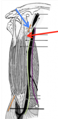
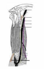
Tell the story of the Ulnar nerve.
|

A terminal branch of the medial branch of the brachial plexus. It travels posteriorly and travels between the olecranon and the medial epicondyle (funny bone space). Travels deep to the Flexor Carpi Ulnaris which it innervates and superficial to FDP half of which it innervates. These are the 1.5 muscles it innervates in the forearm then it continues into the hand where it divides into a superficial and a deep branch. The superficial branch is sensory and the deep branch innervates the intrinsic muscles of the hand.
|
|
|
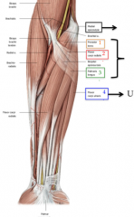
Tell the story of the radial nerve.
|

A terminal branch of the posterior cord of the brachial plexus. Innervates the Triceps Teres and enters the spiral groove of the humerus. It crosses the intermuscular septum and come anterior. Near the cubital region it divides into the superficial and deep branch. The deep dives deep into the extensor region of the forearm. The superficial runs under the brachioradialis and then comes lateral to be sensory to the dorsum of the hand. The deep pierces the supinator.
|
|
|
What are the two divisions of the brachial artery?
|

The radial and ulnar artery. This divides just distal to the cubital fossa.
|
|
|
The common interosseous artery gives rise to an anterior and a posterior. These arteries are a branch from which main artery?
|

The Ulnar artery
|
|
|
The Anterior Interosseus Nerve and Artery are branches from what main source?
|

AIN is a branch of the median nerve and the AIA is a branch of the common interosseous artery which is a branch of the ulnar artery.
|
|
|
True or False: The median nerve supplies muscles of the deep compartment of the flexor forearm?
|

True but incompletely-better way to state is this-the AIN, the major branch coming off the median nerve, supplies the muscles of the deep compartment of the flexor forearm
|
|
|
What are the three deep muscles that are supplied by the AIN?
|
FDP (radial half digits 2-3)
Pronator Quadratus Flexor Pollicis Longus |
|
|
What branches of the radial and ulnar artery supply the elbow joint?
|
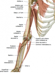
Recurrent and collateral branches.
|
|
|
What is the distal attachment of the triceps brachii?
|

The Olecranon of the ulna
|
|
|
The medial epicondyle was an attachment for many _____________ muscles while the lateral epicondyle is the attachment for many of the _______________ muscles of the forearm.
|
Flexor
Extensor |
|
|
Where is the LIster's Tubercle?
|

The Dorsal tubercle of the radius and a projection from the styloid processes. This is the attachment of the Extensor Pollicis Longus
|
|
|
What artery supplies the extensor compartment of the arm?
|
The Posterior Interosseous Artery-a branch of the Ulnar artery.
|
|
|
What band of connective tissue keeps the extensor muscles from bowing when the wrist is extended?
|
The Extensor Retinaculum
|
|
|
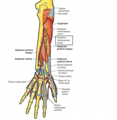
Which nerve pierces the supinator muscle?
|
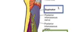
The radial nerve-once it emerges it is the posterior interosseous nerve
|
|
|
Which tendon do the extensor muscles of the forearm originate from?
|
The common extensor tendon. An injury to this tendon is known as tennis elbow
|
|
|
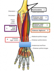
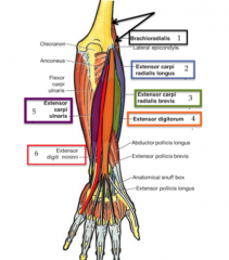
Why is the brachioradialis considered part of the extensor forearm when it is located on the lateral anterior side?
|

It is innervated by the radial nerve and is a powerful flexor of the forearm, but only in the mid-prone position. It only acts upon the elbow joint because it does not cross the wrist
|
|
|
What motions is the extensor carpi radialis longus? capable of?
|

Inserts upon the 2nd metacarpal and extends and laterally deviates the wrist. It is innervated by the radial nerve.
|
|
|
What motions is the extensor carpi radialis brevis responsible for?
|

The extension and lateral deviation of the wrist. Attachments are the CFT and the base of the 3rd metacarpal. This muscle is innervated by the deep radial nerve.
|
|
|
What are the two muscles innervated by the deep radial nerve?
|

The extensor carpi radialis brevis and supinator
|
|
|
What innervates the extensor digitorum?
|

The posterior interosseous nerve. This muscle extends the digits
|
|
|
Describe the extensor carpi ulnaris.
|

This superficial extension muscle is innervated by the PIN (a branch of the radial nerve). It is responsible for extending the wrist and ulnar/medial deviation
|
|
|
Describe the extensor digit minimi
|

A superficial extensor muscle Innervated by the posterior interosseous nerve and attaches to the expansion hood of the fifth digit.
|
|
|
What are the muscles of the superficial layer of the extensor forearm?
|

Brachioradialis
Extensor digiti minimi Extensor digitorum Extensor carpi ulnaris Extensor carpi radialis longus Extensor carpi radialis brevis |
|
|
Why does the supinator capable of its motion if it is part of the extensor compartment of the forearm?
|
It wraps around the radius and therefore is a powerful supinator-innervated by the deep radial nerve
|
|
|
Which inserts most distally:
Abductor pollicis longus Extensor pollicis brevis Extensor pollicis longus |

The Extensor pollicis longus inserts along the base of the distal phalanx of the thumb and therefore is most distal. The brevis attaches at the proximal phalanx and the abductor inserts at the base of the first metacarpal (thumb metacarpal)
|
|
|
What nerve innervates the extensor pollicis brevis?
|

The posterior interosseous nerve (a branch of the radial nerve)
|
|
|
Describe the extensor indicis
|

One of the deep muscles of the extensor forearm that extends the index finger. This blends in with the expansion hood and attaches at the base of the middle phalanx of the second digit. This is innervated by PIN
|
|
|
What blood supply is responsible for the extensor compartment of the forearm?
|

The posterior interosseous artery (a branch of the common IA and a terminal part of the ulnar artery). There are also some branches of the radial nerve responsible for the lateral area.
|
|
|
What is De Quervain's disease?
|

Entrapment tendonitis at the styloid process of the radius of: Abductor pollicis longs and extensor pollicis brevis. Presents with radial wrist pain and difficulty gripping, pinching and located pain above the styloid process
|
|
|
How do you test for De Quervain's Disease?
|
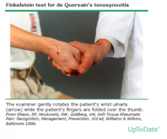
Grasp the thumb in the rest of the fingers while flexing or medially deviating the wrist.
|
|
|
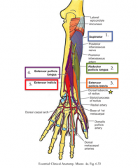
What are the muscles of the deep extensor compartment?
|

Supinator
Abductor Pollicis Longus Extensor Pollicis Longus Extensor Pollicis Brevis Extensor Indicis |