![]()
![]()
![]()
Use LEFT and RIGHT arrow keys to navigate between flashcards;
Use UP and DOWN arrow keys to flip the card;
H to show hint;
A reads text to speech;
49 Cards in this Set
- Front
- Back
|
Anatomical position |
standing upright, palm face upwards(supination), eyes opened, mouth closed, facing forward, arms to the side |
|
|
Planes |
sagittal, coronal/frontal, transverse/horizontal |
|
|
sagittal |
left and right halves (parasagittal – notdivided into equal halves vs. midsagittal divided into equal halves) |
|
|
Coronal/Frontal |
anterior (ventral) andposterior (dorsal) |
|
|
Transverse/Horizontal |
upper and lower halves(superior and inferior) |
|
|
Medial |
towards the midline |
|
|
Lateral |
away from the midline |
|
|
Anterior/ventral |
front |
|
|
Posterior/dorsal |
back |
|
|
Superior/cranial |
above (towards the head) |
|
|
inferior/caudal |
below (towards the "tail") |
|
|
superficial |
closer to the surface |
|
|
deep |
more internal |
|
|
proximal |
closer |
|
|
distal |
further away |
|
|
ipsolateral |
same side |
|
|
contralateral |
opposite sides |
|
|
abduction |
away from the midline |
|
|
adduction |
towards the midline |
|
|
flexion |
bend or flex |
|
|
extension |
straighten |
|
|
pronation |
palms down |
|
|
supination |
palms up (soup) |
|
|
circumduction |
circular motion |
|
|
opposition |
thumb to finger |
|
|
axial skeleton |
skull, neck, trunk/torso (ribs, sternum, vertebrae, sacrum) |
|
|
Appendicular |
Limbs + pectoral and Pelvic girdles (includesclavicle, scapula and pelvis) |
|
|
blood vessels |
arteries (blood from heart) and veins (blood to heart) |
|
|
Nervous system |
CNS and PNS |
|
|
CNS |
brain and spinal chord (everything within) |
|
|
PNS |
Everything external to the brain and spinal cord – spinalnerves, cranial nerves, ganglia etc |
|
|
Neuron |
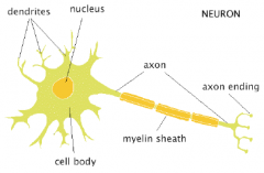
smallest functional unit of the nervous system I. Cell body (soma) – where info is integrated II. Dendrites – receptor segments (receive info)III. Axons – projecting segments (send info) IV. Synapses – where communication between two neurons |
|
|
Axosomatic |
synapse between axon of one neuron and cell bodyof another |
|
|
Axodendritic |
synapse between axon of one neuron and dendriteof another (most common) |
|
|
Axoaxonal |
synapse between axons of two different neurons |
|
|
Nucleus |
collection of cell bodies within the CNS |
|
|
Tract |
collection of cell processes within the CNS |
|
|
ganglion |
collection of cell bodies within the PNS |
|
|
nerve |
collection of cell processes within the PNS |
|
|
afferent signals |
sensory from PNS towards the CNS |
|
|
efferent signals |
motor from the CNS to PNS |
|
|
Basic Neuron forms |
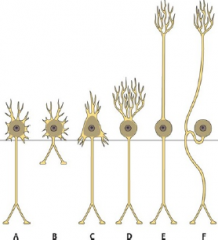
I. Multipolar neuron / bipolar neuron – is a long axonseperated from dendrites by a body (this is the motor neurons) - A in picture II. Pseudounipolar neuron – the dendrites and axons are notseperated by a cell body, it looks like one cell process but isactually two (this is our sensory) ***How can you remember this? M for motor and the S inpSeudounipolar for sensory. |
|
|
Branches of PNS |
Somatic: external environment and voluntary for exmoving muscles Autonomic: internal environment and involuntaryex gland secretion |
|
|
Development of the Nervous system |
neural plate/ neural folds (edge of plate) --> neural tube --> neural crest cells (along length of tube) neural tube --> somites (on either side of tube) branchial arches (pharyngeal arches) |
|
|
neural crest cells |
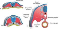
(along length of tube) give rise to cranial, spinal, and autonomic ganglia |
|
|
somites |
(on either side of tube) give rise to vertebra, muscle mass, and spinal nerves |
|
|
branchial arches (pharyngeal arches) |
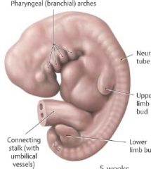
give rise to cranial nerves - each arch contains cartilage, cranialnerve, muscle, and artery. |
|
|
branchial arches and associated nerve |
I. innervated by CNV, trigeminal nerve (specifically the mandibular branch, V3) - Gives rise to muscles of mastication II. innervated byCN VII, facial nerve -Gives rise to muscles of facial expression III. innervated by CN IX, glossopharyngeal nerve. - Gives rise to one muscle called the stylopharyngess IV & VI. innervated by CNX, Vagus nerve - Give rise to a number of muscles V. Does not give rise to any muscles, is vestigial |
|
|
Dermatones |
areas of the skin that are supplied by the same spinalnerve. Spinal nerves innervate our body in stripes. (Dermatones allow oneto predict if there is nerve damage + what nerve is damage). |