![]()
![]()
![]()
Use LEFT and RIGHT arrow keys to navigate between flashcards;
Use UP and DOWN arrow keys to flip the card;
H to show hint;
A reads text to speech;
349 Cards in this Set
- Front
- Back
|
skeletal system consists of |
bones, cartilages, ligamens |
|
|
________ provides flexible strength and support for body structures such as the thoracic cage, the external ear, and the trachea and larynx. -At joints of the body, this can also unite adjacent bones or provide cushioning between them. |
cartilage |
|
|
_______ are the strong connective tissue bands that hold the bones at a moveable joint together and serve to prevent excessive movements of the joint that would result in injury. |
Ligaments |
|
|
Providing movement of the skeleton are the muscles of the body, which are firmly attached to the skeleton via connective tissue structures called _______ |
tendons. |
|
|
in adults there are ____ bones |
206 bones |
|
|
younger people have ____ bones |
more bones bc some fuse thogether |
|
|
bones functions |
- provide support for internal organs - primary storage site for minerals like calcium and phosphate - bone marrow stores fat and houses the blodo cell producing tissue of the body |
|
|
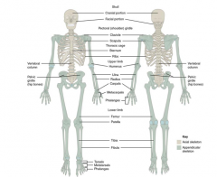
skeleton subdivided into 2 divisions |
axial and appendicular |
|
|
forms the vertical, central axis of the body and includes all bones of the head, neck, chest, and back - protects brain, spinal cord, heart, lungs - attachment site for muscles that move the head, neck, and back, and muscles that act across the shoulder and hip joints |
axial skeleton |
|
|
axial skeleton of adult consists of ___ bones |
80 bones - including skull, vertebral column. thoracic cage |
|
|
skull is formed by ___ bones |
22 bones - 7 additional bones associated with the head (hyoid bone and ear ossicles) |
|
|
vertebral column consists of ___ bones, plus the sacrum and coccyx |
24 bones - called vertebra |
|
|
thoracic cage includes ___ pairs of ribs |
12 pairs of ribs - and the sternum |
|
|
know |
|
|
appendicular skeleton is made up of all |
bones of the upper and lower limbs - also the bones that attach each limb to the axial skeleton |
|
|
___ bones in the appendicular skeleton of an adult |
126 bones |
|
|
skeletal structure of the head that supports the face and protects the brain - facial bones and brain case (or cranial vault) |
cranium (skull) |
|
|
brain case or cranial vault |
protects brain and houses the middle and inner ear structures |
|
|
in adult skull consists of ___ individual bones |
22 individual bones - 21 are immobile and united into a single unit - 22nd bone is the mandible (lower jaw) = only moveable bone of the skull |
|
|
only moveable bone of the skull |
mandible (lower jaw) |
|
|
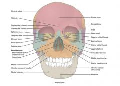
anterior view of skull |
- facial bones, nasal cavity, upper and lower jaws |
|
|
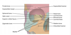
bony socket that houses the eyeball and muscles that move the eyball or open the upper eyelid |
orbit |
|
|
upper margin of the anterior orbit |
supraorbital margin |
|
|
located near the midepoint of the supraorbital margin is a small opening called - which provides for passage of a sensory nerve to the skin of the forehead |
supraorbital foramen |
|
|
below the orbit is the ____ - which is the point of emergence for a sensory nerve that supplies the anterior face below the orbit |
infraorbital foramen |
|
|
know |
|
|
glabella - anterior view of skull |
inbetween eyebrows |
|
|
supraorbital foramen - anterior view of skull |
inner eyebrow |
|
|
supraorbital margin - anterior view of skull |
outer eyebrow |
|
|
sphenoid bone - anterior view of skull |
center back of eyes |
|
|
temporal bone - anterior view of skull |
temple area |
|
|
ethmoid bone - anterior view of skull |
just inner to the syphenoid bone |
|
|
nasal bone - anterior view of skull |
nose bone |
|
|
palatine bone - anterior view of skull |
just below the ethmoid bone |
|
|
nasal septum consists of |
perpendicular plate of ethmoid bond and vomer bone |
|
|
perpendicular plate of ethmoid bone - anterior view of skull - in nasal septum |
upper rod in nasal cavity |
|
|
vomer bone - anterior view of skull - in nasal septum |
lower rod in nasal cavity |
|
|
maxilla - anterior view of skull |
above teeth |
|
|
alveolar process of maxilla - anterior view of skull |
below maxilla above teeth |
|
|
menal foramen - anterior view of skull |
outer chin |
|
|
mandible - anterior view of skull |
center butt chin |
|
|
alveolar process of mandible - anterior view of skull |
above mandible below teeth |
|
|
inferior nasal concha - anterior view of skull |
lower side bone in nasal cavity |
|
|
middle nasal concha - anterior view of skull |
upper side bone in nasal cavity |
|
|
infraorbital foramen - anterior view of skull |
inner cheek |
|
|
zygomatic bone - anterior view of skull |
cheek bone |
|
|
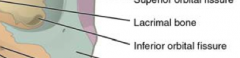
inferior orbital fissure - anterior view of skull |
outer lower fissure in sphenoid bone |
|
|
hypothalamus controls |
homeostasis |
|
|
lacrimal bone - anterior view of skull |
inside orbit and outside nasal bone |
|
|
superior orbital fissure - anterior view of skull |
biggest fissure in orbit |
|
|
optic canal - anterior view of skull |
little inner fissure/hole in cavity |
|
|
orbit - anterior view of skull |
eye socket |
|
|
parietal bone |
above temporal bone and outside of frontal bone |
|
|
smallest of the separate vertebrae and allow the most mobility |
vervical vertebra (C1-C7) |
|
|
C1 - atlas |
no body, superior articulating surface articulate with the occipital condyles of the skull allowing the yes-yes motion of the head |
|
|
C2 - axis |
dens (odontoid process) which, with the transverse ligaments holding it against the anterior arch of the atlas, allows the no- no motion of the head (rotation) |
|
|
C3-C7 |
bifid spinous process, transverse foramina, oval shaped body - c7 is bump at the base of the neck |
|
|
thoracic vertebra (T1-T12) |
these articulate with the ribs they are : - long, slender spinous process - heart shaped body - coastal facets ( on superior and inferior edges of body (centrum)) = these articulate with the head ( end ) of a rib - transverse coastal facets on the transverse processes articulate with the tubercle of the rib |
|
|
lumbar vertebrae (L1-L5) |
largest of the vertebrae, and least movable; they bear the weight of the torso - stout body - broad, short spine - large flat transverse process |
|
|
sacrum identifying features |
- median sacral crest (spinous processes are fused) - sacral foramina (for passage of spinal nerves and blood vessels) - auricular surface (auricle refers to the ear) |
|
|
auricular surface articulates with what bone |
hip bone |
|
|
auricular surface articulation with the hip bone name |
sacroiliac joint |
|
|
- small, wide side to side - short bifid, projects directly posteriorly - vertebral foramen= triangular - transverse process = contain foramina - superior/ inferior artiuclar process = sup facet to sup pos; inf facet to inf ant |
cervical c3-c7 |
|
|
- larger than cervical, heart shaped, bears two costal demfacets - long sharp, prjects inferiorly - vertebral foramen= circular - transverse process = bears facets for the ribs (except T11 and T12) - superior/ inferior artiuclar process = sup facet to pos; inf facet to ant - have sites for rib attachment |
THORACIC |
|
|
- massive, kidney shaped - short, blunt, projects directly posteriorly - vertebral foramen= triangular - transverse process = thin and tapered - superior/ inferior artiuclar process = sup facet to posmed; inf facet to antlat |
lumbar |
|
|
____ holds weight of the head and articulates with the occipital condyles on the occipital bone |
atlas C1 |
|
|
dens is on the axis __ - forms an axis of rotation for the atlas rotating on the axis |
C2 |
|
|
only ____ vertebrae have foramina in their transverse processes - spinous processes are biffid (split at the end) |
cervical |
|
|
spinous processes of the thoracic vertebrae are often |
long and thin and point caudally |
|
|
superior articular processes and facets of the thoracic vertebrae face |
posteriorly |
|
|
posteriorly facing facets alow |
rotation in the thoracic region |
|
|
spinous processes of the lumbar vertebrae are often |
shorter and more square |
|
|
superior articular proceesses and facets of the lumbar vertebrae face |
medially |
|
|
medially facing facets allow |
flexion extesion in the lumbar region |
|
|
vertebrae are seperrated by |
intervertebral discs made of fibrocartilage which absorb shocks - has 2 major regions= central gelatinous nucleus pulposus and outer ring of encircling colagen fibers called the anulus fibrosis that stabilizes teh disc and contains the nucleus pulposus |
|
|
herniated disc |
ruptured disc - anulus fibrosus ruptures and nuceus pulposes protrudes (herniates) through it - compresses adjacent nerves |
|
|
thoracic and sacral curvatures of the spie are |
primary curvatures - bc they are present at birth |
|
|
this becomes prominant when the baby holds its head up |
cervical curvature |
|
|
this becomes prominent when the baby begins to walk |
lumbar curvature |
|
|
fetus vertebrae bone number |
33 |
|
|
adult vertebrae bone number |
24 - 5 sacral and 4 coccysx bones of the fetus fuse together |
|
|
in fetus |
- more bones - carpals and tarsals are not osiied - component parts of the sternum are not fused - frontal bone is split at the midsaggital line at the metopic suture |
|
|
fontanelle |
soft membraneous spot on the head of a baby due to incomplete fusion of the crnial bones - ossification causes the fontanelle to close by 18 to 24 months |
|
|
organ protected by sternum |
lungs and heart |
|
|
what bone articulates at the claviuclar notch |
manubrium of sternum |
|
|
point of attachment for several ligaments and muscles |
xiphoid process - lower part of sternum |
|
|
fetus skull has these fontanels |
has anterior fontaneal, sphenoid fontanel, posterio fontanel, mastoid fontanel |
|
|
nasal cavity is divided into halves by the |
nasal septum |
|
|
upper portion of the nasal septum is formed by the |
perpendicular plate of the ethmoid bone and the lower portion is the vomer bone |
|
|
each side of the nasal cavity is this shape |
triangular in shape, with a broad inferior space that narrows superiorly |
|
|
bony plates prjecting from each lateral wall - these are |
inferior nasal concha - independent bone of the skull |
|
|
above inferior concha - part of the ethmoid bone |
middle and superior (lateral to perpendicular plate) nasal conchae |
|
|
seperating brain case from jaw is |
zygomatic arch |
|
|
bony arch on the side of the skull that spans from cheek to above the ear canal - formed by : temporal process of the zygomatic bone (short anterior component) and zygomatic process of the temporal bone (longer posterior portion) - temporal process anteriorly and zygomatic process posteriorly join together to form arch |
zygomatic arch |
|
|
above the zygomatic arch is a shallow space called the ______ - contain muscles that act on the mandible during chewing |
temporal fossa |
|
|
inferior to the zygomatic arch and deep to the vertical portion of the mandible is ____ - contain muscles that act on the mandible during chewing |
infratemporal fossa |
|
|
cranial cavity |
interior space of the brain case that is occupied by the brain - bound superiorly by the rounded top of the skull (calvaria) and flat bones |
|
|
floor of the brain case |
base of the skull - has openings cranial nerves, blood vessels, and spinal cord - subdivided into 3 large spaces : anterior cranial ossa, middle cranial fossa, posterior cranial fosa - from anterior to posterior the fossae increase in depth |
|
|
ditch |
fossa |
|
|
brain case consists of __ bones - these consist of the paired parietal and temporal bones, plus unpaired frontal, occipital, sphenoid, and ethmoid bones |
8
|
|
|
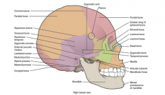
this forms most of the upper lateral side of the skull - paired bones - bound anteriorly by the frontal bone, inferiorly by the temporal bone, and posteriorly by the occipital bone |
parietal bone |
|
|
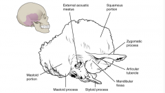
forms hte lower lateral side of the skull - where hair first turns gray - flattened, upper portion is the squamous portion of the temporal bone - projecting anteriorly is the zygomatic process of the temporal bone which forms the posterior portion of the zygomatic arch - posteriorly is the mastoid portion of the temporal bone and projecting inferiorly from here is a large prominence the mastoid process which is a muscle attachment site (behind earlobe) - interior of skull the petrous portion of the temporal bone forms the prominent diagonally oriented petrous ridge (insdie this are small cavities that house the structures of the middle and inner ears) |
temporal bone |
|
|
know |
|
|
- flattened, upper portion is the ______portion of the temporal bone |
squamous |
|
|
- projecting anteriorly is the ________ which forms the posterior portion of the zygomatic arch |
zygomatic process of the temporal bone |
|
|
- posteriorly is the_______ of the temporal bone and projecting inferiorly from here is a large prominence the _______ which is a muscle attachment site (behind earlobe) |
mastoid portion, mastoid process |
|
|
- interior of skull the petrous portion of the temporal bone forms the |
prominent diagonally oriented petrous ridge (insdie this are small cavities that house the structures of the middle and inner ears) |
|
|
temporal bone and its squamous, mastoid, and zygomatic portions |
|
|
This is the large opening on the lateral side of the skull that is associated withthe ear. |
external acoustic meatus (ear canal) - temporal bone |
|
|
This opening is located inside the cranial cavity, on the medial side of the petrous ridge. It connects to the middle and inner ear cavities of the temporal bone. |
internal acoustic meatus - temporal bone |
|
|
This is the deep, oval-shaped depression located on the external base of the skull, just in front of the external acoustic meatus. The mandible (lower jaw) joins with the skull at this site as part of the temporomandibular joint, which allows for movements of the mandible during opening and closing of the mouth. |
mandibular fossa - temporal bone |
|
|
The smooth ridge located immediately anterior to the mandibular fossa. Both the articular tubercle and mandibular fossa contribute to the temporomandibular joint, the joint that provides for movements between the temporal bone of the skull and the mandible. |
articular tubercle - temporal bone |
|
|
Posterior to the mandibular fossa on the external base of the skull is an elongated, downward bony projection called the ______. This structure serves as an attachment site for several small muscles and for a ligament that supports the hyoid bone of the neck. |
styloid process - temporal bone |
|
|
This small opening is located between the styloid process and mastoid process. This is the point of exit for the cranial nerve that supplies the facial muscles. |
stylomastoid foramen - temporal bone |
|
|
The carotid canal is a zig-zag shaped tunnel that provides passage through the base of the skull for one of the major arteries that supplies the brain. Its entrance is located on the outside base of the skull, anteromedial to the styloid process. The canal then runs anteromedially within the bony base of the skull, and then turns upward to its exit in the floor of the middle cranial cavity, above the foramen lacerum. |
carotid canal - temporal bone |
|
|
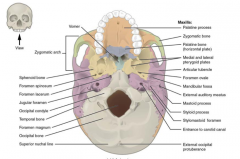
the hard palate is formed anteriorly by the palatine processes of the maxilla bones and posteriorly by the horizontal plate of the palatine bones - inferior view |
|
|
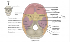
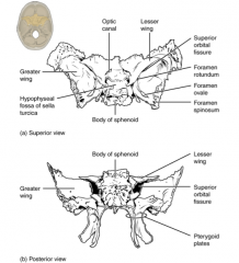
- the lesser wing of the sphenoid bone separates the anterior and middle cranial fossae - petrous ridge (petrous portion of temporal bone) separates the middle and posterior cranial fossae - superior view |
|
|
single bone that forms the forehead |
frontal bone |
|
|
anterior midline of frontal bone between the eybrows is |
glabella (depression) |
|
|
the opening that provides passage for a sensory nerve to the forehead |
supraorbital foramen |
|
|
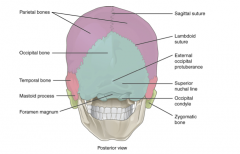
the single bone that forms the posterior base of the cranial cavity |
occipital bone |
|
|
at the posterior midline of the occipital bone is the ____ that serves as an attachment site for a ligament of the posterior neck |
external occipital protuberance |
|
|
lateral to the external occipital protuberance is the ___ which represenet the most superior point at which muscles of hte neck attach to the skull, with only the scalp coveing the skull above these llines |
superior nuchal line |
|
|
foramen magnum |
occipital bone - allows for passage of the spinal cord as it exits the skull |
|
|
on either side of the foramen magnums is an oval shaped ____ that form joints with the first cervical vertebra and thus support the skull on top of the vertebral column |
occipital condyle |
|
|
posterior view of the skull |
|
|
single, complex bone of the central skull - serves as a keystone bone bc it joins with almost every other bone of the skull |
sphenoid bone |
|
|
these form the lip of a prominent ridge that marks the boundary between the anterior and middle cranial fossae inside the cranial cavity |
lesser wings of the sphenoid bone - sphenoid bone |
|
|
located at the midline of the middle cranial fossa - resembles horse saddles used by turks with high back and tall front |
sella turcica - sphenoid bone |
|
|
rounded depression in the floor of the sella turica is the ___ which houses the pituitary (hyophyseal) gland |
hypophyseal (pituitary) fossa - sphenoid bone |
|
|
these extend laterally to either side away from the sella tucica, where they form the anterior floor of the middle cranial fossa - best seen on outside of the lateral skull, forms rectangular area immediately anterior to the squamous portion of the temporal bone |
the greater wings of the sphenoid bone |
|
|
plates of the sphenoid bone |
medial pterygoid plate = form the posterior, lateral walls of the nasal cavity and lateral pterygoid plate = attachment sites for chewing muscles that fill the infratemproal space and act on the mandible |
|
|
sphenoid bone |
|
|
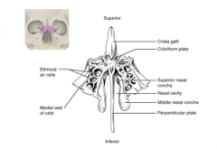
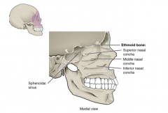
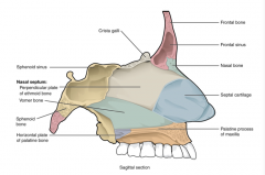
single, midline bone that forms the roof and lateral walls of the upper nasal cavity, te upper portion of the nasal septum, and contributes to the medial wall of the orbit - on interior of the skull it forms a portion of the floow of the anterior cranial cavity |
ethmoid bone |
|
|
in nasal cavity, perpendicular plate of this bone forms the upper portion of the nasal septum |
part of ethmoid bone |
|
|
thmoid bone forms lateral walls of the upper nasal cavity and extending from the lateral walls are the ________ which are thin curved projections that extend into the nasal cavity |
superior nasal concha and middle nasal concha |
|
|
In the cranial cavity, the ethmoid bone forms a small area at the midline in the floor of the anterior cranial fossa. This region also forms the narrow roof of the underlying nasal cavity. - this portion consists of 2 parts= |
cristal galli and the cribriform plates |
|
|
a small upward bony projection located at the midline. It functions as an anterior attachment point for one of the covering layers of the brain. |
crista galli |
|
|
lateral of the crista galli is ____ small, flattened area with many small openings termed olfactory foramina |
cribriform plate |
|
|
small nerve branches from the olfactory areas of the nasal cavity pass through __ to enter the brain |
olfactory foramina in the cribriform plate |
|
|
________ are located between orbit and upper nasal cavity - forms lateral nasal cavity wall and a protion of the medial orbit wall - has paranasal sinus system |
lateral portions of the ethmoid bone |
|
|
midline view of the sagittally sectioned skull |
|
|
ethmoid bone |
|
|
ethmoid bone |
|
|
an immobile joint between adjacent bones of the skull. - filed with dense, fibrous connective tissue - twisty lines interlock adjacent bones |
suture |
|
|
- side to side across the skull - joins the frontal bone the the right and left parietal bones |
coronal suture |
|
|
- posteriorly from the coronal suture, running along the midline at the top of the skull in the sagittal plane of section |
sagittal suture |
|
|
- extend downward and laterally to either side away from its junction with the sagittal suture - joins the occipital bone to the right and left parietal and temporal bones |
lambdoid suture |
|
|
- on the lateral skull - unites squamous portion of the temporal bone with the parietal bone |
squamous suture |
|
|
- small h shaped suture line region that unites the frontal bone, parietal bone, squamous portion of the temporal bone, and greater wing of the sphenoid bone - weakest part of the skull - 2 fingers above zygomatic arch and thumb width posterior |
pterion |
|
|
__% of all injury related deaths in the us are caused by head injuries - most involve falls |
30% |
|
|
most common skull fracture |
linear skull fracture, fracture lines radiate from point of impact |
|
|
comminuted fracture |
where the bone is broken into several pieces ath the point of impact |
|
|
depresed fracture |
when the fractured bone is pushed inward |
|
|
contrecoup (counterblow) fracture |
bone at the point of impact is not broken,, but a fracture occurs on the opposite side of the skull - fracture of the occipital bone can occur like this, making a basilar fracture that can damage the artery that passes through the carotid canal |
|
|
strong blow to the pterion rgion can |
fracture bones around the pterion if artery under is damaged bleedin can cause a hematoma (collection of blood) between the brain and the interior of the skull, as blood accumulates it will put pressure on the brain |
|
|
facial bones of the skull form |
upper and lower jaws, nose, nasal cavity, nasap septum, and the orbit - 14 bones - 6 paired bones = maxilla, palatine, zygomatic, nasal, lacrimal, and inferior nasal conchae - 2 unpaired bones = vomer and mandible bones |
|
|
one of a pair that together form the upper jaw, much of the hard palate, the medial floor of the orbit and the lateral base of the nose |
maxially bone or the mailla |
|
|
the curved, inferior margin of the maxillary bone that forms the upper jaw and contains the upper teath is the |
alveolar process of the maxilla |
|
|
teeth are anhored into socket called |
alveolus |
|
|
on anterior maxilla below orbit is the ____, this is the point of exit for a sensory nerve that supplies the nose, upper lip, and anterior cheek |
infraorbital foramen - maxillary bone |
|
|
inferior skull, this is from maxillary bone and joins together at the midline ot form the anterior 3/4 of the hard palate |
palatine process - maxillary bone |
|
|
the bony plate that forms the roof of the mouth and the floor of the nasal cavity, separating the oral and nasal cavities |
the hard palate - maxillary bone |
|
|
one of a pair of irregularly shaped bones that contribute small areas to the lateral walls of the nasal cavity and the medial wall of each orbit - largest region is the horizontal plate - plates join together at the midline to form the posterior quarter of the hard palate - best seen at the inferior view |
palatine bone |
|
|
1/1000 births, mostly male - partial or complete failure of the right and left poritons of the upper lip to fuse together |
cleft lip |
|
|
cleft palate |
1/2500 births and more common in females - failure of the two halves |
|
|
also known as the cheekbone - lateral wall of orbit - short temporal process of the zygomatic bone projects posteriorly, where it forms the anterior portion of the zygomatic arch |
zygomatic bone |
|
|
- short temporal process of the zygomatic bone projects posteriorly, where it forms the |
anterior portion of the zygomatic arch |
|
|
one of the two small bones that articulate (join) with each other to form the bony base of the nose - support the cartilage that form the lateral walls of the nose |
nasal bone |
|
|
small, rectangular bone that forms the anterior, medial wall of the orbit |
lacrimal bone |
|
|
anterior portion of the lacrimal bone forms a shallow depression called the |
lacrimal fossa |
|
|
inferiorly from the lacrimal fossa is the |
nasolacrimal canal |
|
|
lacrimal fluid drains at the |
medial corner of the eye into the nasolacrimal canal, then to open into the nasal cavity, behind the inferior nasal concha. - normally drains posteriorly but with an increased flow of tears dut to crying or eye iritation, some drain anteriorly, thus causing a runny nose |
|
|
triangular shaped and forms the posterior inferior part of the nasal septum - best seen when looking from behind into the posterior openings of the nasal cavity from here it is seen to form the entire height of the nasal septum |
vomer |
|
|
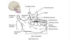
- only movable bone of the skull - first r and l but then fuse each side consists of horizontal body and posteriorly, a vertically oriented ramus |
mandible |
|
|
outside margin of the mandible where the body and ramus come together is |
angle of the mandible |
|
|
more anterior projection is the flattened ______, which provides attachment for one of the biting muscles |
coronoid process of the mandible |
|
|
the posterior projection is the ______, which is topped by the oval shaped condyle |
condylar process of the mandible |
|
|
the condyle of the mandible articulates (joins) with the |
mandibular fossa and articular tubercle of the temporal bone |
|
|
temporomandibular joint |
allows for opening and closing of the mouth |
|
|
broad u shaped curve located between the coronoid ad codylar processes is the |
mandibular notch |
|
|
landmarks for the mandible include |
alveolar process of the mandible, mental protuberance, mental foramen, mylohyoid line, mandibular foramen, lingula |
|
|
upper border of the mandibular body and serves to anchor the lower teeth |
alveolar process of the mandible |
|
|
the forward projection from the inferior margin of the anterior mandible that forms the chin |
mental protuberance |
|
|
the opening located on each side of the anterior lateral mandible, which is the exit site for a sensory nerve that supplies the chin |
mental foramen |
|
|
bony ridge extends along the inner aspect of the mandibular body - the muscle that forms the floor of the oral cavity attaches to the mylohoid lines on both side of the mandible |
mylohyoid line |
|
|
this opening is located on the medical side of the ramus of the mandible. the opening leads into a tunnel that runs down the length of the mandibular body. the sensory nerve and blood vessels that supply the lower teeth enter the mandibular foramen and then follow this tunnel. |
mandibular foramen |
|
|
to numb dental work |
inject into the lateral wall fo the oral cavity at a point prior to where this sensory nerve enters the mandibular foramen |
|
|
small flap of bone located next to mandibular foramen, on the medial side of the ramu - ligament that anchors the mandible during opening and closing of the mouth extends down from the base of the skull and attaches to this |
lingula of the mandible |
|
|
mandible |
|
|
the walls of each orbit include contributions from |
7 skull bones - frontal bone = roof - zygomatic bone = lateral wall and lateral floor - maxilla and palantine = medial floor - thmoid bone and lacrimal bone = medial wall - sphenoid bone = posterior orbit |
|
|
allows for passage of the optic nerve from the retina to the brain |
optic canal |
|
|
lateral to the optic canal is a fissue |
superior orbital fissure - provides passage for the artery that supplies the eyball, sensory nerbes, and the nerves that supply the muscles involved in the eye movements |
|
|
orbit |
|
|
nasal septum - bone and cartilage - upper portion of septum is formed by ____ - lower and posterior parts of septum are formed by ______ |
- upper portion of septum is formed by ethmoid bone - lower and posterior parts of septum are formed by vomer bone |
|
|
a flexible plate that fills in the gap between the perpendicular plate of the ethmoid and vomer bones - extends where it separates the right and left nostrils - not found in dry skull |
septal cartilage |
|
|
attached to lateral wall on each side of the nasal cavity are sup, mid, and inf - bony plates that curve downward - warm and humidify air before it enters the lungs - allows mucus to trap dust, pollen, bac and viruses |
nasal conchae - sup and mid are formed by ethmoid bone - inf is independent bone |
|
|
saggital nasal septum |
|
|
fossae |
spaces |
|
|
this is shallowest of 3 crainal fossae - orbits and frontal lobes of the brain - bunded by the frontal bone - lesser wings of sphenoid bone form the prominent ledge that marks the boundary between the anterior and middle cranial fossae - at midline floor is ethmoid bone with crista galli and on the sides cribriform plates |
anterior cranial fossa |
|
|
- from lesser wings of sphenoid bone to petrou ridges of temporal bones - temporal lobes of the brain - divided at the midline by the sella turcica, part of the sphenoid bone - has openings for passage of blood vessels and cranial nerves |
middle cranial fossa |
|
|
- large opening into posterior orbit on anterior wall of middle cranial fossa, lateral to optic canal and under lesser wing of the sphenoid bone - nerves to eyeball, and sensory nerves to the forehead pass through this oening |
superior orbital fissure |
|
|
opening at anterior lateral corner of the sells turcica - passage for optic nerve into the orbit |
optic canal - middle cranial fossa |
|
|
openings in the middle crania fossa |
- optic canal - superior orbital fissure - forament rotundum - foramen ovale of the middle cranial fossa - foramen spinosum - carotid canal - foramen lacerum |
|
|
This rounded opening is located in the floor of the middle cranial fossa, just inferior to the superior orbital fissure. It is the exit point for a major sensory nerve that supplies the cheek, nose, and upper teeth. |
foramen rotundum |
|
|
This large, oval-shaped opening in the floor of the middle cranial fossa provides passage for a major sensory nerve to the lateral head, cheek, chin, and lower teeth. |
foramen ovale of the middle cranial fossa |
|
|
This small opening, located posterior-lateral to the foramen ovale, is the entry point for an important artery that supplies the covering layers surrounding the brain. The branching pattern of this artery forms readily visible grooves on the internal surface of the skull and these grooves can be traced back to their origin at the foramen spinosum. |
foramen spinosum |
|
|
this is the zig zag passageway through which a major artery to the brain enters the skull. The entrance to the carotid canal is located on the inferior aspect of the skull, anteromedial to the styloid process. From here, the canal runs anteromedially within the bony base of the skull. Just above the foramen lacerum, the carotid canal opens into the middle cranial cavity, near the posterior-lateral base of the sella turcica. |
carotid canal |
|
|
irregular opening in the base of the skull inferior to the exit of the carotid canal - it is filled with cartilage in life - nothing passes through it |
foramen lacerum |
|
|
lacerum |
raged or torn |
|
|
- cerebellum of brain - bounded anteriorily by the petrous ridges (which hs internal acoustic meatus), occipital bone is floor and posterior wall - divided at midline by the large framen magnum |
posterior cranial foss |
|
|
this opening provides passage of the nerve from the hearing and equilibium organs of the inner ear an dnerve that suplies the muscles of the face |
internal acoustic meatus - posterior cranial fossa |
|
|
at anterior lateral margin of the foramen magnum is the - inferior of skull at base of the occipital condyle and provide passage for an important nerve to the tongue |
hypoglossal canal
- posterior cranial fossa |
|
|
inferior to the internal acoustic meaturs is this where cranial nerves from the brain exit the skull via this opening - exit point through the base of the skull ofr all the venous return blood leaving the brain - determined by the venous structures that carry blood inside the skull which form large grooves on the inner walls of the posterior cranial fossa which end at this |
jugular foramen - posterior cranial fossa |
|
|
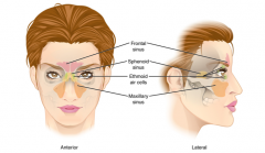
- these are hollow air filled spaces in bone of the skull - all communicate with the nasal cavity and are lined with nasal mucosa - serve to reduce bone mass and lighten the skull, add resonance to the voice - produce excess mucus |
paranasal sinuses |
|
|
located above eyebrows within the frontal bone - divided or fused in single space - most anterior of paranasal sinuses |
frontal sinus |
|
|
larges sinus - paired and located within the right and left maxiallary bones under the orbits - sinus infections - bc their connection to nasal cavity is high on their medial wall they are difficult to drain |
maxillary sinus |
|
|
single midline sinus located in body of sphenoid bone anterior and inferior to the sella tucica - most posterior paranasal sinuses |
sphenoid sinus |
|
|
small spaces seperated by thing bony walls in the ethmoid bone - on both sides of ethmoid bone between upper nasal cavity and medial orbit behind superior nasal conchae |
ehtmoid air cell |
|
|
paranasal sinuses |
|
|
independent bone that does not contact other bones and is not part of the skull - u shpaed in upper neck near the inferior mandible - tips of u face posteriorily - help by small muscles that move up down or forward back - movement is coordinated with movements of the tongue, larynx, pharynx during swallowing and speaking |
hyoid bone |
|
|
The ______ bone is located in the upper neck and does not join with any other bone. It provides attachments for muscles that act on the tongue, larynx, and pharynx. |
hyoid bone |
|
|
- vertebrae seperated and united by intervertebral discs - flexible column that supports head, neck, and body and allows movements - protects spinal cord, which is through openings int he vertebrae |
vertebral column, spinal column, or spine |
|
|
the vertebral column of an adult |
- 24 vertebrae, plus the sacrum and coccyx. - divided into three regions: cervical C1–C7 vertebrae, thoracic T1–T12 vertebrae, and lumbar L1–L5 vertebrae. - two primary curvatures (thoracic and sacrococcygeal curves) - two secondary curvatures (cervical and lumbar curves). |
|
|
vertebral column orginally has this many vertbrae |
33 but reduce to 24 |
|
|
vertebral column 5 regions |
|
|
|
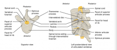
- 7 cerveical vertebrae - C1 articulates (forms a joint) with occipital condyles of the skull - C1 artuclates with C2 and so on |
Cervical C1-C7 vertebrae |
|
|
below cervical vertebrae |
throacic T1-T12 vertebrae |
|
|
lower back - below thoracic vertebrae |
Lumbar L1-L5 vertebrae |
|
|
formed by the fusion of the 5 sacral vertebrae |
sacrum - pelvis and vertebrae |
|
|
formed by fusion of 4 small coccygeal vertebrae |
coccyx or tailbone |
|
|
sacral and coccygeal fusion do not start until age |
20 -middle age |
|
|
almost all mammals have |
7 cervical vertebrae regardless of body size |
|
|
curvatures in vertebral column |
- strength, flexibility, and ability to absorb shock - primary ( retained from original fetal curvature) and secondary (developed after birth) |
|
|
primary curvatures (retained from original fetal curvature of the spine) - anteriorily |
thoracic curve and sacrococcygeal curve (formed by sacrum and coccyx) |
|
|
secondary curvatures (develop after birth, concave posteriorly, opposite in direction to original fetal curvature) |
cervical curve (hold head) and lumbar curve (stand and walk) |
|
|
a disorder characterized by an excessive posterior curvature of the thoracic region - humpback - osteoporosis causes weakening and gradual collapse occurs |
kyphosis |
|
|
a disorder characterized by an excessive anterior curvature of the lumbar region - swayback - obesity or late pregnancy - anterior tilt of pelvis |
lordosis |
|
|
a disorder characterized by an abnormal lateral curvature accompanied by twisting of the vertebral column - most common vertebral abnormality among girls - bend forward and r l side will not be level |
scoliosis |
|
|
typical vertebra will consist of a |
body, vertebral arch, and 7 processes |
|
|
The ______ is the anterior portion of each vertebra and is the part that supports the body weight. Because of this, they progressively increase in size and thickness going down the vertebral column. The ____ of adjacent vertebrae are separated and strongly united by an intervertebral disc. |
body |
|
|
this forms the posterior portion of each vertebra - has 4 parts : RL pedicles and RL laminae |
vertebral arch |
|
|
- these each form on eof the lateral sides of the vertebral arch - anchored to the posterior side of the vertebral body |
pedicle |
|
|
each of these forms the part of the posterior roof of the vertebral arch |
lamina |
|
|
large opening between the vertebral arch and body is the ___ which contains the spinal cord |
vertebral foramen |
|
|
vertebral foramina of all vertebrae align to form the ____, which serves as the bony protection and passageway for the spinal cord down the back |
vertebral (spinal) canal |
|
|
notches in the margins of the pedicles of the aligned vertebrae form an ____, the opening where a spinal nerve exits form the vertebral column |
intervertebral foramen |
|
|
these project laterally and arise from the junction point between the pedicle and lamina |
transverse process - vertebral arch process |
|
|
this projects posteriorly at the midline of the back |
spinous process (vertebral spine) - vertebral arch process |
|
|
transverse and spinous processes serve as |
muscle attachment sites |
|
|
______ extends upward and ______ projects downward on each side of a vertebrae |
superior articular process is upward and inferior articular process is downward on each side of a vertebrae |
|
|
parts of vertebrae |
|
|
intervertebral disc |
|
|
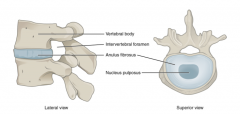
The bodies of adjacent vertebrae are separated and united by an______, which provides padding and allows for movements between adjacent vertebrae. |
intervertebral disc -The disc consists of a fibrous outer layer called the anulus fibrosus and a gel-like center called the nucleus pulposus. |
|
|
The intervertebral foramen is the |
opening formed between adjacent vertebrae for the exit of a spinal nerve. |
|
|
cervical vertebrae are smaller than lumbar vertebrae due to |
differences in the proportion of body weight that each supports |
|
|
- small body, carry least weight - bifid (y shaped) spinous process - C3-C6 spinous processes are short - C7 spine is longer (base of neck) - u shaped transverse processes to allow passage of the spinal nerves, each contain transverse foramen this has artery that supplies the neck - superior and inferior articular processes are flattened and largely face upward or downward |
cervical vertebrae |
|
|
1st cervical vertebrae C1 or atlas |
- supports the skull - no body or spinous process - ring shaped and has anterior arch dn posterior arch - transverse processes are longer and extend more laterally - superior articular processes face upward and are deeply curved for articulation with the occipital condyles on hte base of the skull - interior articular processes are flat and face down to join with superior articular processes of the c2 vertebrae |
|
|
2nd cervical vertebra or axis |
- rotation RL
- distinguished by dens (odontoid process), a bony projection that goes up from vertebral body and joins the inner anterior arch of the atlas by transverse ligament |
|
|
bodies of this are larger than cervical vertebrae - spinous process is long and downward angle that causes it to overlap the next inferior vertebra - superior articular process face anteriorily and inferior process faces posteriorly - have facets ( articulation site where a rib is attached), costal facet, and facet on transverse process for articulation with the tubercle of a rib |
thoracic vertebrae |
|
|
thoracic vertebrae articulation sites where a rib is attached |
facet |
|
|
in thoracic vertebrae the 2 facets on the lateral sides of the body each is called a ___, these are for articulation with the head (end) of the rib |
costal facet |
|
|
thoracic vertebra |
|
|
this carries the greatest amount of body weight and have large thick vertebrae - short transverse processes and short, rounded spinous process that projects posteriorly - articular processes are long, superior process facing backward and inferior facing forward |
lumbar vertebrae |
|
|
triangular shaped bone, thick, wide, tapers into inferior, apex - formed by fusiono f 5 sacral vertebrae, after 20 yrs |
sacrum |
|
|
anterior surface lines of vertebral fusion can be seen as 4 transverse ridges - on posterior surface the midline is ____, a bumpy ridge that is the remnant of the fused spinous processes - the fused transverse processes form the ______ |
- on posterior surface the midline is median sacral crest, a bumpy ridge that is the remnant of the fused spinous processes - the fused transverse processes form the lateral sacral crest |
|
|
the anterior lip of the superior base of the sacrum |
sacral promontory |
|
|
lateral to sacral promontory is roughened auricular surface which joins |
with ilium portion of the hipbone to form immobile sacroiliac joints of hte pelvis |
|
|
inferiorily through the sacrum is a bony tunnel ____, which ends at the sacral hiatus near inferior rip of sacrum |
sacral canal |
|
|
sacral foramina |
ant and post surfaces of sacum which have paired openings that connect to the sacral canal - openings called posterior (dorsal) sacral foramen and anterior (ventral) sacral foramen - these openings allow for the sacral spinal nerves to exit the sacrum |
|
|
- found on either side of the superior opening of the sacral canal, articulates with the inferior articular porcess from the L5 vertebra |
superior articular process of the sacrum |
|
|
- derived from the fusion of four very small coccygeal vertebrae - It articulates with the inferior tip of the sacrum. - It is not weight bearing in the standing position, but may receive some body weight when sitting. |
The coccyx, or tailbone, |
|
|
______ is formed from the fusion of five sacral vertebrae, whose lines of fusion are indicated by the transverse ridges. The fused spinous processes form the median sacral crest, while the lateral sacral crest arises from the fused transverse processes. |
The sacrum |
|
|
this is formed by the fusion of the four small coccygeal vertebrae |
coccyx |
|
|
vertebral column stability due to |
intervertebral discs and ligaments |
|
|
a fibrocartilaginous pad that fills the gap between adjacent vertebral bodies - provide padding - thin in cervical region and thickest in lumbar region - 25 percent of height between top of pelvis and base of skull - flexible and can change shape - 2 parts : anulus fibrosus and nucleus polposus |
intervertebral disc |
|
|
tough fibrous outer layer of the intervertebral disc - circle |
anulus fibrosus |
|
|
softer, gel like material. high water content that resists compression and is important for weight bearing - older=less water content in this causing the disc to become thinner, decreasing total body height, and reduce flexibility and range of motion of the disc |
nuceus polposus |
|
|
most common sites for disc herniation (rupture) due to weak anulus fibrosis are |
L4/L5 or L5/S1 intervertebral discs - can cause sciatica (pain that radiates from lower back to thigh and into the leg) |
|
|
Weakening of the anulus fibrosus can result in |
herniation (protrusion) of the nucleus pulposus and compression of a spinal nerve, resulting in pain and/or muscle weakness in the body regions supplied by that nerve. |
|
|
Adjacent vertebrae are united by |
ligaments that run the length of the vertebral column along both its posterior and anterior aspects |
|
|
- anterior side of the entire vertebral column - resists excessive backward bending of the spinal cord - important in the neck |
anterior longitudinal ligament |
|
|
- posterior side of teh vertebral column - interconnects the spinous process of the thoracic and lumbar vertebrae - forward bending |
supraspinous ligament |
|
|
in the posterior neck, the supraspinous ligament expands to become |
nuchal ligmant |
|
|
this is attached ot the cervical spinous proceess and extends upward and posteriorly and attaches to the midline base of teh skull, out to the external occipital protuberance - supports the skull and prevents its from falling forward - stronger in 4 legged animals - limits the anterior bending of the head and neck |
nuchal ligament |
|
|
anterior to the spinal cord, attached to the posterior sides of the vertebral bodies - important in anterior bending |
posterior longitudinal ligament |
|
|
posterior to the spinal cord is _____ which are short paired ligaments that each interconnect that lamina regions of adjacent vertebrae - elastic fibers - important in anterior bending |
ligamentum flavum (yellow ligament) |
|
|
Chiropractors are health professionals who use |
nonsurgical techniques to help patients with musculoskeletal system problems that involve the bones, muscles, ligaments, tendons, or nervous system. |
|
|
thoracic cage (rib cage) forms the |
thorx (chest) portion of the body, which consists of 12 pairs of ribs with their costal cartilages and the sternum
- ribs are anchored posteriorly to the 12 thoracic vertebrae - this protects the heart and lungs |
|
|
The thoracic cage is formed by the |
(a) sternum and (b) 12 pairs of ribs with their costal cartilages. |
|
|
The ribs are anchored |
posteriorly to the 12 thoracic vertebrae. |
|
|
The sternum consists of the |
manubrium, body, and xiphoid process. |
|
|
The ribs are classified as |
true ribs (1–7) and false ribs (8–12). The last two pairs of false ribs are also known as floating ribs (11–12). |
|
|
wider superior portion of the sternum |
manubrium |
|
|
top of manubrium u shaped border called |
jugular (suprasternal) notch |
|
|
shallow depression on either side at superior lateral margins of the manubrium, this is the site of the sternoclavicular joint |
clavicular notch |
|
|
first ribs also attach to the |
manubrium |
|
|
the manubrium and body join together at the - sternum - second rib attaches here |
sternal angle |
|
|
the______ is the highest rib that can be identified by palpation |
second rib |
|
|
ribs 3-7 attach to the |
sternal body |
|
|
inferior tip of the sternum is the |
xiphoid process - cartiliagous early in life |
|
|
the ribs articulate posteriorly with the |
T1-T12 thoracic vertebrae |
|
|
ribs articulate anteriorly via their |
costal cartilages to the sternum |
|
|
posterior end of a rib is - articulates primarily with the costal facet on the body of the next higher vertebra |
head of the rib |
|
|
lateral to the head of the rib is the |
neck of the rib |
|
|
small bump on the posterior rib surface is the - articulates with the facet located on the transverse process of the same numbered vertebra |
tubercle of the rib |
|
|
lateral to the tubercle of the rib is the - the point at which the rib has its greatest degree of curvature |
angle of the rib |
|
|
these form the most posterior extent of hte thoracic cage |
the angles of the ribs |
|
|
in anatomical postition the angles of the ribs |
angles align with medial border of hte scapula |
|
|
for passage of blood vessels and nerve is found in inferior margin of each rib |
costal groove |
|
|
each rib ends in a - made of hyaline cartilage - then attached to sternum |
costal cartilage |
|
|
rib classifications |
true ribs, false ribs, and floating ribs |
|
|
ribs 1-7 are classified as |
true ribs (vertebrosternal ribs) - costal region attach directly to the sternum |
|
|
ribs 8-12 are classified as |
false ribs (vertebrochondral ribs) - costal cartilages do not attach directly - ribs 8-10 costal cartilages are attached to the cartilage of the next higher rib |
|
|
ribs 11-12 are classified as |
floating ribs (vertebral ribs) - short - costal cartilages terminate within the musculature of the lateral abdominal wall |
|
|
- 3rd week of embryonic development - rod like structure - dorsal along the length of the embryo |
notochord |
|
|
tissue over the notochord forms the nerual tube which gives rise to the |
brain and spinal cord |
|
|
enlarged mesoderm tissue on sides of the notochord - 4th week |
somite |
|
|
most medial somite part is - consist of an embryonic tissue called mesenchyme which gives rise to fibrous connectives tissues, cartilages, and bones |
sclerotome |
|
|
_______ gives rise to fibrous connectives tissues, cartilages, and bones |
mesenchyme |
|
|
soft spot on infant head - dense connective tissue - allow for growth and expansion of the skull - largest is junction of the frontal and parietal bones |
fontanelle |
|
|
endochronal ossification |
cartilage to bone |
|
|
changes that contribute to the growth and enlargement of the face during childhood |
mastoid process enlarges, two halves of hte mandible and frontal bone fuse together to form single bones, and paranasal sinuses enlarge |
|
|
at time of birth _____ has not yet formed |
mastoid process |
|
|
Development of the vertebrae begins with the |
accumulation of mesenchyme cells from each sclerotome around the notochord. These cells differentiate into a hyaline cartilage model for each vertebra, which then grow and eventually ossify into bone through the process of endochondral ossification. As the developing vertebrae grow, the notochord largely disappears. However, small areas of notochord tissue persist between the adjacent vertebrae and this contributes to the formation of each intervertebral disc. |
|
|
The ribs and sternum develop from |
mesenchyme. |
|
|
The ribs initially develop as |
part of the cartilage model for each vertebra, but in the thorax region, the rib portion separates from the vertebra by the eighth week. The cartilage model of the rib then ossifies, except for the anterior portion, which remains as the costal cartilage. |
|
|
The sternum initially forms as |
paired hyaline cartilage models on either side of the anterior midline, beginning during the fifth week of development. The cartilage models of the ribs become attached to the lateral sides of the developing sternum. Eventually, the two halves of the cartilaginous sternum fuse together along the midline and then ossify into bone. The manubrium and body of the sternum are converted into bone first, with the xiphoid process remaining as cartilage until late in life. |
|
|
premature closure (fusion) of a suture line - due to ossification or failure of brain to properly enlarge - more common in men - primary - early fusion of one cranial suture |
craniosynostosis |
|
|
Which of the following is part of the axial skeleton? a. shoulder bones b. thigh bone c. foot bones d. vertebral column |
d. vertebral column |
|
|
8. Which of the following is a function of the axial skeleton? a. allows for movement of the wrist and hand b. protects nerves and blood vessels at the elbow c. supports trunk of body d. allows for movements of the ankle and foot |
c. supports trunk of body |
|
|
9. The axial skeleton ________. a. consists of 126 bones b. forms the vertical axis of the body c. includes all bones of the body trunk and limbs d. includes only the bones of the lower limbs |
a. consists of 126 bones |
|
|
10. Which of the following is a bone of the brain case? a. parietal bone b. zygomatic bone c. maxillary bone d. lacrimal bone |
a. parietal bone |
|
|
the lambdoid suture joins the parietal bone to the |
occipital bone |
|
|
12. The middle cranial fossa ________. a. is bounded anteriorly by the petrous ridge b. is bounded posteriorly by the lesser wing of thesphenoid bone c. is divided at the midline by a small area of theethmoid bone d. has the foramen rotundum, foramen ovale, andforamen spinosum |
d. has the foramen rotundum, foramen ovale, andforamen spinosum - bounded POSTERIORLY by petrous ridge - bounded ANTERIORLY by lesser wing of spenoid boneis divided at the midline by a small area of the sphenoid bone |
|
|
13. The paranasal sinuses are ________. a. air-filled spaces found within the frontal, maxilla,sphenoid, and ethmoid bones only b. air-filled spaces found within all bones of theskull c. not connected to the nasal cavity d. divided at the midline by the nasal septum |
a. air-filled spaces found within the frontal, maxilla,sphenoid, and ethmoid bones only |
|
|
14. Parts of the sphenoid bone include the ________. a. sella turcica b. squamous portion c. glabella d. zygomatic process |
a. sella turcica |
|
|
15. The bony openings of the skull include the ________. a. carotid canal, which is located in the anterior cranial fossa b. superior orbital fissure, which is located at the superior margin of the anterior orbit c. mental foramen, which is located just below the orbit d. hypoglossal canal, which is located in the posterior cranial fossa |
d. hypoglossal canal, which is located in the posterior cranial fossa |
|
|
16. The cervical region of the vertebral column consists of ________. a. seven vertebrae b. 12 vertebrae c. five vertebrae d. a single bone derived from the fusion of five vertebrae |
a. seven vertebrae |
|
|
17. The primary curvatures of the vertebral column ________. a. include the lumbar curve b. are remnants of the original fetal curvature c. include the cervical curve d. develop after the time of birth |
b. are remnants of the original fetal curvature |
|
|
18. A typical vertebra has ________. a. a vertebral foramen that passes through the body b. a superior articular process that projects downward to articulate with the superior portion of the next lower vertebra c. lamina that spans between the transverse process and spinous process d. a pair of laterally projecting spinous processes |
c. lamina that spans between the transverse process and spinous process |
|
|
19. A typical lumbar vertebra has ________. a. a short, rounded spinous process b. a bifid spinous process c. articulation sites for ribs d. a transverse foramen |
a. a short, rounded spinous process |
|
|
20. Which is found only in the cervical region of the vertebral column? a. nuchal ligament b. ligamentum flavum c. supraspinous ligament d. anterior longitudinal ligament |
a. nuchal ligament |
|
|
21. The sternum ________. a. consists of only two parts, the manubrium and xiphoid process b. has the sternal angle located between the manubrium and body c. receives direct attachments from the costal cartilages of all 12 pairs of ribs d. articulates directly with the thoracic vertebrae |
b. has the sternal angle located between the manubrium and body |
|
|
22. The sternal angle is the ________. a. junction between the body and xiphoid process b. site for attachment of the clavicle c. site for attachment of the floating ribs d. junction between the manubrium and body |
d. junction between the manubrium and body |
|
|
23. The tubercle of a rib ________. a. is for articulation with the transverse process of a thoracic vertebra b. is for articulation with the body of a thoracic vertebra c. provides for passage of blood vessels and a nerve d. is the area of greatest rib curvature |
a. is for articulation with the transverse process of a thoracic vertebra |
|
|
24. True ribs are ________. a. ribs 8–12 b. attached via their costal cartilage to the next higher rib c. made entirely of bone, and thus do not have a costal cartilage d. attached via their costal cartilage directly to the sternum |
d. attached via their costal cartilage directly to the sternum |
|
|
25. Embryonic development of the axial skeleton involves ________. a. intramembranous ossification, which forms the facial bones. b. endochondral ossification, which forms the ribs and sternum c. the notochord, which produces the cartilage models for the vertebrae d. the formation of hyaline cartilage models, which give rise to the flat bones of the skull |
b. endochondral ossification, which forms the ribs and sternum |
|
|
26. A fontanelle ________. a. is the cartilage model for a vertebra that later is converted into bone b. gives rise to the facial bones and vertebrae c. is the rod-like structure that runs the length of the early embryo d. is the area of fibrous connective tissue found at birth between the brain case bones |
d. is the area of fibrous connective tissue found at birth between the brain case bones |