![]()
![]()
![]()
Use LEFT and RIGHT arrow keys to navigate between flashcards;
Use UP and DOWN arrow keys to flip the card;
H to show hint;
A reads text to speech;
247 Cards in this Set
- Front
- Back
|
bone or osseous tissue |
hard, dense connective tissue that forms most of the adult skeleton, the support structure of the body. |
|
|
areas of the skeleton where bone move (ribcage, joints)
- semi-rigid form of connective tissue, flexibile and smooth surfaces |
cartilage |
|
|
body system composed of bones and cartilage and performs - support body - facilitates movement - protects internal organs - produces blood cells - stores and releases minerals and fat |
skeletal system |
|
|
a doctor who specilizes in diagnosing and treating disorders and injuries related to the muscoskeletal system |
orthopedist |
|
|
bone tissue function |
- reservoir for minerals (calcium and phosphorous) and can be released back into the bloodstream to maintain levels needed to support physiological processes - fat storage and blood cell production |
|
|
soft connective tissue that fills the interior of most bone is |
bone marrow - yellow marrow = stores fat - red marrow = responsible for hematopoiesis |
|
|
this marrow contains adipose tissue, the triglycerides stored in the adipocytes of the tissue can be a source of energy |
yellow marrow |
|
|
marrow where hematopoiesis (production of blood cells, rbc, wbc, platelets) takes place |
red marrow |
|
|
206 bones in adult skeleton divided into 5 catagories based on shapes and functions |
- sesamoid bone - short bone - long bone - flat bone - irregular bone |
|
|
this bone is cylindrical in shape, longer than wide - shape not size - humerus, ulna, radius, femur, tibia, fibula, metacarpals, phalanges, metarsals, phalanges - function as levers; move when muscles contract |
long bones |
|
|
- cube like in shape, equal in length, width, thickness - only in = carpals(wrist), tarsals(ankle) - provide stability and support and limited motion |
short bones |
|
|
- thin, curved - cranial , scapulae (shoulder blades), sternum (breastbone), ribs - points of attachment for muscles and often protect internal organs |
flat bone |
|
|
- no easily characterized shape - complex shape - vertebrae, facial bones ones that contain sinuses |
irregular bones |
|
|
- small, round bone, shaped like a sesame seed - form in tendons (sheaths of tissue that connect bones to muscles) where a great deal of pressure is generated in a joint -protect tendons by overcome compressive forces - typically found in tendons associated with feet, hands, knees - patellae only ______ bones in common with every person |
sesamoid bones |
|
|
_______ only sesamoid bones in common with every person |
patellae |
|
|
cylinder, longer than wide - function = leverage |
long bones |
|
|
cube like shape - funciton= stability, support, motion |
short bones |
|
|
these bones are complex shaped and protect internal organs |
irregular bones |
|
|
osseous tissue |
bone tissue |
|
|
anatomy of bone |
- diaphysis = ~is the middle region between proximal and distal ends. ~hollow region in diaphysis called medullary cavity (filled with yellow marrow) ~walls are made of compact bone - epiphysis= ~wider section at the end of bone ~filled with spongy bone (which is filled in with red marrow) |
|
|
epiphysis meets the diaphysis at the |
metaphysis |
|
|
- narrow area that contains the epiphyseal plate (growth plate), a line of hyaline (transparent) cartilage in a growing bone, this cartilage is replaced by osseous tissue and the epiphyseal plate becomes an epiphyseal tissue in early adulthood |
metaphysis |
|
|
medullary cavity |
- has yellow bone marrow - has membranous lining called endosteum where bone growth, repair, and remodeling occur |
|
|
outer surface of the bone is covered with fibrous membrane that contains blood vessels, nerves, lymphatic vessels that nourish compact bone - tendants and ligaments also attach to bones here - covers the entire outer surface except where the epiphyses meet other bones to form joints ( this region is covered with articular cartilage) |
periosteum |
|
|
articular cartilage |
- convers where the epiphyses meet other bones to form joints - thin layer of cartilage that reduces friction and acts as a shock absorber |
|
|
periosteum forms the |
outer surface of bone |
|
|
endosteum lines |
meduallary cavity |
|
|
flat bones like cranium have layer of |
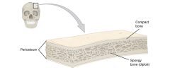
diploe (spongy bone) linked on either side by a layer of compact bone - 2 layers of compact bone and the interior spony bone work together to protect the internal organs |
|
|
diploe |
spongy bone - in flat bones - linked by layer of compact bone |
|
|
3 general classes of bone markings |
1. articulations ( processes that produce joints ) 2. projections 3. holes ( passageway for blood vessels and nerves) |
|
|
where two bone surfaces come together - conform to one another |
an articulation - bone marking |
|
|
an area of a bone that projects above the surface of the bone - attachment points for tendons and ligaments - size and shape is an indication of the forces exerted through the attachment to the bone |
a projection - bone marking |
|
|
an opening or groove in the bone that llows blood vessels and nerves to enter the bone - size reflect size of vessels and nerves that penetrate the bone |
hole - bone marking |
|
|
articulations |
- where two bones meet - ex. knee joint |
|
|
head |
- prominant rounded surface - ex. head of femur |
|
|
facet |
- flat surface - ex. vertebrae |
|
|
condyle |
- rounded surface - ex. occipital condyles |
|
|
projections |
- raised markings - ex. spinous process of the vertebrae |
|
|
protuberance |
- protruding - ex. chin |
|
|
process |
- prominence feature - ex. transverse process of vertebra |
|
|
spine |
- sharp process - ex. ischial spine |
|
|
tubercle |
- small, rounded process - ex. tubercle of humerus |
|
|
tuberosity |
- round surface - ex. deltoid tuberosity |
|
|
line |
- slight, elongated ridge - ex. temporal lines of the parietal bones |
|
|
crest |
- ridge - ex. iliac crest |
|
|
holes |
- holes and depressions - ex. foramen ( holes which blood vessels can pass through ) |
|
|
fossa |
- elongated basin - ex. mandibular fossa |
|
|
fovea |
- small pit - ex. fovea capitis on the head of the femur |
|
|
sulcus |
- groove -ex. sigmoid sulcus of the temporal bones |
|
|
canal |
- passage in bone - ex. auditory canal |
|
|
fissure |
- slit through bone - ex. auricular fissure |
|
|
foramen |
- hole through bone - foramen magnum in the occipital bone |
|
|
meatus |
- opening into canal -ex. external auditory meaturs |
|
|
sinus |
- air-filled space in bone - ex. nasal sinus |
|
|
examples of processes formed where tendons or ligaments attach |
- fovea capitis, head, tubercle, sulcus |
|
|
examples of processes formed to articulate with adjacent bones |
condyles, tubercle, facet, fossa, condyle |
|
|
examples of openings |
canal, sinus, foramen, fissure, protuberance |
|
|
______ give bones their hardness and ______ give them flexibility so they are not brittle |
hydroxyapalite crystals, collagen fibers |
|
|
hydroxyapatite |
which is from inorganic salt crystals, attaching on the collagen fibers, and is formed by the combination of calcium phosphate and calcium carbonate - incorporates other inorganic salts like magnesium hydroxide, fluoride, and sulfate as it cyrstallizes or calcifies on the collagen fibers |
|
|
4 types of cells found in bone tissue |
osteoblasts, osteocytes, osteogenic cells, osteoclasts |
|
|
osteocyte |
- maintains bone tissue - bone cell |
|
|
osteoblast |
- forms bone matrix - bone cell |
|
|
osteogenic cell |
- stem cell - bone cell |
|
|
osteoclast |
- resorbs bone - bone cell |
|
|
osteogenic cells are undifferentiated and develop into |
osteoblasts |
|
|
when osteoblasts get trapped in calcified matrix, their structure and function changes and become |
osteocytes |
|
|
osteoclasts devlop from |
monocytes and macropphages and differ in appearance from other bone cells |
|
|
this is the bone cell responsible for forming new bone and is found in growing proteions of bone; periosteum and endosteum - do not divide - synthesize and secrete the collagen matrix and calcium salts |
osteoblast |
|
|
the primary cell of mature bone and most common type of bone cell - located in lacunas and surrounded by bone tisue - maintain the mineral concentration of the matrix via the secretion of enzymes - lack mitotic activity ( do not divide ) - communicate and receive nutrients via channels through canaliculi within the bone matrix |
osteocytes |
|
|
canaliculi |
channels within the bone matrix |
|
|
these cells are undifferentiated with high mitotic activity and are only bone cells that divide - immature are found in deep layers of periosteum and the marrow - differentiate and develop into osteoblasts |
osteogenic cell |
|
|
old bone is dissolved for |
repair or calcium release |
|
|
cell responsible for bone resorption or breakdown is the - found on bone surfaces - multinucleated - originate from monocytes and macrophages(2 types of wbc) not from osteogenic cells - constantly breaking down old bone while osteoblasts are forming new bone |
osteoclast |
|
|
balance between osteoblasts(form new bone) and osteoclasts (break down old bone) |
responsible for constant subtle reshaping of bone |
|
|
osteogenic cells develop into |
osteoblasts |
|
|
osteogenic cells location |
deep layers of the periosteum and the marrow |
|
|
osteoblasts funciton |
bone formation |
|
|
osteoblasts location |
growing portions of bone, including the periosteum and endosteum |
|
|
osteocytes function |
maintain mineral concnetration of matrix |
|
|
osteocytes location |
entrapped in matrix |
|
|
osteoclasts function |
bone resorption |
|
|
osteoclasts location |
bone surfaces and at sites of old, injured or unneeded bone |
|
|
compact bone |
- dense - foudn under periosteum and in diaphyses of long bones - core |
|
|
osteon or haversian system |
- microscopic unit of compact bone - composed of concentric rings of calcified matrix called lamellae - central canal = has blood vessels, nerves, and lymphatic vessels, branch off through perforating canal(volkmanns canals) to extend to the periosteum and endosteum |
|
|
concentric rings of calcified matrix called |
lamellae |
|
|
osteocytes are located |
in lacunae, found at borders of adjacent lamellae |
|
|
canaliculi connect with |
canaliculi of other lacunae and evenutally with the central canal - this allows nutrietns to osteocytes an wastes removed |
|
|
cancellous bone |
spongy bone |
|
|
this contains osteocytes housed in lacunae but are not arranged in concentric circles - lacunae in lattice netwrok of matrix spikes called trabeculae |
spongy bone |
|
|
trabeculae |
spongy bone - where lacunae is - makes bones lighter so that muscles can move them more easily - protect red marrow |
|
|
pagats disease |
- over 40 - overactive osteoclasts - moer bone resorbed than made - pelvis, skull, spine, legs - akaline phosphatase elevated |
|
|
spongy bone and medullary cavity receive noursihment from |
arteries that pass through the compact bone - enter through nutrient foramen ( small openings in the diaphysis ) |
|
|
osteocytes in spongy bone are nourished by |
blood vessels of the periosteum that penetrate spongy bone and blood that ciruclates int eh marrow cavities - then collected by veins which then pass out of the bone through the foramina |
|
|
blood vessels and nerves are concentrated |
concentrate in metabolically active regions of the bone - bc they regulate blood supplies and bone growth - both enter bone through the nutrient foramen |
|
|
2 osteogenic pathways |
intramembranous ossification and encdochndral ossification - bone is same regardless of the pathway that produces it |
|
|
process of bone development |
ossification (osteogenesis) |
|
|
cartilage |
produced by chondroblasts and consists of hyaluronic acid, chondroitin sulfate, collagen fibers, and water - as mineral matrix surround chondroblasts they are called chondrocytes - avascular - nutrients and waste are facilitated by diffusion through the matrix |
|
|
avascular |
no blood vessels supplying nutrients and removing metabolic wastes |
|
|
intramembranous ossification |
compact and spongy bone develops directly from sheets of mesenchymal (undifferentiated) connective tissue - flat bones of face, cranial bones, clavicles (collarbones) are formed via this |
|
|
early osteoblasts appear in a cluster called |
ossification center |
|
|
osteoblasts secrete |
osteoid = uncalcified matrix, which calcifies(hardens) within a few days as mineral salts are depsoited on it |
|
|
uncalcified matrix, which calcifies(hardens) within a few days as mineral salts are depsoited on it, and trapping the osteoblasts within, they then become osteocyte, osteogenic cells in surrounding connective tissue differentiate into new osteoblasts |
osteoid |
|
|
osteoid (unmineralized bone matrix) secreted around the capillaries results in a |
trabecular matrix |
|
|
osteoblasts on the surface of the spongy bone become |
periosteum |
|
|
trabecular bone crowds newarby blod vessels, which eventually condenses into |
red marrow |
|
|
Intramembranous ossification begins in utero and continues on into adolescence. At birth, the skull and clavicles are not fully ossified nor are the sutures of the skull closed. This allows the skull and shoulders to |
deform during passage through the birth canal. The last bones to ossify via intramembranous ossification are the flat bones of the face, which reach their adult size at the end of the adolescent growth spurt. |
|
|
in this ossification bones develop by replacing hyaline cartilage - cartilage does not become bone but is replaced by bone - longer than intramembranous ossification - base of skull and long bones via this |
endochondral ossification |
|
|
a membrane that covers the cartilage |
perichondrium |
|
|
endochondrial ossification 5 steps |
1. mesenchymal cells differentiate into chondrocytes 2. cartilage model of future bone and perichondrium form 3. capillaries penetrate cartilage. perichondrium transforms into periosteum. periosteal collar develops. primary ossification center develops 4. cartilage and chondrocytes continue to grow at ends of the bone 5. cartilage remains at epiphyseal plate and at joint surface as articular cartilage |
|
|
capillaries penetrating the cartilage initiates the transformation of |
perichondrium into bone producing periosteum |
|
|
primary ossification center |
region deep in the periosteal collar where ossification begins - 2 or 3 month of fetal life |
|
|
secondary ossification center |
- after birth - matrix mineralization, death of chondrocytes, invasion of blood vessels from the periosteum, and seeding with osteogenic cells that become osteoblasts - occurs in epiphyseal regions |
|
|
epiphyseal plate is |
area of growth in long bone - layer of hyaline carilage where ossification occurs in immature bones - on epiphyseal side of epiphseal plate, cartilage is formed - on the diaphyseal side, cartialge is ossified and the diaphysis grows in length |
|
|
reserve zone |
- zone of epiphyseal plate - matrix production - region closest to the epiphyseal end of the plate and has small chondrocytes (that dont act in bone growth but hold the epiphyseal plate to the osseous tisue of the epiphysis) within the matrix |
|
|
proliferative zone |
- zone of epiphyseal plate - next layer inward after the reserve zone ( more thward the diaphysis) - contains stacks of larger chondrocytes - makes new chondrocytes (via mitosis) |
|
|
zone of maturation and hypertrophy |
- zone of epiphyseal plate - next layer inward after the proliferative zone - chondrocytes are older and larger than those in the proliferative zone - lipids, glycogen, alkaline phosphatase accumulate; matrix calcifies |
|
|
more matured cell are _____ to the diaphyseal end of the epiphyseal plate |
closer |
|
|
longitudinal growth of bone is result of |
cell division in proliferative zone and maturation of cells in zone of maturation and hypertrophy - happens when osseous tissue is added to the diaphysis - rate of growth is controlled by hormones |
|
|
zone of calcified matrix |
- zone of epiphyseal plate - zone closest to the diaphysis - cell death - most chondrocytes are dead bc matrix has calcified - capillaries and osteoblasts from diaphysis penetrate this zone and osteoblasts secrete bone tissue on the remaining calcified cartilage - connects the epiphyseal plate the the diaphysis |
|
|
when chondrocytes in episphyseal plate cease their proliferation and bone replaces the cartilage |
longitudinal bone growth stops - all that remains of the epiphyseal plate is the epiphyseal line |
|
|
as a bone matures the epiphyseal plate turns into a |
epiphyseal line |
|
|
growth in diameter can continue even after longitudinal growth ceases this is called |
appositional growth |
|
|
appositional growth |
- osteoclasts resorb old bone that lines the medullary cavity while osteoblasts, via intramembranous ossification, produce new bone tisssue beneath the periosteum - increases the diameter of the diaphysis and diameter of the medullary cavity - called modeling |
|
|
osteoclasts resorb old bone that lines the medullary cavity while osteoblasts, via intramembranous ossification, produce new bone tisssue beneath the periosteum - increases the diameter of the diaphysis and diameter of the medullary cavity |
called modeling |
|
|
process in which matrix is resorbed on one surface of a bone and deposited on another is |
bone modeling - takes place during bones growth |
|
|
resorption of old or damaged bone takes place on the same surface where osteoblasts lay new bone to replace that which is resorbed - in adult life - injury, exercise - 5-10% remodeling without injury or exercise |
remodeling |
|
|
osteogenesis imperfect OI |
genetic disease where bones do not form properly so they break easily - also called brittle bone disease - affects production of collagen - research for bisphosphonates to treat OI |
|
|
broken bone - it will heal whether or not a physician resets it in its anatomical position - but if not reset correctly, the healing process will keep the bone in its deformed position |
fracture |
|
|
when a broken bone is manipulated and set into its natural position without surgery, the procedure is called a |
closed reduction |
|
|
_________ requires surgery to expose the fracture and reset the bone. |
Open reduction |
|
|
types of fractures |
- closed - open - transverse - spiral - comminuted - impacted - greenstick - oblique |
|
|
transverse fracture |
occurs straight across the long axis of the bone |
|
|
oblique fracture |
occurs at an angle that is not 90 degrees |
|
|
spiral fracture |
bone segments are pulled apart as a result of twisting motion |
|
|
comminuted fracture |
several breaks result in many small pieces between two large segments |
|
|
impacted fracture |
one fragment is driven into the other, usually as a result of compression |
|
|
greenstick fracture |
partial fracture where only one side of the bone is broken |
|
|
open (or compound) fracture |
a fracture in which at least one end of the broken bone tears through the skin; causes a high risk of infection |
|
|
closed (or simple) fracture |
a fracture in which the skin remains intact |
|
|
when a bone breaks blood flows form any vessel torn by the fracture - could be periosteum, osteons, or medullary cavity |
- blood begins clotting and 6-8 hours after the fracture the clotting blood has formed a fracture hematoma |
|
|
blood begins clotting and 6-8 hours after the fracture the clotting blood has formed a _______ - the disruption of blood flow to the bone results in death of bone cells around the fracture |
fracture hematoma |
|
|
stages in fracture repair |
1. fracture hematoma forms 2. internal and external calli form 3. cartilage of the calli is replaced by trabecular bone 4. remodeling occurs |
|
|
Within about 48 hours after the fracture, chondrocytes from the endosteum have created an ___________ by secreting a fibrocartilaginous matrix between the two ends of the broken bone |
internal callus (plural = calli) |
|
|
Within about 48 hours after the fracture, the periosteal chondrocytes and osteoblasts create an ________ of hyaline cartilage and bone, respectively, around the outside of the break |
external callus |
|
|
Over the next several weeks of a fracture, |
osteoclasts resorb the dead bone; osteogenic cells become active, divide, and differentiate into osteoblasts. The cartilage in the calli is replaced by trabecular bone via endochondral ossification |
|
|
mechanical stress stimulates |
deposition of mineral salts and collagen fibers - why people who exercise regularly have thicker bones |
|
|
vitamin d |
- calcium absorption in small intestine, calcium reabsorption from the kidneys - remodeling |
|
|
magnesium |
- more than 60 percent of it in the body is in the skeleton |
|
|
fluoride |
- form fluorapatite - helps stabilize and strengthen bone mineral - can enter spaces within hydroxyapatite crystal and increase their density |
|
|
omega 3 |
- reduce inflammation in various parts of the body (osteoblasts) |
|
|
calcium |
Needed to make calcium phosphate and calcium carbonate, which form the hydroxyapatite crystals that give bone its hardness |
|
|
vitamin d |
Needed for calcium absorption |
|
|
Vitamin K |
Supports bone mineralization; may have synergistic effect with vitamin D |
|
|
growth hormone (pituitary gland |
hormone that influences osteoblasts and or maintains the matrix - controls bone growth - triggers chondrocyte proliferation in epiphyseal plates, growth in length of long bones - increases calcium retention, enhances mineralization and stimulates osteoblastic activity, improves bone density |
|
|
thyroxine (thyroid gland) |
hormone that influences osteoblasts and or maintains the matrix promotes osteoblastic activity and synthesis of bone matrix |
|
|
sex hormones estrogen and testosterone |
hormone that influences osteoblasts and or maintains the matrix - promote osteoblatic activity and production of bone matrix and responsible for growth spurt in adolescence - promote conversion of epiphyseal plate to epiphyseal line(end of longitudinal growth) |
|
|
calcitrol (kidneys) |
hormone that influences osteoblasts and or maintains the matrix active form of vitamin d - stimulates abosrption of calcium and phosphate from the digestive tract |
|
|
osteoporosis |
- decrease in bone mass that occurs when rate of bone resportion exceeds the rate of bone formation - in pagets disease = new bone is formed in an attempt to keep up with the resorption by the overactive osteoclasts - osteoporosis does not have the elevated blood levels of alkaline phosphatase found in pagets disease - proximal ends of femur, vertebrae, and wrist - histologically, a reduction in the thickness of compact bone and the number and size of trabeculae in cancellous bone. - vitamin d, calcium, and weight bearing exercise is encouraged - bisphosphonates (same as in pagets), calcitonin, and estrogen (women only) |
|
|
hormone that influences osteoclasts |
parathyroid hormone and calcitronin |
|
|
parathyroid hormone |
hormone that influences osteoclasts - stimulates osteoclast proliferation and activity - calcium is released from bones into circulation 0 increase calcium ion concentration in blood - promotes reabsorption of calcium by the kidney tubules - stimulate synthesis of vitamin d (promotes intestinal absorption of calcium) |
|
|
calcitonin |
- horomone secreted by the thyroid gland - inhibits osteoclast activity and stimulates calcium uptake by the bones - reduce concentration of calcium ions in blood - parathyroid hormone and cacitonin are not secreted at the same time because of calcium homeostasis |
|
|
this hormone that affects the skeletal system increases the length of long bones, enhances mineralization, and improves bone density |
growth hormone |
|
|
this hormone that affects the skeletal system stimulates bone growth and promotes synthesis of bone matrix |
thyroxine |
|
|
this hormone that affects the skeletal system promotes osteoblastic activity and production of bone matrix; responsible for adolescent growth spurt; promote conversion of peiphyseal plate to epiphyseal line |
sex hormones |
|
|
this hormone that affects the skeletal system stimulates absorption of calcium and phosphate from digestive tract |
calcitrol |
|
|
this hormone that affects the skeletal system stimulates osteoclast proliferation and resorption of bone by osteoclasts; promotes resabsorption of calcium by kidney tubules; indeirectly increases calcium absortpion by small intestine |
parathyroid hormone |
|
|
this hormone that affects the skeletal system inhibits osteoclast activity and stimulates calcium uptake by bones |
calcitonin |
|
|
most abundant mineral in bone and human body - tooth health, bone minearlization, regulation of the heart rate and strength of contraction, blood coagulation, contraction of smooth and skeletal muscle cells, and regulation of nerve impulse conduction - 10 mg/dL is normal |
calcium |
|
|
a condition characterized by abnormally low levels of calcium, can have an adverse effect on a number of different body systems including circulation, muscles, nerves, and bone. - blood has difficulty coagulating, the heart may skip beats or stop beating altogether, muscles may have difficulty contracting, nerves may have difficulty functioning, and bones may become brittle. - cause can range from hormonal imbalances to an improper diet. Treatments vary according to the cause, but prognoses are generally good. |
hypocalcemia |
|
|
a condition characterized by abnormally high levels of calcium, the nervous system is underactive, which results in lethargy, sluggish reflexes, constipation and loss of appetite, confusion, and in severe cases, coma. |
hypercalcemia |
|
|
body systems that work together to maintain a normal caclium level in the blood |
skeletal, endocrine, and digestive systems, kidneys |
|
|
normal calcium level |
10 mg/dL |
|
|
increased ca level |
- hyroid gland releases calcitonin, osteoclasts activity is inhibited, ca reabsorption in the kidneys decreases, ca levels in blood decrease |
|
|
decreased ca level |
parathyroid glands release pth, osteoclasts release ca from bone, calcium is reabsorbed from urine by the kidneys, calcium absorption in the small intestine increases via vitamin d syntheis, ca level in blood increases |
|
|
only way calcium enters the body |
diet |
|
|
bones act as this for calcium |
storage site |
|
|
body does this with calcium when blood levels get too high |
deposits calcium in the bones - regulated by parathyroid PTH hormone, vitamin d, calcitonin |
|
|
body does this with calcium when blood levels get too low |
releases calcium when blood levels drop too low - regulated by parathyroid PTH hormone, vitamin d, calcitonin |
|
|
cells of parathyroid PTH gland have plasma membrane receptors for calcium, when ca not binding the cells release PTH which stimulates osteoclast proliferation and resoption of bone by osteoclasts - the demineralization releases ca into the blood - pth promotes reabsorption of calcium from the urine by the kidneys so calcium returns to the blood - pth stimulates synthesis of vitamin d, which stimulates calcium absorption from any digested food in the small intestine |
pathway in calcium homeostasis where ca levels are too low |
|
|
thyroid gland releases calcitonin, inhibits osteoclast activity and stimulates calcium uptake by the bones, decreases reabsorption of calcium by the kidneys |
pathway in calcium homeostasis where ca levels are too high |
|
|
1. Which function of the skeletal system would be especially important if you were in a car accident? a. storage of minerals b. protection of internal organs c. facilitation of movement d. fat storage |
b. protection of internal organs |
|
|
2. Bone tissue can be described as ________. a. dead calcified tissue b. cartilage c. the skeletal system d. dense, hard connective tissue |
a. dead calcified tissue |
|
|
3. Without red marrow, bones would not be able to ________. a. store phosphate b. store calcium c. make blood cells d. move like levers |
c. make blood cells |
|
|
4. Yellow marrow has been identified as ________. a. an area of fat storage b. a point of attachment for muscles c. the hard portion of bone d. the cause of kyphosis |
a. an area of fat storage |
|
|
5. Which of the following can be found in areas of movement? a. hematopoiesis b. cartilage c. yellow marrow d. red marrow |
b. cartilage |
|
|
6. The skeletal system is made of ________. a. muscles and tendons b. bones and cartilage c. vitreous humor d. minerals and fat |
b. bones and cartilage |
|
|
7. Most of the bones of the arms and hands are long bones; however, the bones in the wrist are categorized as ________. a. flat bones b. short bones c. sesamoid bones d. irregular bones |
b. short bones |
|
|
8. Sesamoid bones are found embedded in ________. a. joints b. muscles c. ligaments d. tendons |
c. ligaments |
|
|
9. Bones that surround the spinal cord are classified as ________ bones. a. irregular b. sesamoid c. flat d. short |
a. irregular |
|
|
10. Which category of bone is among the most numerous in the skeleton? a. long bone b. sesamoid bone c. short bone d. flat bone |
a. long bone |
|
|
11. Long bones enable body movement by acting as a ________. a. counterweight b. resistive force c. lever d. fulcrum |
c. lever |
|
|
12. Which of the following occurs in the spongy bone of the epiphysis? a. bone growth b. bone remodeling c. hematopoiesis d. shock absorption |
c. hematopoiesis |
|
|
13. The diaphysis contains ________. a. the metaphysis b. fat stores c. spongy bone d. compact bone |
b. fat stores |
|
|
14. The fibrous membrane covering the outer surface of the bone is the ________. a. periosteum b. epiphysis c. endosteum d. diaphysis |
a. periosteum |
|
|
15. Which of the following are incapable of undergoing mitosis? a. osteoblasts and osteoclasts b. osteocytes and osteoclasts c. osteoblasts and osteocytes d. osteogenic cells and osteoclasts |
c. osteoblasts and osteocytes |
|
|
16. Which cells do not originate from osteogenic cells? a. osteoblasts b. osteoclasts c. osteocytes d. osteoprogenitor cells |
d. osteoprogenitor cells |
|
|
17. Which of the following are found in compact bone and cancellous bone? a. Haversian systems b. Haversian canals c. lamellae d. lacunae |
c. lamellae |
|
|
18. Which of the following are only found in cancellous bone? a. canaliculi b. Volkmann’s canals c. trabeculae d. calcium salts |
c. trabeculae |
|
|
19. The area of a bone where the nutrient foramen passes, forms what kind of bone marking? a.a hole b. a facet c. a canal d. a fissure |
a.a hole |
|
|
20. Why is cartilage slow to heal? a. because it eventually develops into bone b. because it is semi-solid and flexible c. because it does not have a blood supply d. because endochondral ossification replaces all cartilage with bone |
d. because endochondral ossification replaces all cartilage with bone |
|
|
21. Why are osteocytes spread out in bone tissue? a. They develop from mesenchymal cells. b. They are surrounded by osteoid. c. They travel through the capillaries. d. Formation of osteoid spreads out the osteoblaststhat formed the ossification centers. |
d. Formation of osteoid spreads out the osteoblasts that formed the ossification centers. |
|
|
22. In endochondral ossification, what happens to the chondrocytes? a. They develop into osteocytes. b. They die in the calcified matrix that surrounds them and form the medullary cavity. c. They grow and form the periosteum. d. They group together to form the primary ossification center. |
b. They die in the calcified matrix that surrounds them and form the medullary cavity. |
|
|
23. Which of the following bones is (are) formed by intramembranous ossification? a. the metatarsals b. the femur c. the ribs d. the flat bones of the cranium |
d. the flat bones of the cranium |
|
|
24. Bones grow in length due to activity in the ________. a. epiphyseal plate b. perichondrium c. periosteum d. medullary cavity |
a. epiphyseal plate |
|
|
25. Bones grow in diameter due to bone formation ________. a. in the medullary cavity b. beneath the periosteum c. in the epiphyseal plate d. within the metaphysis |
b. beneath the periosteum |
|
|
26. Which of the following represents the correct sequence of zones in the epiphyseal plate? a. proliferation, reserved, maturation, calcification b. maturation, proliferation, reserved, calcification c. calcification, maturation, proliferation, reserved d. calcification, reserved, proliferation, maturation |
c. calcification, maturation, proliferation, reserved |
|
|
27. A fracture can be both ________. a. open and closed b. open and transverse c. transverse and greenstick d. greenstick and comminuted |
b. open and transverse |
|
|
28. How can a fractured diaphysis release fat globules into the bloodstream? a. The bone pierces fat stores in the skin. b. The yellow marrow in the diaphysis is exposed and damaged. c. The injury triggers the body to release fat from healthy bones. d. The red marrow in the fractured bone releases fat to heal the fracture. |
b. The yellow marrow in the diaphysis is exposed and damaged. |
|
|
29. In a compound fracture, ________. a. the break occurs at an angle to the bone b. the broken bone does not tear the skin c. one fragment of broken bone is compressed intothe other d. broken bone pierces the skin |
d. broken bone pierces the skin |
|
|
30. The internal and external calli are replaced by ________. a. hyaline cartilage b. trabecular bone c. osteogenic cells d. osteoclasts |
b. trabecular bone |
|
|
31. The first type of bone to form during fracture repair is ________ bone. a. compact b. lamellar c. spongy d. dense |
c. spongy |
|
|
32. Wolff’s law, which describes the effect of mechanical forces in bone modeling/remodeling, would predict that ________ a. a right-handed pitcher will have thicker bones in his right arm compared to his left. b. a right-handed cyclist will have thicker bones in her right leg compared to her left. c. a broken bone will heal thicker than it was before the fracture. d. a bed-ridden patient will have thicker bones than an athlete. |
a. a right-handed pitcher will have thicker bones in his right arm compared to his left. |
|
|
33. Calcium cannot be absorbed from the small intestine if ________ is lacking. a. vitamin D b. vitamin K c. calcitonin d. fluoride |
a. vitamin D |
|
|
34. Which one of the following foods is best for bone health? a. carrots b. liver c. leafy green vegetables d. oranges |
c. leafy green vegetables |
|
|
35. Which of the following hormones are responsible for the adolescent growth spurt? a. estrogen and testosterone b. calcitonin and calcitriol c. growth hormone and parathyroid hormone d. thyroxine and progesterone |
a. estrogen and testosterone |
|
|
36. With respect to their direct effects on osseous tissue, which pair of hormones has actions that oppose each other? a. estrogen and testosterone b. calcitonin and calcitriol c. estrogen and progesterone d. calcitonin and parathyroid hormone |
d. calcitonin and parathyroid hormone |
|
|
37. When calcium levels are too high or too low, which body system is primarily affected? a. skeletal system b. endocrine system c. digestive system d. nervous system |
d. nervous system |
|
|
38. All of the following play a role in calcium homeostasis except a. thyroxine b. calcitonin c. parathyroid hormone d. vitamin D |
a. thyroxine |
|
|
39. Which of the following is most likely to be released when blood calcium levels are elevated? a. thyroxine b. calcitonin c. parathyroid hormone d. vitamin D |
b. calcitonin |
|
|
adult bones |
206 bones
- 80 axial - 126 appendicular |
|
|
newborn has this many bones |
275-300 bones - bones fuse together in maturations |
|
|
oval opening = |
foramen |
|
|
lacrimall fossa allows _____ to drain into nasal cavity |
tears |
|
|
zygomatic process of ____ bone forms the zygomatic arch |
temporal bone |
|
|
what function does the superior and middle nasal conchae serve |
to increase surface area of nasal cvities and to warm and humidify air as it passes to the lungs |
|
|
together with the _____ bone forms the zygomatic arch (cheekbone) |
temporal bone |
|
|
occipital condyles articulate with what |
first cervical vertebra |
|
|
foramen magnum - what passes through this opening |
the spinal cord |
|
|
suture = |
line in skull |
|
|
fossae = |
shallow depresion or hollow |
|
|
hyoid bone |
tongue and swallowing muscles attach |
|
|
temporomandibular joint |
articulation of the mandibular condyle with the glenoid fossa of the temporal bone - top of jaw where connects with temporal bone |
|
|
supraorbital foramen |
eyebrow |
|
|
mandibular angle |
point of jaw |
|
|
adult vertebral column extending form the skull to the pelvis is composed of ___ bones, including __ individual vertebrae and the fused vertebre that form both the sacrum and the coccyx |
26 bones, 24 individual vertebrae |
|
|
each vertebrae ( except the first and the last ) articulatees with |
one superior vertebra and one inferior vertebra |
|
|
the vertebral column has several functions |
- provides verticl support for the body and supporting weight of the head - helps maintain upright body position - helps transfer axial skeletal weight to the appendicular skeleton of the lower limbs - houses and protecting the delicate spinal cord - provides a passageway for spinal nerves taht connect to the spinal cord |
|
|
medium and superior conchae are (NOT INFERIOR) |
filaments of ethmoid bone |
|
|
cervical vertebrae |
breakfast at 7 |
|
|
thoracic vertebrae |
lunch at 12 noon |
|
|
lumbar vertebrae |
dinner at 5 |
|
|
sinus |
cavity within bones lined by mucous secreting cells |
|
|
types of sinuses |
- frontal - ethmoid - sphenoidal - maxillan (largest) ( 2 RL) |
|
|
the opening which the spinal cord passes through |
vertebral foramen |
|
|
allows the spinal cord to branch off laterally |
intervertebral foramina |
|
|
upper jaw |
maxilla |
|
|
maxiallary sinus |
cavity in bone of maxilla |