Reading...
![]()
Play button
![]()
Play button
![]()
Use LEFT and RIGHT arrow keys to navigate between flashcards;
Use UP and DOWN arrow keys to flip the card;
H to show hint;
A reads text to speech;
51 Cards in this Set
- Front
- Back
|
Define Mesentery
|
A double layer of peritoneum through which nerves, vessels and lymphatics travel to/from organs. The mesentery anchors jejunum and ileum to posterior body wall.
|
|
|
The small intestines are located in what space?
|
The peritoneal cavity
|
|
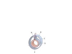
NAme
|

A. Mesentery
B. Peritoneal Cavity C. Peritoneum, parietal layer D. Peritoneum, visceral layer E. Intraperitoneal organ |
|
|
What is peritoneal ligament composed of?
|
Two layers of peritoneum anchoring an organ to the body wall or to another organ (not bone)
|
|
|
Define intraperitoneal organs
|
Organs covered in peritoneum and suspended from body wall by mesentery. An example would be a piece of intestine.
|
|
|
Define retroperitoneal
|
Organs/structures the develop and always remain posterior to the peritoneal sac (kidneys, aorta, IVC)
|
|
|
Define secondarily retroperitoneal
|
Organs that develop intraperitoneally but become compressed against the body wall and appear to be retroperitoneal. These organs are separated from the body wall by a bloodless plane.
|
|
|
Prof said we need to know intraparitoneal, retroperitoneal and secondarily retroperitoneal. Define them
|
Got it?
|
|
|
When lying supine, what are the two lowest portions of the peritoneal cavity where the most fluid will accumulate?
|
1. Hepatorenal recess/pouch (AKA Morrison's Pouch).
2. Rectovesicle/rectouterin pouch (Pouch of Douglas) |
|
|
Where are the paracolic gutters?
|
The are next to the ascending and descending colons.
|
|
|
What tissue structure splits the body into right and left halves?
|
The dorsal mesentary.
|
|
|
How do you get into the lesser sac? What is it also known as?
|
Put fingers deep to duodenum and anterior to pancreas. AKA Foramen of Winslow
|
|
|
What is the name of the foregut artery? What structures does it send blood to?
|
Celiac sends blood to the liver, stomach, spleen, and gall bladder.
|
|
|
What does the spleen do?
|
It stores and recycles blood.
|
|
|
Which parts of the duodenum are intra- or retroperitoneal?
|
The 1st and 4th part are intraperitoneal. The 2nd and 3rd part are retroperitoneal.
|
|
|
What defines the boundries between the foregut and midgut?
|
Defined by arteries that supply them. The foregut is supplied by the celiac artery (which is composed of the 1st and 2nd parts of duodenum). The midgut is supplied by the superior mesenteric artery (which is composed of 3rd and 4th part of duodenum)
|
|
|
What ligament supports the 4th part of the duodenum?
|
It's a suspensory ligament called the Ligament of Trietz that connects the diaphragm to the fourth part of the duodenum.
|
|
|
What ligament supports the 4th part of the duodenum?
|
It's a suspensory ligament called the Ligament of Trietz that connects the diaphragm to the fourth part of the duodenum.
|
|
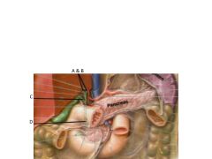
Name em
|

A. Right Hepatic Ducts
B. Left Hepatic Ducts C. Cystic Duct D. Common Bile Duct |
|
|
What three structures make up the Portal Triad?
|
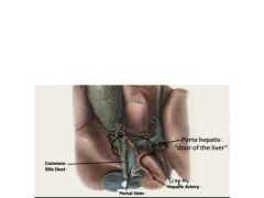
1. Common Bile Duct
2. Portal Vein 3. Proper Hepatic Artery |
|
|
Name the five parts of the pancreas
|
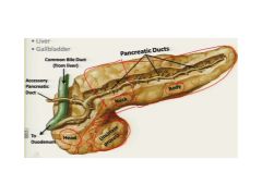
1. Head
2. Uncinate Process 3. Neck 4. Body, 5. Tail |
|
|
What are the four lobes of the liver?
|
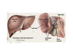
1. Caudate lobe
2. Quadrate lobe 3. Left Lobe 4. Right lobe |
|
|
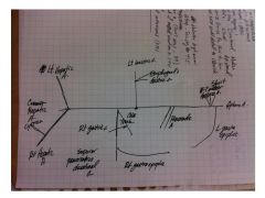
Can you draw the celiac trunk vasculature? See back side
|
|
|
|
The lesser omentum is composed of two ligaments - what are they?
|
Hepatodueodenal and hepatogastric ligament.
|
|
|
By placing your finger and pinching off the structures in the foremen of Winslow, what can you control?
|
1. Common Bile Duct
2. Hepatic Portal Vein 3. Proper Hepatic Artery |
|
|
What structure separates the common hepatic artery from the proper hepatic artery? (In lecture, he said, "If you were in an exam and... how would you figure this out?)
|
The gastroduodenal artery (see N284 or 9/19 1p at 1:18:30)
|
|
|
As the R gastric artery runs along the lesser curvature of the stomach, which artery does it anastomose with?
|
The L gastric artery.
|
|
|
Lecture Q: If the L gastric artery was blocked right at the aorta, how is it possible that the esophagus could still receive blood? Trace the arteries.
|
Celiac trunk --> common hepatic artery --> proper hepatic a --> right gastric a --> up around to esophageal a.
|
|
|
For your own benefit, how does blood reach the R gastro-omentum?
|
Celiac T --> common hepatic --> Gastroduodenal --> R gastro-epiploic
|
|
|
The gastroduodenal artery splits into (3 arteries):
(BTW - N283/4 is worth reviewing over and over again) |
1. R gastro-omentum
2. Supraduodenal a. 3. superior pancreaticoduodenal (ant & post) |
|
|
Where is the division for the midgut/hind gut? All of the small intestine is in the..?
|
The first 2/3 of colon is the midgut and the later 1/3 is the hind gut (this is established primarily by vascular supply). The small intestine is in the mid gut.
|
|
|
What structures make up the mid gut?
|
Parts 3&4 of the duodenum, jejunum, and ilium.
|
|
|
What artery supplies the small intestines (sp Jejunum and Ileum)? What about the large intestines?
|
Sm = Superior Mesenteric Artery
Lg = Inferior mesenteric artery |
|
|
In lecture, Hoagland mentioned three major aa coming off of the superior mesenteric. What are they and what do the supply? (And what's the fourth general ones?)
|
1. Inferior pancreaticoduodenal a - it supplies the duodenum and pancreas (it's the "watershed" a that divides the midgut from the hindgut)
2. Middle Colic - it serves the "middle" of the transverse colon 3. Iliocolic - serve the iliocolic region including the appendicular region that serves the appendix. The general ones include the jejunal and ileal (intestinal) arteries. |
|
|
What is the major artery that serves the Hindgut?
|
The Inferior Mesenteric Artery (the distal 3rd of the transverse colon)
|
|
|
What are the three major aa coming off of the inferior mesenteric a?
|
1. Left colic a
2. Sigmoid branches 3. Superior rectal artery |
|

Name em
|
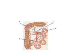
1. Superior mesenteric a
2. Middle colic a 3. Inferior pancreaticoduodenal a 4. Iliocolic a |
|
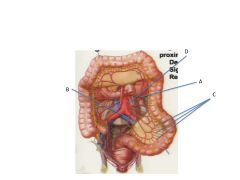
Name em
|

A. Left Colic
B. Superior Rectal C. Sigmoid Branches D. Inferior mesenteric artery |
|
|
Where do the glomeruli, the functional unit of the kidney, reside?
|
The cortex
|
|
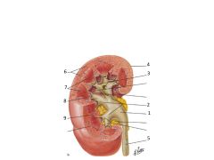
Name
|

1. Renal pelvis
2. major calyces 3. minor calyces 4. renal capsule 5. ureter 6. cortex 7. medulla (pyramids) 8. renal papilla 9. renal column (of Bertin) |
|
|
What is almost always directly superior to the L renal v?
|
Superior mesenteric a
|
|
|
The adrenal gland on the left is called (1)? The adrenal gland on the right is called (2)?
|
1. Left lunar adrenal gland
2. R pyramid adrenal gland |
|
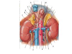
Name
|

1. L testicular (ovarian) a & v
2. R testicular (ovarian) a & v 3. L renal a and v 4. superior mesenteric 5. R superior suprarenal a 6. R middle suprarenal a 7. R inferior suprarenal a 8. R suprarenal v 9. R and L inferior phrenic a 10. L inferior phrenic v |
|
|
The R testicular v drains into the ? The L testicular v drains into the ? This difference can sometimes cause what? Why?
|
R = IVC; L = L renal v. The L renal v has a lower flow rate and this can cause pressure that leads to an engorged testicle - aka a Varicocele - a hemrhoidalization of Left testicle
|
|
|
AS in lecture, "If I were to shoot you, where would the bullet go through?"...in regard to the renal vessels?
|
Renal v, Renal a, then ureter.
|
|
|
Note: the ureter is a muscular tube capable of peristalses. What muscle does it run along? What vessels does it run behind? What vessels does it run anterior to?
|
Runs along psoas major muscle. Runs behind testicular vessels. It runs anterior to the iliac vessels.
|
|
|
Describe origin, insertion and under what ligament the psoas major muscle runs. Its action and innervation.
|
Origin is T12-L5; Insertion is lesser trochanter of femur; Runs under inguinal ligament; It flexes the hip; Innervation is lumbar plexus (L1 book) L2, 3, & 4.
|
|
|
Describe origin, insertion and under what ligament does the psoas minor muscle run
|
Origin is T12 to L1; Inserts at pectineal line; runs under inguinal ligament; has no action (really); and about 50% actually have one
|
|
|
Iliacus muscle originates from; inserts where and with; action is; nerve supply is
|
Originates from the iliac crest/fossa; inserts with psoas major muscle at lesser troachanter of femur; action is to flex the hip; nerve supply is L1 to L3; action flexes hip
|
|
|
Quadradus lumborum originates? Inserts; Its action; nerve supply
|
Originates at iliac crest and iliolumbar ligament, inserts at the 12th rib; helps bend trunk, nerve supply is from lumbar plexus (subcostal n (T12) and L1-4)
|
|
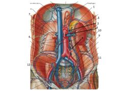
name
|

1. Inferior phrenic
2. Celiac trunk 3. Middle adrenal a 4. SMA 5. Ovarian (testicular aa) 6. Four pairs of lumbar aa 7. Renal aa 8. IMA 9. Internal Iliac 10. Common Iliac 11. External iliac 12. Median sacral |