Reading...
![]()
Play button
![]()
Play button
![]()
Use LEFT and RIGHT arrow keys to navigate between flashcards;
Use UP and DOWN arrow keys to flip the card;
H to show hint;
A reads text to speech;
69 Cards in this Set
- Front
- Back
|
The acetabular fossa is made deeper by the..
|
acetabular labrum
|
|
|
What ligament bridges the acetabular notch?
|
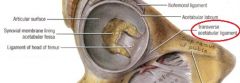
transverse acetabular ligament
|
|
|
The ____________________ attaches to the margins of the acetabular notch and to the transverse acetabular ligament.
|

ligamentum capitis femoris
|
|
|
The main function of the ligamentum capitis femoris is to...
|

carry the artery to the head of the femur, a branch of the obturator artery
|
|
|
The head of the femur is covered by hyaline cartilage except for the small central depression called the ________________, in which you can find the _________________.
|
-fovea capitis femoris
-ligamentum capitis femoris |
|
|
The normal angle of inclination between the femoral neck and shaft is...
|
about 126 degrees (from 115 to 140)
|
|
|
Abnormal femoral angle of inclination: coxa vara...
|
is a decrease in the angle (less than 120 degrees, looks like an "r")
|
|
|
Abnormal femoral angle of inclination: coxa valga...
|
is an increase in the angle (more than 135 degrees, looks like an "l")
|
|
|
shortened leg, limp; pain free gait abnormality; congenital or acquired (infection, tumor, post-traumatic fracture, metabolic)--coxa vara or coxa valga?
|
coxa vara
|
|
|
slipped epiphysis of femoral head; genu varum (bow legged)--coxa vara of coxa valga?
|
coxa valga
|
|
|
The angle of torsion/declination is normally ____ in females and ____ in males.
|
12 degrees females
7 degrees males |
|
|
_______________ is an abnormal increase in the angle of torsion; equals _________________.
|
-anteversion
-internal femoral torsion |
|
|
_______________ is an abnormal decrease in angle of torsion; equals ______________
|
-retroversion (retro - decrease)
-external femoral torsion (ER - external:retro) |
|
|
What is the main blood supply to the head of the femur?
|
retinacular branches of the medial femoral circumflex artery
|
|
|
What is the secondary blood supply to the head of the femur?
|
retinacular branches of the lateral femoral circumflex artery
|
|
|
What artery supplies blood to the head of the femur and is important in children, but not so much in adults?
|
artery to the head of the femur (remember, it's enclosed in the ligamentum capitis femoris)
|
|
|
What are the trochanteric anastomoses?
|
Lat. fem. circumflex a.
Inferior gluteal a. Medial fem. circumflex a. Superior gluteal a. (LIMS) |
|
|
What is a consequence of a femoral neck fracture?
|
torn retinacular branches of femoral circumflex arteries, causing avascular necrosis of the femoral head
|
|
|
What are the 4 causes of avascular necrosis to the femoral head?
|
-chronic alcohol
-steroid use -femoral neck fracture -posterior hip dislocation |
|
|
The _______________ fracture runs from the greater trochanter of the femur to the lesser trochanter and (does/does not) involve the femoral neck. It (does/does not) cause avascular necrosis of the femoral head.
|
-intertrochanteric
-does not involve neck -does not cause avascular necrosis |
|
|
_______________ is a line drawn from the neck of the femur through the _______________ and is important in monitoring changes in the head/neck of the femur (ex. femoral neck fracture).
|
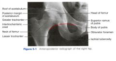
-Shenton's line
-lower margin of the superior pubic ramus |
|
|
What three ligaments form the fibrous capsule around the hip joint?
|
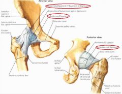
-iliofemoral ligament ("ilYofemoral")
-pubofemoral ligament -ischiofemoral ligament |
|
|
Where does the iliofemoral ligament attach?
What action does it prevent? |
-anterior inferior ilaic spine and acetabular rim to the intertrochanteric line
-prevent hyperextension |
|
|
Where does the pubofemoral ligament insert?
What action does it prevent? |
-obturator crest and blends with iliofemoral ligament
-limits extension and abduction |
|
|
What separates the pubofemoral ligament and the iliofemoral ligament?
|
-gap covered by the iliopsoas muscle and iliopsoas bursa
|
|
|
Where does the ischiofemoral ligament attach?
What action does it prevent? |
-spirals from the ischial part of the acetabular rim to the neck of the femur medial to base of greater trochanter
-prevents hyperextension (NOTE: Both the ischiofemoral and iliofemoral ligaments prevent hyperextension. Pubofemoral prevents extension and abduction). |
|
|
Posterior hip luxation (dislocation) is easiest in what position?
|
-during flexion and adduction of thigh (ligaments prevent extension, so they are relaxed when thigh is flexed)
|
|
|
90% of hip dislocations is in what direction?
|
posterior
|
|
|
Anterior dislocation of hip joint are caused by what action?
This accounts for what percent of hip dislocations? |
-forceful abduction and external rotation
-5% |
|
|
Congenital hip dislocation/developmental dysplasia of the hip occurs in 1.5/1000 live births and is more common in ____
Some of the risk factors include: |
-girls
Risk factors: -family history -generalized ligamentous laxity -breech birth |
|
|
This dislocation, which is occurs prior to skeletal maturity, causes the head and neck to separate where they are located at the epiphyseal plate
|
slipped capital femoral epiphysis
|
|
|
The trochanteric bursa is between what two structures?
|
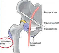
gluteus maximus and the greater trochanter
|
|
|
A person with trochanteric bursitis may produce pain where?
|
pain usually radiates distally along iliotibial tract
|
|
|
What bursa is found under the iliopsoas muscle
|
iliopsoas bursa
|
|
|
The iliopsoas bursa may be inflamed (iliopsoas bursitis) in cases of ____, causing pain in the ____ area
|
-rheumatoid arthritis or overuse injury
-anteromedial thigh pain |
|
|
Branches of what two nerves innervate both the hip and knee joints, allowing hip pain to be referred to the knee and vertebral/sacroiliac/prostate pain to the hip?
|
femoral and obturator nerves
|
|
|
Hip pain is often referred to the ____
|
knee
|
|
|
Vertebral column, sacroiliac joint, or prostate pain may be referred to the ____
|
hip
|
|
|
The gluteus maximus does what actions?
|
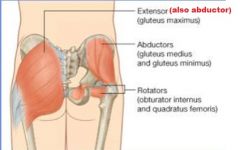
-powerful extensor
-laterally rotates thigh -abducts thigh |
|
|
What muscles are innervated by the superior gluteal nerve?
|
-tensor fascia lata
-gluteus medius -gluteus minimus (everything except the gluteus maximus) |
|
|
What muscles are innervated by the inferior gluteal nerve?
|
gluteus maximus
|
|
|
What is the function of the tensor fascia lata?
|
stabilizes knee during extension
|
|
|
What is the function of the gluteus medius and gluteus minimus?
|
-most important action - stabilize pelvis
-abducts and medially rotates femur at hip joint |
|
|
What are the deep muscles of the gluteal region that cause lateral rotation?
|
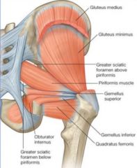
-Piriformis (+abd)
-Gemellus superior (+abd) -Obturator internus (+abd) -Gemellus inferior (+abd) -Obturator externus (not abd) -Quadratus femoris (not abd) The attachments of the last two muscles are a bit more distal on the femur--does not cause abduction. However, all muscle laterally rotate the thigh as they attach on the posterior surface of the femur |
|
|
Clarify slide about nerve
|
slide 27
|
|
|
What are the two arteries of the gluteal region?
|
-superior gluteal artery
-inferior gluteal artery |
|
|
The superior and inferior gluteal arteries are a branch of _____
|
internal iliac artery
|
|
|
The superior gluteal artery enters the gluteal region through the _____ and is ____ to the piriformis muscle
|
-upper part of the greater sciatic foramen
-superior |
|
|
The inferior gluteal artery enters the gluteal region through the ____ and is _____ to the piriformis muscle
|
-lower part of the greater sciatic foramen
-inferior |
|
|
The cruciate anastomoses provide alternate blood supply to the lower limbs due to occlusion of the ______________.
|
femoral artery
|
|
|
The cruciate anastomoses are:
|
Inferior gluteal artery
Medial femoral circumflex artery Lateral femoral circumflex artery First perforating branch of profunda femoris artery "I Got My First and Last Chia Pet" |
|
|
The pudendal canal is formed by the _____ in the _____
|
-obturator interus fascia
-ischioanal fossa |
|
|
What are the nerves of the gluteal muscles?
|
-superior gluteal nerve
-inferior gluteal nerve -nerve to piriformis -nerve to obturator internus -nerve to quadratus femoris |
|
|
A patient has a hard time rising from a seated position, climbing stairs, and running and jumping. What nerve may be damaged? What muscles does this nerve innervate?
|
-inferior gluteal nerve (L5-S2)
-gluteus maximus |
|
|
A patient comes in with weakness in abducting his thigh. Tests show trendelenburg sign and gluteus medius gait. What nerve may be damaged? What muscle does this innervate?
|
-Superior gluteal nerve (L4-S1)
-gluteus medius and minimus |
|
|
Paralysis of the gluteus medius and minimus result in a positive ____ test
|
Tredelenburg
|
|
|
Describe the trendelenburg sign
|
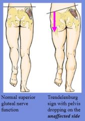
-patient is asked to raise knee
-the "normal side" drops when its knee is lifted and "affected side" supports the body *when standing with both feet, patient leans towards affected side |
|
|
Sciatic nerve damage may be due to:
|
-badly placed gluteal intramuscular injection
-posterior hip dislocation -traction on baby's legs during difficult childbirth |
|
|
Damage to sciatic nerve can cause:
|
-hamstring muscle paralyzed
-all muscles below the knee paralyzed -flail foot |
|
|
One or more roots of the sciatic nerve may be damaged in the vertebral canal due to:
This can cause damage to what nerve? |
-protruding or herniated intervertebral disc
-common fibular (peroneal) nerve - foot drop |
|
|
Damage to the common fibular nerve can cause:
|
-FOOTDROP
-paralysis of muscles in anterior and lateral leg compartments |
|
|
Sciatic nerve may be compressed by what muscle, resulting in...
This may cause: |
-piriformis muscle
-pain and/or paresthesia in gluteal region and posterior thigh |
|
|
Piriformis syndrome is more likely to happen if:
|
-if the common fibular nerve pierces the piriformis
|
|
|
What are the actions of the semimembranosus and semitendinosus muscles?
|
-flex knee
-extend hip -medially rotate lower limb seMi's are Medial rotators |
|
|
What are the actions of the biceps femoris muscles?
|
-flex leg at knee
-extend thigh at hip (only long head) -laterally rotate lower limb (remember, the seMi's Medially rotate) |
|
|
What are the hamstring muscles?
Where do the originate? What never innervates them? |
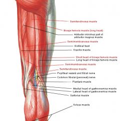
-Semitendinosus
-Semimembranosus -Biceps femoris long head -Adductor magnus, hamstring part -Ischial tuberosity -tibial nerve of sciatic nerve (except short head of bicep femoris) -short head of bicep femoris - common fibular nerve |
|
|
What artery supplies the posterior thigh?
|
profunda femoris artery (Posterior = Profunda)
*also largest branch of femoral artery |
|
|
The posterior thigh muscles (biceps femoris, semitendinosus, semimembranosus) divide into what two divisions in the distal thigh (popliteal fossa)?
|
-tibial division
-common fibular division |
|
|
Trochanteric anastomoses provide collateral circulation to the head of the femur when the _________________ are blocked.
|
femoral or medial femoral circumflex arteries
|