![]()
![]()
![]()
Use LEFT and RIGHT arrow keys to navigate between flashcards;
Use UP and DOWN arrow keys to flip the card;
H to show hint;
A reads text to speech;
226 Cards in this Set
- Front
- Back
|
Functions of the Skeletal System |
|
|
|
Support Function |
Structural support for the body. Framework for the attachment of organs and tissues. |
|
|
Protection Function |
Ribs, Cranium and Vertebrae protect soft organs |
|
|
Movement Function |
Bones provide leverage to provide movement |
|
|
Storage Function |
Minerals and lipids are stored |
|
|
Blood Cell Production |
Red and white blood cells are produced in the bone marrow |
|
|
5 Bone Shapes |
|
|
|
Long Bone Shape/ Location |
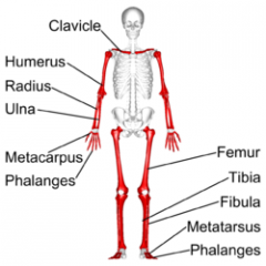
Long and Slender Bones |
|
|
Flat Bone/ Location |
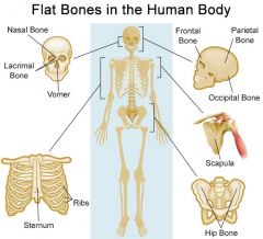
Thin, parallel surfaces |
|
|
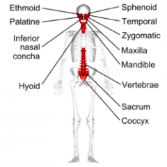
Irregular Bone/ Location |
|
|
|
Short Bone Location |
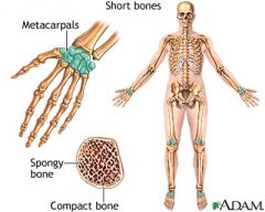
Small and Boxy |
|
|
Sesamoid Bone/ Location |
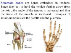
Flat, like sesame seeds |
|

Diaphysis |
1- The shaft; made of compact bone |
|

Epiphysis |
3/2- Expanded ends of the bone; made of cancellous (spongy) bone |
|

Metaphysis |
7- Connects the Epiphysis with the Diaphysis |
|
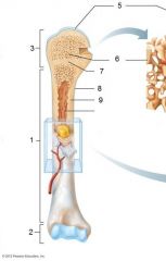
Medullary Cavity |
Inside the compact bone of the diaphysis; contains bone marrow |
|
|
Process |
Any Projection |
|
|
Trochanter |
A process where tendons attach; large, rough projection |
|
|
Tuberosity |
A process where tendons attach; small, rough projection |
|
|
Head |
Processes where tendons attach; expanded articular end of an epidermis separated by a neck |
|
|
Neck |
Processes for articulations with adjacent bones; narrow connection |
|
|
Condyle |
Processes for articulations with adjacent bones; smooth, rounded articular process |
|
|
Fossa |
Depression; a shallow depression |
|
|
Sulcus |
Depression; a narrow groove |
|
|
Foramen |
Opening; A passageway for blood vessels or nerves |
|
|
Canal or Meatus |
Opening; Passage through the substance of a bone |
|
|
Bone Tissue |
Bone is a supportive connective tissue |
|
|
Bone Cells/ Matrix |
|
|
|
Cells of the Skeletal System |
|
|
|
Osteocytes |
Mature bone cells, maintain matrix, and occupy lacuna. |
|
|
Lacuna |
Pockets between layers of matrix which are called lamellae |
|
|
Canaliculi |
Narrow passages that interconnect lacunae |
|
|
Osteoblasts |
Immature bone cells that new matrix |
|
|
Osteoprogenitor |
Stem cells that maintain the osteoblast population |
|
|
Osteoclasts |
Multinucleated cells that remove and recycle bone matrix, OPPOSE the activity of osteoblasts |
|
|
Two Types of Bone Based on Histology |
|
|
|
Osteon |
(Haversian System) The unit of mature compact bone, osteocytes are arranged in concentric layers around the central canals (Haversian Canals) |
|
|
Volkmann's (Perforating) Canals |
Run perpendicular and supply blood to deeper osteons and medulla |
|
|
Concentrate Lamella |
Around central canal |
|
|
Interstitial Lamella |
Between osteons |
|
|
Circumferential Lamella |
At the outer and inner surface of the bone |
|
|
Periosteum |
A membrane with a fibrous outer layer and a cellular inner layer |
|
|
Endosteum |
An incomplete cellular layer containing osteoblasts, osteoprogenitor cells, and osteoclasts |
|
|
Spongy Cancellous Bone |
|
|
|
Ossification |
The replacement of other tissues with bone tissue Two Types: Endochondral & Intramembraneous |
|
|
Endochondral |
|
|
|
Intramembraneous |
|
|
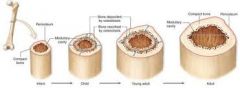
Appositional Growth (In Diameter) |
The growth at the outer surface, cells of the inner layer of the periosteum differentiate into osteoblasts and deposit layers of the bone matrix. Osteoclasts also remove matrix in the inner surface making the medulla larger. |
|
|
Factors That Affect Bone Growth |
|
|
|
Nutrition |
Osseous tissue requires a lot of nutrients and thus is highly vascularized
|
|
|
Hormones |
|
|
|
Bone Remodeling |
Bones are in constant remodeling, a balance between osteoclasts (constantly removing matrix) and osteoblasts (adding matrix); this dynamic cycle allows bone repair |
|
|
Close Fracture |
Completely internal and easy to treat |
|
|
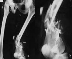
Open Fracture |
Fracture projects through the skin. These are dangerous due to potential infection or excessive bleeding |
|
|
Complete Fracture |
If the bone is completely separated in two pieces |
|
|
Incomplete Fracture |
Bone is not completely separated |
|
|
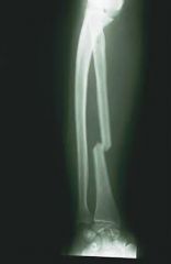
Transverse Fracture |
Break at right angle to the long axis of the bone |
|
|
Comminuted |
Bony fragments are produced |
|
|
Spiral |
Produced by twisting stresses |
|
|
Spiral Fracture |
|
|
Comminuted Fracture |
|
|
Transverse Fracture |
|
|
Bone Disorders |
|
|
|
Osteomyelitis |
Bacterial Infection of the bone marrow |
|
|
Osteopenia |
Deficiency in ossification due to aging, bones becomes thinner and weaker |
|
|
Osteoporosis |
Normal function is compromised |
|
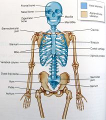
The Axial Skeleton |
1. Skull 2. Vertebral Column 3. Thoracic Cage |
|
|
Skull |
|
|
|
Facial Bones... |
Facial bones protect and support entrances to the digestive and respiratory systems, provide attachment of muscles that control facial expressions
|
|
|
Sutures |
The connections between skull bones. Bones are tied firmly together with dense fibrous connective tissue |
|
|
Superior View of the Skull/ Main Bones |
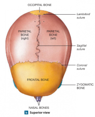
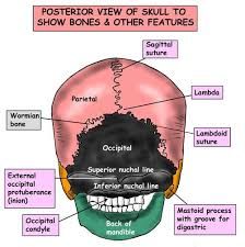
Occipital Bone, Parietal Bone (Left and Right), Frontal Bone, and Nasal Bones |
|
|
Associated Skull Bones |
|
|
|
Cranium Bones |
Eight Bones Total
|
|
|
Facial Bones |
Fourteen Bones Total
|
|
|
Posterior View of the Skull |
|
|
|
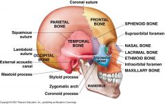
Lateral View of the Skull |
|
|
|
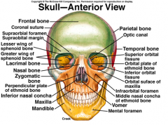
Anterior View of the Skull |
|
|
|
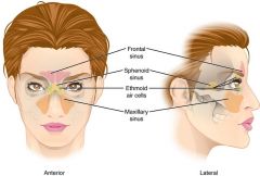
Paranasal Sinuses |
|
|
|
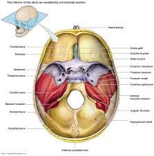
Interior View of the Skull |
|
|
|
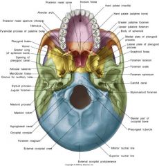
Inferior View of the Skull |
|
|
|
Functions of the Vertebral Column |
|
|
|
Four Major Curves of the Vertebral Column |
|
|
|
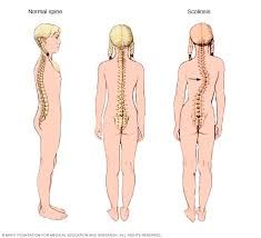
Disorders of the Vertebral Column |
|
|
|
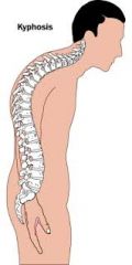
Kyphosis |
|
|
|
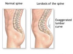
Lordosis |
|
|
|
Scoliosis |
|
|
|
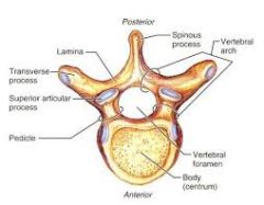
Vertebra |
Body (transfers weight along the axis), arch, processes (spinous, transversal, superior and inferior articular) lamina, and pedicle. |
|
|
Interverbal Discs |
Pads of fibrous cartilage |
|
|
Vertebral Foramen |
|
|
|
Cervical |
|
|
|
Thoracic |
|
|
|
Lumbar |
|
|
|
Sacrum |
|
|
|
Coccyx |
|
|
|
Thoracic Cage consists... |
of the thoracic vertebrae, ribs, and sternum |
|
|
Thoracic Cage Functions |
|
|
|
Ribs |
|
|
|
Vertebrosternal |
Ribs 1-7 |
|
|
Vertebrochondral |
Ribs 8-10 |
|
|
Floating Ribs |
Ribs 11 and 12 |
|
|
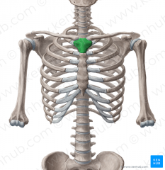
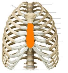
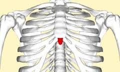
Sternum |
A flat bone that forms in the anterior midline of the thoracic cavity |
|
|
Three Thoracic Cage Components |
|
|
|
The Appendicular Skeleton |
|
|
|
Manubrium
|
|
|
Sternal Body |
|
|
Xiphoid Process
|
|
|
The Pectoral (Shoulder Girdle) |
|
|
|

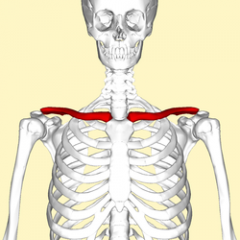
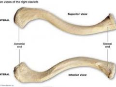
The clavicles are "S" shaped bones that originate at the manubrium of the sternum and articulate with the acromion process of the scapula -sternal and acromial ends |
|
|
|
|
|
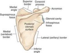
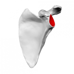
Glenoid Cavity (Fossa)
|
|
|
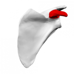
Coracoid Process
|
|
|
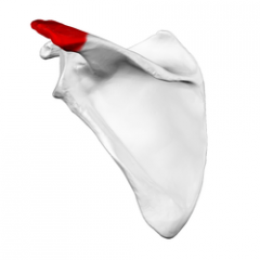
Acromion Process
|
|
|
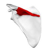
Scapular Spine
|
|
|
Acromial end of the clavicle |
Facet for articulation with acromion; lateral end of the clavicle |
|
|
Sternal end of the clavicle |

Medial end of the clavicle |
|
|
Upper Limb |
Bones of the arm, forearm, wrist, and hand |
|
|
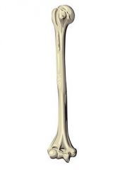
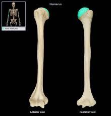
Humerus Bone: Bone of the upper arm |
|
|
Humerus Head |

Articulates with scapula |
|
|
Humerus Greater and Lesser Tubercle |
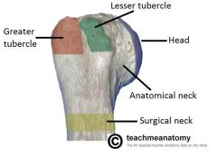
Round projections, important for muscle attachment |
|
|
Humerus Intertubercular Groove |
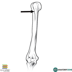
Between two tubercles (tendon of the latissimus dorsil) |
|
|
Humerus Trochlea |
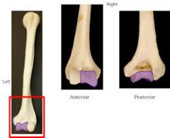
"Pulley," medial portion of condyle |
|
|
Humerus Capitulum |
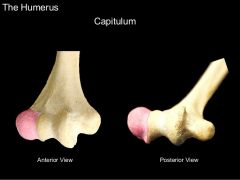
Lateral "ball" like portion of condyle |
|
|
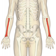
Forearm |
The ulna and radius run parallel in the forearm |
|
|
Forearm- Ulna |
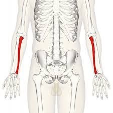
is medial |
|
|
Forearm- Olecranon |
(Posterior) point of the elbow |
|
|
Forearm- Trochlear Notch |
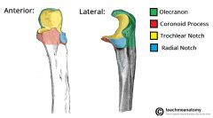
Anterior; articulates with the humerus |
|
|
Forearm- Ulna Head |
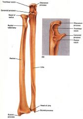
(distal end) articulates with radius |
|
|
Forearm- Radius Head |

Articulates with the capitulum of humerus |
|
|
Forearm- Radius |
Lateral |
|
|
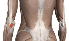
Forearm- Radial Tuberosity |
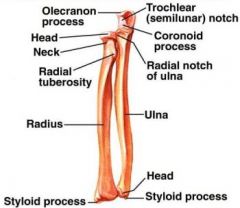
Where biceps attach |
|
|
Forearm- Styloid Process |

(distal) stabilizes articulation of radius with wrist |
|
|
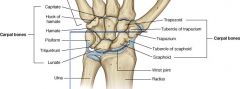
Wrist- Carpal Bones |
|
|
|
Hand |
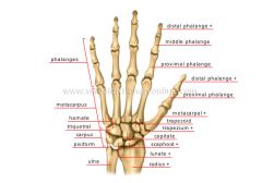
Contains five metacarpal bones, labeled I-V starting from lateral (thumb) -14 Phalanges (digit bones); every finger contains a proximal, middle, and distal phalanx (except the thumb which only contains a proximal and distal bone) |
|
|
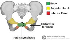
The Pelvic Girdle contains the... |
Coxal bones, acetabulum, iliac crest, and obturator foramen |
|
|
Coxal Bones |
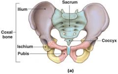
Formed by fusion of three bones: Ilium, Ischium, and pubis |
|
|
Acetabulum |
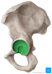
Concave socket, articulates with the head of the femur
|
|
|
Iliac Crest |
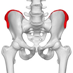
Most superior ridge |
|
|
Obturator Foramen |
|
|
|
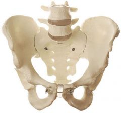
Female Pelvis |
|
|
|
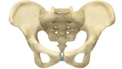
Male Pelvis |
|
|
|
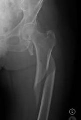
Femur |
Longest and heaviest bone in the body |
|
|
Femur- Head |
Articulates with the acetabulum of the coxal bone |
|
|
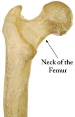
Femur- Neck |
|
|
|
Femur- Greater and Lesser Trochanter |
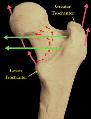
Attach to large tendons |
|
|
Femur- Medial and Lateral Condyles |
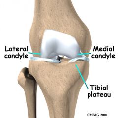
Participate in the knee joint |
|
|
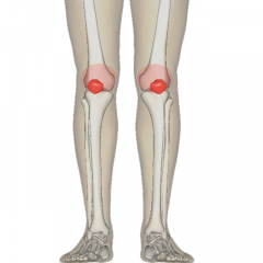
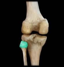
Patella- Forms within the tendon of the quadriceps femoris |
|
|
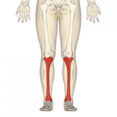
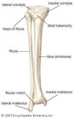
Tibia- the large medial bone of the lower leg |
|
|
Tibia- Anterior Margin (Anterior Crest) |
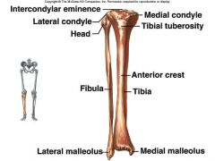
Begins at the tibial tuberosity and extends along the surface |
|
|
Tibia- Tibial Tuberosity |
|
|
|
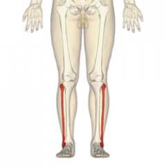
Fibula- Slender bone that parallels the tibia |
|
|
Fibular Head |
Articulates with the tibia |
|
|
Lower Leg- Lateral and Medial Malleolus |

Lateral (fibula) and Medial (tibia) malleolus provide lateral stability to the ankle |
|
|
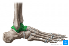
Ankle |
Contains seven tarsal bones |
|
|
Talus- transfers the weight from the tibia to the toes |
|
|
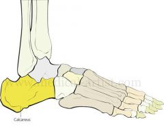
Calcaneus- heel bone; longest of the tarsal bones |
|
|
Achilles Tendon |
Attaches to the calcaneus |
|
|
Foot Contains... |
Five long metatarsal bones and fourteen phalanges |
|
|
Five Long Metatarsal Bones |
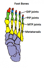
Identified by I-V starting from medial to lateral |
|
|
Fourteen Phalanges |
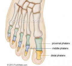
Proximal, middle, and distal except the hallux (big toe) |
|
|
Ways the skeleton can be used to tell age |
|
|
|
Articulations |
Joints where two bones interact |
|
|
Articulation Functions |
ALWAYS A COMPROMISE BETWEEN STRENGTH AND MOBILITY; classified according to function (range of motion) or structure |
|
|
Synarthrosis Movement |
No movement |
|
|
Amphiarthrosis Movement |
Little Movement |
|
|
Diarthrosis Movement |
Free Movement |
|
|
Bony Fusion |
Between two bones; Synarthrosis (No movement) |
|
|
Fibrous Joint |
Fibrous tissue between bones; Synarthrosis (No movement) / Amphiarthrosis (Little Movement) |
|
|
Cartilaginous Joint |
Two bones with cartilage between them; Synarthrosis (No movement) / Amphiarthrosis (Little Movement) |
|
|
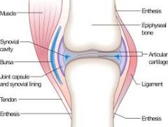
Synovial Joint |
Have a capsule; complex structure; Diarthroses (free movement) |
|
|
Four Types of Synarthrosis |
|
|
|
Sutures |
Located only in the skull; bones are interlocked and bound with fibrous connective tissue. Examples: Coronal, sagittal, and lambdoid sutures |
|
|
Frontanels |
Soft spots on the head of babies that allow for natural birth |
|
|
Gomphosis |
Binds the teeth to bony sockets of maxilla and mandible; bound by a fibrous connective tissue called periodont |
|
|
Synchondrosis |
Rigid, Cartilaginous bridge between two articulating bones; ex. epiphyseal cartilage, vertebrosternal ribs and sternum |
|
|
Synostosis |
Immovable joint created when two bones fuse and the boundary disappears. Ex. frontal or metopic suture |
|
|
Amphiarthrosis |
Much stronger than a freely movable joint |
|
|
Two Types of Amphiarthrosis Joints |
|
|
|
Diarthrosis (synovial) |
Typically at the end of long bones |
|
|
Diarthrosis Components- Articular Capsule |
|
|
|
Diarthrosis Components- Synovial Fluid |
A clear viscous solution that fills the capsule and serves for lubrication, nutrition, and padding |
|
|
Diarthrosis Components- Articular Cartilage |
Covers the bone to reduce friction (functions like a sponge) |
|
|
Linear Movement |
Gliding motion; carpal bones |
|
|
Angular Movement |
Point stationary but shaft changes angle relative to the surface; most common in the human body |
|
|
Rotational Movement |
Point remains stationary while the shaft, held at less than a 90-degree angle, can move in a complete circle ex. shoulder |
|
|
Extension |
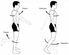
Straightening movement that increases the angle between body parts. When a joint can move forward or backward, extension refers to movement in the posterior direction. |
|
|
Flexion |

Opposite of extension |
|
|
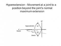
Hyperextension |
|
|
|
Abduction |

Movement away from the midline of the body |
|
|
Adduction |
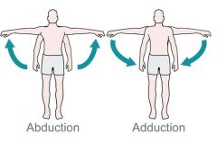
Movement toward the midline of the body |
|
|
Supination |

Rotation of the forearm turning the palm of the hand outwards so that it faces away from the body
|
|
|
Pronation |
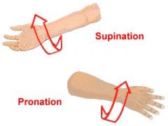
The forearm is a rotational movement where the hand and upper arm are turned inwards
|
|
|
Eversion vs. Inversion |
Inversion and eversion refer to movements that tilt the sole of the foot away from (eversion) or towards (inversion) the midline of the body.
|
|
|
Depression vs. Elevation |
Depression is the downward movement of structures of the body; Elevation is the upward movement of structures in the body
|
|
|
Retraction vs. Protraction |
Protraction is movement of a body part in the anterior direction, i.e. being drawn forwards. The movement of protraction is the opposite of the movement of retraction (backwards)
|
|
|
Opposition |
Brings the thumb and the little finger together |
|
|
Lateral Flexion |
Movement of the head or trunk laterally away from the midline of the body |
|
|
Gliding Synovial Joint |
Planar, relatively flat surfaces slide across one another, limited movement. Slight nonaxial or multiaxial movement; example: intercarpal and intertarsal joints, vertebrocostal joints |
|
|
Hinge Joint |
Permits angular movement in a single plane. Monaxial movement; example: elbow joint, knee joint, ankle joint |
|
|
Pivot Joint |
Monoaxial joint that allows only rotation. Monaxial (rotation) movement; Examples: Atlas/ axis |
|
|
Ellipsoidal Joint |
Oval articular face nestles in a depression, allows biaxial movement. ex. metatarsophalangeal joints |
|
|
Saddle Joint |
Each articular is concave in along one axis and convex along the other; allows biaxial angular motion, but not rotation. Example: first carpometacarpal joint |
|
|
Ball and Socket Joint |
Round head of one bone rests within a cup-shaped depression of another; allows both angular and rotational movement; example: shoulder joint, hip joint |
|
|
Shoulder Joint |
|
|
|
Elbow Joint |
|
|
|
Hip Joint |
|
|
|
Knee Joint |
|
|
|
Joint Disorders |
Common due to heavy "wear and tear," especially in older individuals |
|
|
Arthritis |
All diseases that affect synovial joints (always involves damage to the articular cartilage) |
|
|
Osteoarthritis |
(degenerative arthritis) usually occurs above 60 years of age. The leading cause of chronic disability |
|
|
Rheumatoid Arthritis |
Inflammation of the joints due to autoimmune diseases, bacteria, viruses, allergies, and genetic factors |
|
|
Gouty Arthritis |
Formation of crystals of uric acid in the synovial joints which interfere with function |
|
|
Treatment of Joints |
|
|
|
Cartilaginous Joints |
|
|
|
Synovial Joint- Articular Cartilage |

A thin layer of hyaline cartilage that covers the articulating surfaces of the bones reducing friction and absorbing shock |
|
|
Synovial Joint- Articular Capsule |
Surrounds each synovial joint consisting of two layers, the fibrous outer layer and the synovial membrane |
|
|
Synovial Joint- Fibrous Outer Layer |
Dense irregular connective tissue with abundant collagen fibers and is continuous with the periosteum of the articulating bones |
|
|
Synovial Joint- Synovial Membrane |
Composed of areolar connective tissue with abundant elastic fibers |
|
|
Synovial Joint- Synovial Cavity |

A small space filled with synovial fluid (a thick colorless liquid that lubricates the joint surfaces and absorbs shock. Also supplies oxygen and nutrients to and removes carbon dioxide and metabolic wastes from the chondrocytes of the articular cartilage |
|
|
Menisci |
Also called articular disks, fibrocartilage pads that act as shock absorbers and provide a better fit between articulating surfaces |
|
|
Types of Synovial Joints |
|
|
|
Plane Joint |
(Gliding joint) consists of flat articular surfaces that slide over one another, nonaxial; intercarpal joints, intertarsal joints, and the sacroiliac joints |
|
|
Hinge Joint |
One concave articular surface and one convex articular surface articulate to allow movement around one axis, uniaxial; elbow, the knee, interphalangeal joints and the temporomandibular joint |
|
|
Pivot Joint |
The rounded or conical articular surface of one bone articulates with a shallow depression on another bone, uniaxial movement; atlantoaxial joint and the proximal radioulnar joint |
|
|
Condylar Joint |
An oval-shaped condyle articulates with an elliptical cavity, biaxial; metacarpophalangeal joints and the atlanto-occipital joint |
|
|
Saddle Joint |
In which two saddle-shaped articulating surfaces articulate with each other, biaxial movement; only exists between the metacarpal of the thumb and the trapezium of the wrist |
|
|
Ball- and- Socket Joint |
The ball-shaped head of one bone, multiaxial movement; the shoulder and hip joints |