![]()
![]()
![]()
Use LEFT and RIGHT arrow keys to navigate between flashcards;
Use UP and DOWN arrow keys to flip the card;
H to show hint;
A reads text to speech;
76 Cards in this Set
- Front
- Back
|
4 Basic Tissue Types |
Epithelium, Connective, Muscular, Nervous |
|
|
Where can you find Epithelium |
Covering and lining, and glands It is the interface between 2 different environments -skin: air -gut tube: gut lumen -trachea:air |
|
|
Where can you find Connective tissue |
Bone, cartilage |
|
|
Classification criteria for epithelium |
Number of cells in the layer- simple(one) stratified(more then one) Shape of the cells- squamous, cuboidal, columnar |
|
|
Specific functions of Epithelium tissue |
Protection, secretion and absorption, excretion, sensory reception |
|
|
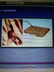
Simple squamous |
|
|
|
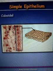
Simple cuboidal |
|
|
|
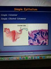
Simple columnar and simple ciliated columnar |
|
|
|
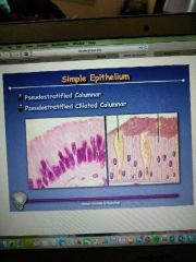
Psuedostratified columnar and psuedostratified ciliated columnar |
|
|
|
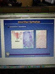
Stratified squamous |
|
|
|
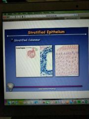
Stratified columnar |
|
|
|
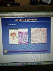
Transitional |
|
|
|
Apical Specializations |
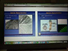
Microvilli (do not move) Cillia (move back and forth) |
|
|
Juctions |

Junctional complexes -hold cells together all the way around the cell Gap junctions-exist between two cells. They are like a tunnel between them that allow things to travel from one cell to the next |
|
|
Basement membrane |
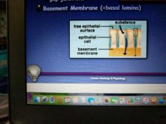
Basal lamina. Exist below the cells. It is what the cells attach to |
|
|
Exocrine glands |
Secrete enzymes into ducts that lead to target tissue Unicellular Ex: goblet cells |
|
|
Endocrine |
Secretes products such as hormones directly into the blood |
|
|
Common characteristics of connective tissue |
Extensive intercellular matrix Derived from mesenchyme( embryonic tissue) |
|
|
Locations of connective tissues |
-virtually all other tissues are embedded in it -underlies all epithelium -surrounds all muscles -lies within muscles -connects individual muscle fibers together -surrounds all blood vessels -surrounds all nerves that lie outside the CNS-surrounds and penetrates all glands NS-surrounds and penetrates all glands -surrounds and penetrates all glands |
|
|
Functions of connective tissue |
-support -convey body fluids -defense |
|
|
Loose connective tissue proper structures used for support |
-collagen fibers(keep from being pulled apart) -reticular fibers(provide support for things traveling through connective tissue) -elastic fibers(bend ex is ear) -fibroblasts( produce other fibers) |
|
|
Loose connective tissue proper structures used to convey body fluids |
Ground substance (hold tissue fluid in place) |
|
|
Loose connective tissue proper structures used for defense |
-histiocytes (eat and destroy) -plasma cells(antibodies) -mast cells (heparin and histamine) Eosinophils and neutrophils) |
|
|
Irregular Dense connective tissue proper |
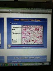
|
|
|
Regular dense connective tissue proper |
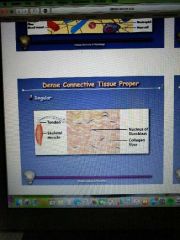
|
|
|
Fatty connective tissue proper |
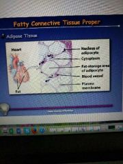
Adipose tissue |
|
|
Cartilage characteristics |
-firm intercellular matrix -chondrocytes (need oxygen and nutrients that are transported by diffusion through the capillaries that come up to the perichon) -lacunae -perichondrium( encloses cartilage) -avascular -no nerves |
|
|
Types of cartilage |
-hyaline cartilage -elastic cartilage -fibrocartilage |
|
|
Hyaline cartilage |
We have a lot more of it when we are younger The entire fetal skeleton vs. adult trachea,medial ends of ribs, and larynx(voice box) |
|
|
Elastic cartilage |
The ear can bend and stretch and returns back |
|
|
Fibrocartilage |
More collagen Found in the vertebrae to act as shock absorbers, in articulation between left and right pubic bone |
|
|
Bone |
Matrix -65% inorganic crystals -35% organic crystals Gross structure -compact bone(make up outer wall) -spongy bone(inside of bone) -articular cartilage -medullary cavity -periostium(around the bone falls off when dry) -endostium |
|
|
Microscopic structures of bones |
-osteocytes -lacunae -canaliculi -osteoblasts -osteoclasts |
|
|
Bone development |
Intramembranous(simple) -membrane bone(mesenchyme, woven bone,lamellar) Endochondral ossification(complex) -endochondral bone |
|
|
Bone growth |
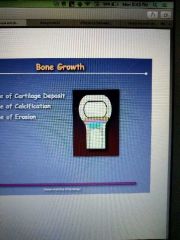
-zone of cartilage deposit -zone of calcification -zone of erosion |
|
|
Vascular tissue |
Plasma Formed elements -erythrocytes -leucocytes -thrombocytes(platelets) |
|
|
Embryonic development |
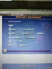
Differentiation |
|
|
Body tube |
-skin -coelom -serous membrane -body wall |
|
|
Gut tube |
Serous membrane. Runs down middle of body |
|
|
Dorsal body wall midline |
-vertebrae -nerve cord |
|
|
Embryonic period |
By 8 weeks outlines a person but nothing is functioning |
|
|
Fetal period |
Baby grows and develops systems closer to function |
|
|
Dorsal body wall lateral |
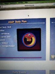
Kidneys Retroperitoneal |
|
|
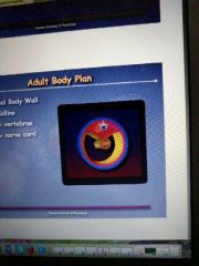
Adult body plan limbs |

Skin Muscle Bones |
|
|
Early events in embryonic development |
Ovulation Fertilization Cleavage Implantation |
|
|
Ovulation |
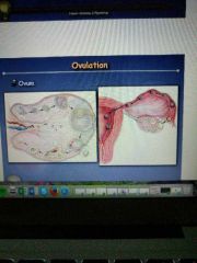
Ovum |
|
|
Fertilization |
Sperm -head(dna) -tail(flagellum) Zygote -2 cell -4 cell -8 cell -16 cell |
|
|
Late cleavage |
Morula(solid ball or cells) Blastocyst(hallow ball of cells) -trophoblast -blastocoel (the space in the middle) -inner cell mass (ICM) |
|
|
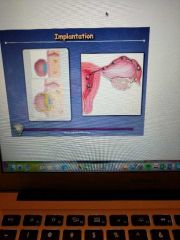
Implantation |
|
|
|
What is the integument system made of |
Epidermis -ectoderm Dermis -mesoderm Hypodermis |
|
|
Integument functions |
Protection Temperature regulation Vitamin D production Protection from UV Excretion Sensory reception |
|
|
Layers of the epidermis |
5 layers. Oldest on top youngest on bottom. All are produced by the bottom layer -stratum corneum( constantly shedding these cells) -stratum lucidum -stratum grandulosum( layer where cells die) -stratum spinosum -stratum basale |
|
|
Melanocytes |
Melanin Neutral crest |
|
|
Tanning |
More melanin/melanocytes |
|
|
Dermis |
2 layers -papillary layer(dermal ridges, finger prints) -reticular layer(lots of collegin) Muscles -arrector pili( goose bumps) -striated muscles |
|
|
Epidermal derivatives |
Hair follicle Sebaceous gland -sebum Sweat gland Nails -stratum corneum -stratum lucidum |
|
|
Articulation |
Any place where 2 or more bones meet May be freely moveable, slightly moveable, or immovable Classifications -functional -structural |
|
|
Fibrous joint |
No movement, joints are locked together Ex: suture |
|
|
Cartilaginous joint |
Held together with cartilage Ex: synchondrosis(hyaline cartilage in ribs) symphysis (fibrocartilage intervertebral disks) |
|
|
Synovial joint |
Allows for controlled movement Ex: nonaxial (gliding), axial (uni, bi, tried) |
|
|
Uniaxial |
Hinge joints. Move in one direction |
|
|
Biaxial |
2 directions Can move in a circle(circumduction) Ex:fingers, wrist |
|
|
Triaxial joint |
Ball and socket 3 directions Ex: hip, shoulder |
|
|
Structure of synovial joints |
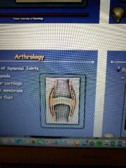
Joint capsule Articular cartilage Synovial membrane Synovial fluid |
|
|
Blood and Nerve supply to joint |
Very rich in blood supply Proprooreception(sensory info about positioning and pressure in joints. It's how your brain knows how to move limbs where you want them to go) Bursae -bursitis( most common in places like the knee) |
|
|
What are muscles |
Move bones and are found lying alongside and attached to bones Move hollow organs and are found embedded in the walls of hollow organs |
|
|
What do muscles consist of |
-connective tissue -blood vessels -nerves -muscle tissue |
|
|
Myofilaments |
Thick filament -myosin Thin filament - actin |
|
|
Different types of muscle tissue |
Striated muscle(skeletal muscle) Cardiac muscle Smooth muscle |
|
|
Smooth muscle fibers arrangement |
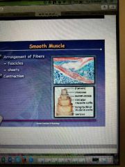
-fascicles -sheets |
|
|
Smooth muscle fibers size |
1-40 mm in length 1-40 mm in diameter |
|
|
Sarcomere |
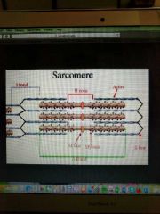
Z- line I- band A- band |
|
|
Sarcomere contraction |
|
|
|
Sarcoplasmic reticulum |
Calcium ions (Ca -2) -chemical for the reaction Transverse tubules (t- tubules) are in a 1/2 ratio with the lateral sacs Triad No energy is used to start muscle contraction An electrical impulse is sent, travels along t-tubules, which changes permiability for calcium and contraction happens Energy is needed to stop contraction since you are moving backwards along the concentration gradient |
|
|
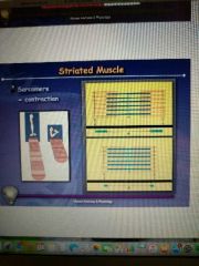
Arrangement of fibers in striated muscle |
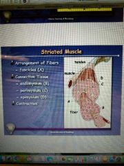
|
|
|
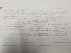
Cardiac muscle |
|