Reading...
![]()
Play button
![]()
Play button
![]()
Use LEFT and RIGHT arrow keys to navigate between flashcards;
Use UP and DOWN arrow keys to flip the card;
H to show hint;
A reads text to speech;
104 Cards in this Set
- Front
- Back
- 3rd side (hint)
|
The first 7 ribs are named?
|
vertebrosternal ribs
|
|
|
|
Ribs 8-10 are named?
|
vertebrochondral ribs
|
|
|
|
Ris 11 & 12 are named
|
floating ribs
|
|
|
|
ribs 8-12 are collectively named?
|
false ribs
|
|
|
|
The diameter of the spinal cord and size of the neural arch decreases as the spinal cord runs _____?
|
inferior
|
|
|
|
Axial skeleton includes?
|
Skull
Ossicles Hyoid Vertebra Ribs Sternum |
|
|
|
Vertebral recombination is when?
|
Each sclerotome splits into cranial and caudal segments. As the segmental spinal nerves grow out to innervate the myotomes, the cranial segment unites with the caudal segment of the next superior sclerotome to form a rudiment.
|
|
|
|
The notochord enclosed to become the _____?
|
nucleus pulposus
|
|
|
|
The annulus fibrosis is formed from?
|
coalesce of remaining cells from the splitting of sclerotomes.
|
|
|
|
the order of bone development is?
|
1-somite
2-sclerotome 3-cartilage 4-bone |
|
|
|
Primary ossification centers each vertebra is?
|
neural arch
*lamina and pedicle *fuse at age 2 years -neuro-central synchondrosis *fibrous joint *hyaline cartilage *fuse at age 7 years -centrum= body |
|
|
|
Secondary ossification centers of each vertebra is?
|
anular epiphysis
superior and inferior *tip of transverse process *tip of spinous process *fuse by 18 years |
|
|
|
What is the development of the ribs?
|
Costal processes begin to elongate in the fifth week. Late in the 6th week the costovertebral joints form and separate into ribs and vertebrae.
|
|
|
|
What is the development of the ribs? (2-16)
|
*Paired mesenchymal condensations called sternal bars form in the ventral body wall at the end of week 6.
*fuse at cranial end; *zip together cranio-caudally; *ossification centers appear at 60 days; *xiphoid process is not present until birth |
|
|
|
Dermamyotomes develop into?
|
dermatomes and myotomes
|
|
|
|
Dermatome
|
dorsolateral-lateral plate mesoderm which becomes the dermis.
|
|
|
|
myotomes develop into
|
dorsal epimere -> deep mm of the back (epaxial)
ventral hypomere -> other muscles (hypaxial) |
|
|
|
adaptations to secondary curvatures are?
|
Change in muscle attachments
Change in pelvis shape Change in magnum foramen Change in gait Short neck: head centre on vertebrae proprioception Minute tail: 3-5 coccygeal somites fuse into one coccyx |
|
|
|
Secondary curvatures are?
|
cervical and lumbar lordosis
|
|
|
|
primary curvatures are?
|
Thoracic, Sacral and coccyx kyphotic curvatures
|
|
|
|
Differences in the cervical vertebra
|
20-bifed spinous process
25-transverse foramen 23-anterior tubercle 24-posterior tubercle |
|
|
|
Differences in the thoracic vertebra?
|
9-inferior costovertebral facet
10-superior costovertebral facet 22-costotransverse facet |
|
|
|
Differences in the lumbar vertebra?
|
30-accesory process
31-mamillary process |
|
|
|
where is the sacral base angle?
|
between L5 and sacral base
|
|
|
|
cervical superior auricular facet (SAF) orientation?
|
superior-posterior
|
|
|
|
cervical inferior auricular facet orientation?
|
inferior-anterior
|
|
|
|
Cervical osteology
|
rectangular body with uncinate process
- pedicles: short - lamina: long and thin -spinous process: bifid -transverse process: short, grooved with foramen for vertebral artery -Intervertebral Foramen: round -disc height to body ratio: 2:5- most movement in the spine |
|
|
|
In which plane do the cervical faces lye?
|
coronal
|
|
|
|
Thoracic osteology
|
- rectangular body with vertebral-costal facet
- pedicles: short and strong - lamina: angled inferior -spinous process: thin and long, inbrication T5-8 -transverse process: short, angulated with articular process for rib -Intervertebral Foramen: small and circular -disc height to body ratio: 1:5-less movement |
|
|
|
thoracic superior auricular facet (SAF) orientation?
|
superior-posterior-lateral
|
|
|
|
Thoracic inferior auricular facet orientation?
|
inferior-anterior-medial
|
|
|
|
In which plane are the thoracic facets orientated?
|
coronal
|
|
|
|
Lumbar osteology
|
rectangular (square) body
-short and strong pedicles -short and broad lamina -spinous process: thick and broad, points posterior -transverse process: long and slender -Intervertebral Foramen: large and triangular -disc height to body ratio: 1:3 |
|
|
|
Lumber superior auricular facet (SAF) orientation?
|
concave, face posterior-medial
|
|
|
|
Lumbar inferior auricular facet orientation?
|
convex, face anterior-lateral
|
|
|
|
Where is the mammillary process located?
|
mammillary process on superior- posterior edge of (SAF) of lumbar vertebra.
|
|
|
|
In which plane are the lumbar facets orientated?
|
lie in sagittal plane becoming more coronal at L-S junction
|
|
|
|
Scoliosis?
|
Lateral curvature of the spine
Predominately in the thoracic vertebrae |
|
|
|
Hemivertebra?
|
when only right or left half develops
*usually in the thoracic vertebrae |
|
|
|
Spondylolisthesis/ spondylolysis
|
laminae and spinous process being a separate unit
*pars interarticularis |
|
|
|
Lumbarisation
|
when S1 is separate and appears as a lumbar
*may count 6 separate lumbar vertebra |
|
|
|
sacarisation
|
when L5 is joined and appears attached to the sacrum
*will count 4 separate lumbar vertebra |
|
|
|
Butterfly vertebra
|
Problem with centrum
Thoracic Kyphosis or scoliosis |
|
|
|
Agenesis of the atlas
|
Neural arch
AKA posterior arch Neural arch not present Anterior arch Usually normal |
|
|
|
Spina bifida occulta
|
Tuft of hair
Non-union of right and left neural arches No neural tube involvement Usually lumbars, can also be at C1 No clinical manifestations (can be 10% of population) |
|
|
|
Spina bifida meningocele
|
Neural tube defect along with neural arch defect
Fluid filled meninges protrusion Only dura protrusion |
|
|
|
Spina bifida meningomyelocele
|
Neural tube defect with neural arch defect
Neural tube tissue and dura protrusion |
|
|
|
Where is the carotid tubercle often compressed?
|
the anterior tubercle of the transverse process of C6
|
|
|
|
Axis
|
dens
*vertebral body- *spinous process- *transverse process-very short *transverse foramen *facet for C1 is on the dens |
|
|
|
Atlas
|
anterior tubercle
(mid body) facet for the dens is posterior *posterior tubercle not called the spinous process (mid laminar) *superiorly articulates with Occiput longer, more oblong facet found anteriorly *inferiorly articulates with axis smaller, rounder facet found anteriorly *large transverse process |
|
|
|
cervical rib
|
a supernumerary rib articulating with a cervical vertebra, usually 7th but not reaching the sternum anteriorly. In about 1% of the population - clinically important because they may compress C8 and T1 nerves or the inferior trunk - causing tingling and numbness along the medial aspect of the forearm. They may also compress the subclavian artery causing resulting in ischemic muscle pain
|
|
|
|
Cervical Rib Syndrome
|
(a) arterial thoracic outlet syndrome in which the subclavian artery is compromised by a fully formed cervical rib;
(b) true neurogenic thoracic outlet syndrome, in which the proximal lower trunk of the brachial plexus is compromised by a translucent band extending from the rudimentary cervical rib to the first rib |
|
|
|
Lushka’s joints
|
a series of joint like structures at the lateral edges of the vertebral bodies from vertebra C3 to T1, forming small spur like lips at the upper surface, covered with cartilage, and containing a capsule filled with fluid. They are considered by some to be diarthrodial joints and by others to be degenerative spaces of the intervertebral disks filled with extra cellular fluid and lined by a membrane formed by fibrocystes. They are frequent sites of spur formation.
Stedman: uncovertebral joint |
|
|
|
Ossification of ribs begins near ____ and spreads ____ and _______
|
angle
anterior posterior |
|
|
|
The sternum body articulates with ribs
|
2-7
|
|
|
|
The xiphoid process articulates with ribs?
|
does not articulate with ribs
|
|
|
|
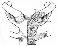
Clavicle
20-articular disc 19-articular capsule 24-interclavicular ligament 21-anterior sternoclavicular ligament 23-costoclavicular ligament |
|
|
|
longissimus thoracis muscles attach to?
|
posterior, inferior of TP to lamina
|
|
|
|
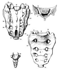
18 Base of the sacrum
19 Superior articular process 20 Anterior lip 22 Superior articular facet 23 Intermediate sacral crest 25 Anterior sacral foramena 27 Posterior sacral foramena 28 Median sacral crest 30 Lateral sacral crest 31 Sacral horns 33 Sacral hiatus 36 Cornua of coccyx |
|
|
|
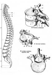
2-Cervical
20-bifed spinous process 25-transverse foramen 23-anterior tubercle 24-posterior tubercle 4-Thoracic 9-inferior costovertebral facet 10-superior costovertebral facet 22-costotransverse facet 5-Lumbar 30-accesory process 31-mamillary process 6-Sacrum 5 fused 7-Coccyx 3-5 fused |
|
|
|
what type of joint is the sacro-iliac joint
|
Diarthrodial: fibrous and synovial
|
|
|
|
Vertebral body and disc are Concave posterior? Tor F
|
True
|
|
|
|
what is the only part of the vertebral disc that is vascular?
|
their most peripheral parts - supported by diffusion through the spongy bone of vertebral bodies
|
|
|
|
Which portion of the vertabral disc repairs faster? the peripheral vascular portion or the avascular regions?
|
peripheral vascular
|
|
|
|
What portion of the vertebral column do the IVD's account for?
|
1/5th
|
|
|
|
Nucleus pulposus better developed in cervical thoracic or lumbar regions?
|
cervical and lumbar regions
|
|
|
|
can a vertebral end plate fracture be seen on radiography?
|
No. (due to compression)
|
|
|
|
Schmorl’s nodes are?
|
invagination of disc material into vertebral endplate
|
|
|
|
herniated disc
|
Annular ring disruption (circumferential tears)
Radial tears (leads to coelescening of radial tears) Can lead to disc protrusion (posterior protrusion with intact PLL) tend to be posterior-lateral where there are no longitudinal ligaments (Moore). Localized back pain from stretching of the longitudinal ligaments and annulus; but also presses on nerve roots. |
|
|
|
can sciatica be caused by IV disc that compresses and compromises L5 or S1 root?
|
Yes. IV foramina decrease in size while the nerves increase
|
|
|
|
the nuchal ligament is derived from?
|
supraspinous ligament (in cervical region)
|
|
|
|
the nuchal ligament is derived from?
|
supraspinous ligament (in cervical region)
|
|
|
|
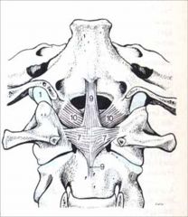
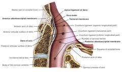
Cruciform ligament: “Cross”
*7= transverse ligament of atlas (part of cruciform) *9= longitudinal cruciform lig. (superior and inferior) *10=alar ligament Apical ligament is deep: from most superior tip of dens to occiput |
|
|
|
|
|
|
|
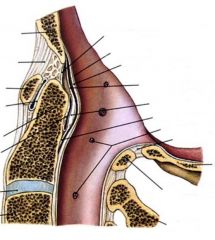
ribs- 1, 11 and 12: single head articulation
Ribs 11 and 12: no TP articulation 19 = lateral costotransverse lig 17 = costotransverse lig 12 = joint capsule 11 = head of rib 10 = costovertebral articulation 13 = radiate lig of the head of the rib 16 = costotransverse joint |
|
|
|
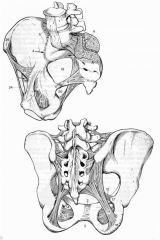
1-symphysis
2-superior pubic ligament 3-inferior pubic ligament 4-articulation of auricular surface of hip bone and sacrum 5-interosseous ligament 6- dorsal lig 7- sacroiliac ligament 8- iliolumbar ligament 11-sacrotuberous lig 12- sacrospinous lig 13-obturator membrane 14-obturator canal 19- greater sciatic foramen 20- lesser sciatic foramen 24-inguinal ligament 25-ASIS 26- pubic tubercle (Page 87 atlas) |
|
|
|
Axis of rotation
|
based on plane of articulation
*more horizontal=more rotation |
|
|
|
Coupled motion
|
in flexion demonstrate translation and rotation
|
|
|
|
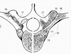
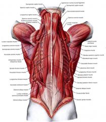
Vertebral processes for muscle attachments
|
spinous process (20)
*transverse process (29) *accessory process: lumbar (30) on the TP, near the pedicle on the posterior surface mm attachment for Intertransversarii dorsales *mammillary process: lumbar (31) on the posterior edge of the Superior Articular Process *articular process superior articular process (SAP) inferior articular process (IAP) *pars interarticularis the part of the lamina between the SAP and the IAP |
|
|
|
epaxial are ______musculature and the hypaxial are _____
|
intrinsic
extrinsic |
|
|
|
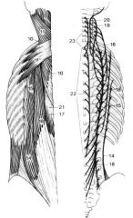
epaxial muscles
|
14-16:Iliocostalis - -M&D pg 538
=lumborum, thoracicus, cervicis, 17-20:Longisimus -M&D pg 538 =lumborum, thoracicus, cervicis, capitis 21-24:Spinalis -M&D pg 538 =thoracicus, cervicis, capitis 9:Superior posterior serrate (not epaxial) Innervated by T2-5 intercostal nerve 10:Splenius cervicis-M&D pg 537 11:Splenius capitis -M&D pg 537 |
|
|
|
|
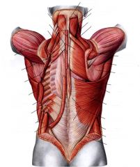
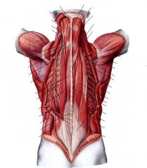
Epaxial muscles-superficial on right, intermediate on left: know Origin, insertion, action and innervations
|
|
|
|
|
|
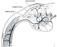
Epaxial (intrinsic) muscle innervation?
|
Dorsal rami of spinal nerves
|
|
|

Vertebral facets are innervated by?
|
articular branch
|
|
|
|
Sub occipital triangle
|
- the gap between inferior and superior oblique muscles and the major posterior rectus muscle of the head.
|
|
|
|
pre-vertebral muscles
|
Rectus capitis anterior
*flexion *branches from loop between C1-2 Longus capitis *flexion *anterior rami of C3-6 to occiput Longus colli *flexion/rotation *anterior rami of C3-6 to T3 |
|
|
|
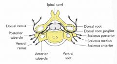
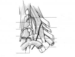
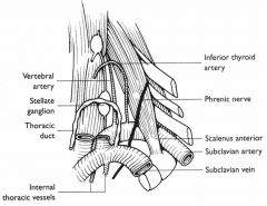
scalene muscles & Origin & Insertion
|
Anterior
*anterior tubercles of C4-6 to 1st rib *cervical spinal nerves C4-6 Middle *posterior tubercles of TP of C4-6 *superior surface of 1st rib Posterior *posterior tubercles of TP of C4-6 *external border of 2nd rib |
|
|
|
|
|
|
|
|
|
|
|
muscle attachments of sternum
|
sternohyoid
sternocleidomastoideus rectus abdominis pectoralis major |
|
|
|
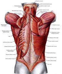
dorsal lumbar muscles
|
Lumbosacral fascia
Iliocostalis lumborum Multifidus lumborum Rotatores lumborum Intertransversarii lumborum Semispinalis in the lumbar |
|
|
|
Thoracolumbar fascia:
gives rise to |
Post serratus inferior
Latisimus dorsi Glut max |
|